nueva página del texto (beta)
 Inglés (pdf)
 Artículo en XML
 Referencias del artículo
 Enviar artículo por email
 Citado por SciELO
  Similares en
SciELO
Similares en
SciELO 
 Permalink
Introduction
In Yucatán, México, ancient Mayan knowledge has provided remedies for the treatment of diverse ailments, especially among rural populations. Inflammation is a response induced by pathogens, damaged tissue or autoimmune and inflammatory diseases. Physiologically, inflammation operates in a restricted environment to erradicate infected or devitalized tissue. Although usually beneficial to the host, inflammation is intrinsically destructive to the immediate surroundings (Ahmed et al., 1995). Rheumatism is a term used to describe the inflammation or degeneration of the connective tissues of the body, and this condition affects primarily the muscles, joints and tendons, among other tissues. Rheumatism can also refer to the autoimmune disease that leads to the deformity of muscles or joints (joint destruction) (Zuther, 2009). Severalmolecules are involved in the inflammatory response, including prostaglandins and nitric oxide (NO) (Gallin and Snyderman1999; Koeberle and Werz 2015). NO production by activated macrophages has been shown to mediate immune functions, including antimicrobial and antitumor activities. However, excess NO production has been implicated in inflammatory processes, septic shock, asthma and autoimmune diseases such rheumatoid arthritis (Mac Mickinget al., 1995; Jerzynskaet al., 2014). Therefore, the inhibition of NO production has become a therapeutic target for the treatment of inflammatory diseases (Azadmehr, 2009; Wen-Jun. et al., 2015).
Inflammation affects many people worldwide, and homemade remedies prepared from single plants or mixtures of plants are used in several cultures to treat these symptoms. Two common plants used in the Yucatán Peninsula to treat inflammation and rheumatism are Critoniaaromatisans(DC.) R.M. King & H. Rob. and Montanoa grandiflora DC., members of the Asteraceae family. These plants are commonly used alone, together or in different plant mixtures to prepare anti-inflammatory and anti-arthritis remedies. C. aromatisans(formerly Eupatorium hemipteropodum) is known in Mexico as “Chiople” and in Cuba as “Trébol de olor”. Mayan people traditionally apply the leaves to the painful area; for example, the leaves are applied to the forehead to treat migraines. The leaves are also used to prepare decoctions to treat oedema and can be used as a poultice or in ointments elaborated with petrolatum (Méndez-González et al., 2012). The production of such ointments involves pulverizing the leaves and boiling them with petrolatum. The mixture is filtered and placed in containers. Then, it is ready touse topically on the affected area of the body. The leaves have also been used to aromatize tobacco (Roig, 1974). This species has never been chemically or biologically evaluated. Montanoa grandiflora (Eurycomahartwegiana) is known as the “daisy tree,” and its flowers are used ornamentally. This plant possesses a resinous fragrance, and in traditional Mayan medicine, the leaves are used to treat rheumatism (alone or as a poultice) and stomach aches (Méndez-González et al., 2012). There are no reports on the biological properties of this plant. Therefore, the aim of this investigation was to evaluate the anti-inflammatory activities of the polar (methanolic/acetonitrile) and non-polar (hexanic) extracts of these two plants that are traditionally used to alleviate symptoms of inflammation.
Methods
Abbreviations
NO (nitric oxide), CA (Critoniaaromatisans), MG (Montanoa grandiflora), MeOH (methanol), acn (acetonitrile), hx (hexane), DMSO (dimethyl sulfoxide), TPA (12-O-tetradecanoylphorbol-13-acetate), DMEM (Dulbecco’s Modified Eagle Medium), MTT ( 3-(4, 5-dimethylthiazol-2-yl)-2,5-diphenyl tetrazolium bromide), CAacn (MeOH/acn extract from C. aromatisans), cahx (hx extract from C. aromatisans), MGacn (MeOH/ acn extract from M. grandiflora), MGhx (hexanic extract from M. grandiflora).
Plant material
Leaves from C. aromatisans(871.2 g) and M. grandiflora (127.3 g) were collected from the botanical garden of the Traditional Medical Centre of Yaxcabá, Yucatán, México. At1 km N of the road to “Libre Unión” town. Both species were collected in June 2010 and were identified by taxonomist professor Mendez-González. A voucher specimen of each plant was deposited at the Herbarium “Roger Orellana” of the Centro de InvestigaciónCientífica de Yucatán (CICY) under the collection numbers A. Dorantes 134 (C. aromatisans) and M. Méndez 1754 (M. grandiflora).
Preparation of the extracts
Powdered leaves of the plants were extracted with methanol (MeOH) at room temperature (three times) to obtain the C. aromatisans(243 g) and M. grandiflora (21 g) MeOH extracts. A portion of each methanolic extract (1 g) was dissolved in MeOH, and then acetonitrile (acn) was added (1:3). This mixture was extracted with hexane, and then, both phases were separated to obtain the non-polar hexanic (CAhx and MGhx) and polar MeOH/acn fractions (CAacn and MGacn).
In vitro assays
Isolation and cell culture of peritoneal macrophages
Macrophages were isolated from the peritoneal cavities of male ICR mice. Briefly, macrophages were harvested immediately by lavage with 10 mL of cold sterile PBS injected into the peritoneal cavity of each mouse, and the resultant exudates were immediately collected, washed two times with PBS, re-suspended and adjusted to 1x10⁶ cells/mL in DMEM containing 10% inactivated foetal bovine serum, 100 U/mL penicillin and 100 μL streptomycin (referred to as complete DMEM). Fifty-microliter aliquots of the cell suspension (5 x 10⁴ cells/well) were cultured in 96-well plates with 200 μL of complete DMEM for 24 h at 37°C in a humidified 5% CO2 incubator. Non-adherent cells were removed by gentle washing with DMEM, and then, freshly prepared medium containing the plant extracts was added.
Treatment of peritoneal macrophages
Purified peritoneal macrophages (5×10⁴ cells/well) were incubated with the plant extracts at different concentrations (1, 10, 100, 500 and 1000 μg/mL). A control group treated with DMEM was included. The plates were maintained at 37oC in a humidified 5% CO2 incubator. After incubation, the supernatants were removed, and LPS (1 μg/mL) was added to the culture. The supernatants were collected after 72 h of LPS treatment for nitrite determination.
Measurement of nitrite production
Nitrite production, an indicator of NO synthesis, was determined by the Griess reaction (Green, 1982). Briefly, 50 μL of supernatant from each well of the 96-well plates were incubated with an equal volume of Griess reagent (1% sulphanilamide/0.1% naphthalene diamine dihydrochloride/2.5% H3PO4) for 10 min at room temperature to quantify the level of nitrite (Ding, 1988). The absorbance was determined at 550 nm using a microplate reader. The relationship between the absorbance and the concentration of NO was determined using dilutions of sodium nitrite in culture medium with known concentrations (0-100 μM) as standards.
Determination of cell viability
Cell viability was assessed using the mitochondrial-dependent reduction of 3-(4,5-dimethylthiazol-2-yl)-2,5-diphenyl tetrazolium bromide (MTT) to purple formazan. Cells were obtained as described previously and were plated in a 96-well culture dish at a concentration of 5×10⁴ cells/well in complete DMEM. After an overnight incubation, the cells were washed with DMEM, and subsequently, various concentrations of the tested extracts at 1, 10, 100, 500 and 1000 μg/mL were added in a total volume of 200 μL of medium. The cells were then cultured for 24 h at 37oC in a humidified 5% CO2 incubator. The supernatants were removed, and the cells were incubated with MTT (5 mg/mL in PBS) for 4 h at 37oC and 5% CO2. After this incubation, the formazan crystals formed were dissolved in DMSO. The extent of the reduction of MTT was quantitated by measuring the absorbance at 550 nm using a microplate reader. The proliferation percentage was calculated using the following equation:
In vivo assays
Animals
Male and female Balb/C mice (20-25 g) were purchased from the Animal Breeding laboratories of CIBO-IMSS (Guadalajara, Jalisco, Mexico). ICR mice (20-25 g) were provided from the Animal House of Centro de InvestigacionesRegionales “Dr. HideyoNoguchi”-UADY (Mérida, Yucatán, México). The animals were provided a standard pellet diet and water ad libitum. Five animals were used in each group. The animals were processed according to the ethical guidelines for Mexican procedures (Norma oficial Mexicana NOM-062-ZOO, 1999).
Preparation of test samples
Mice were weighed and then treated per os with 200 mg/kg of extract previously dissolved in a solution of 5% Tween 80 in water. The control group received 100 μL of 5% Tween 80 in pure water and received the same experimental handling as the test groups. The anti-inflammatory drug indomethacin (10 mg/kg) was used as a positive control (Boughton-Smith, 1993).
Carrageenan-induced hind paw oedema in Balb/c mice
The in vivo anti-inflammatory activity was evaluated using the carrageenan-induced hind paw oedema model with slight modifications (Winter, 1962). Briefly, four experimental groups and two control groups (positive and negative) were formed. Five Balb/c mice were included in each group and 30 min after the oral administration (200 mg/kg) of the test sample or vehicle, a suspension of carrageenan (Sigma, St. Louis, MO, USA) at 1% (0.25 μL/ paw) in sterile physiological saline solution (0.9%) was injected into the subplantar tissue of the right hind paw of each mouse. The thickness of the hind paw was measured before oral administration and 3 h after injection with carrageenan. The difference in footpad thickness was measured using a precision micrometer. The mean values of treated groups were compared with the mean values of the untreated control group (negative control), and the percentage inhibition of inflammation was calculated.
TPA-induced mouse ear oedema
Four experimental groups and two control groups (positive and negative) were formed, with five Balb/c mice in each group. Briefly, 2.5 μg of TPA (12-O-tetradecanoylphorbol-13-acetate) dissolved in 20 μL of acetone was applied to each side of the right ear of each mouse in the experimental groups. This thickness of the ear was measured before oral administration and 3 h after the TPA was applied. The difference in ear thickness was measured using a precision micrometer. The mean values of the treated groups were compared with the mean values of the untreated control group, and the percentage inhibition of inflammation was calculated.
Acute oral toxicity
Acute oral toxicity was assessed in a shortterm observation period of 72 h over morbidity and mortality of the animals (OECD, 2001). The mice were treated with a single dose of 200 mg/kg of the polar and nonpolar extracts of C. aromatisansand M. grandiflora. The mice were observed for 4 h after the administration of the samples, paying special attention to the changes and alterations in behaviour and appearance of the fur. The observations were continued for 1 h each day. The following indicators of toxicity were assessed and recorded: breathing, pruritus, lethargy, appearance of the fur, excessive consumption of water, changes in eyes and mucous membranes, walking alterations, position, manipulation, drowsiness, self-cleanliness, convulsions, salivation, diarrhoea, auto mutilation, coma and death.
Gc-ms Analysis
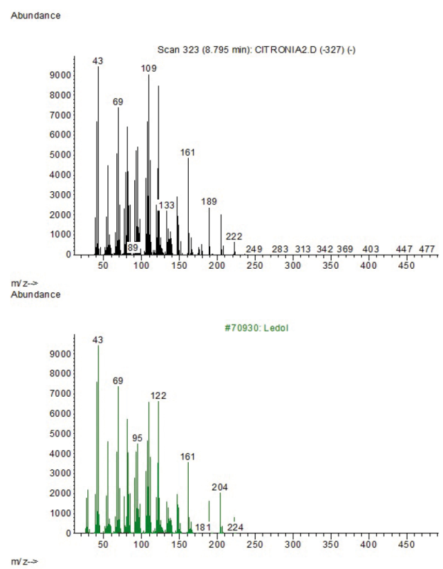
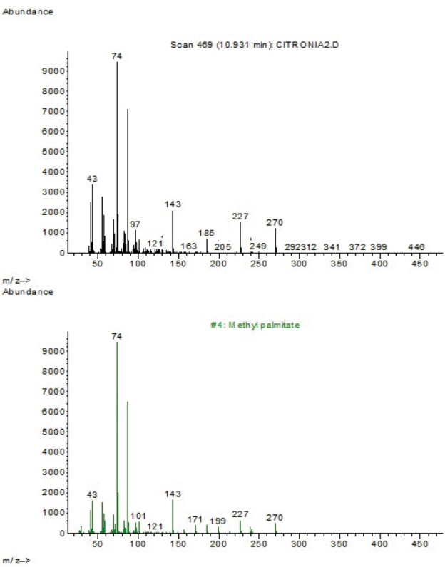
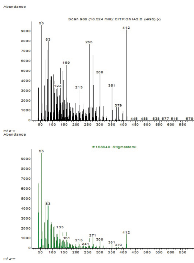
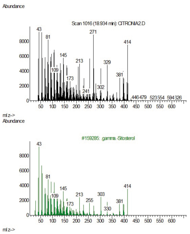
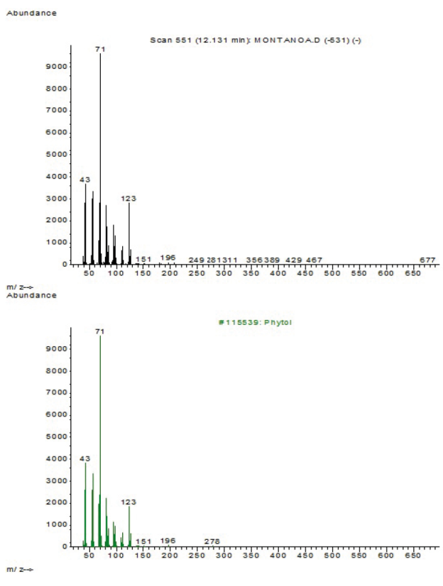
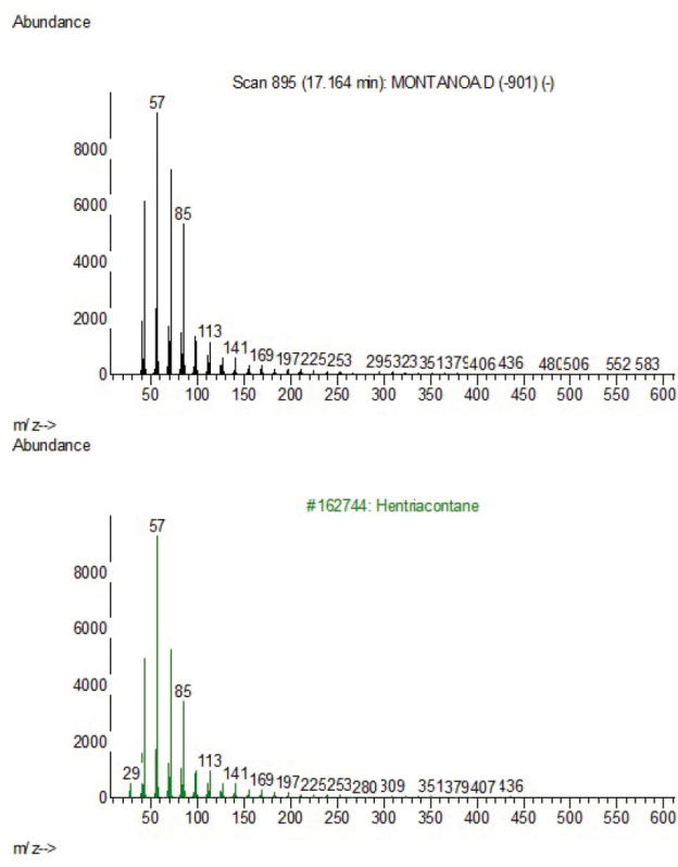
Preliminary analyses of the n-hexanic extracts were performed by gas chromatography/mass spectrometry with an Agilent technologies 6890N system equipped with an HP-5 MS capillary column (30 m × 25 mm x 0.25 μm). Helium was used as the carrier gas; the injection volume was one microliter, and the temperature program used was as follows: 50°C (3 min); an initial temperature of 80°C (5 min.); an increase to 215°C at 4°C/min; and a hold at 215°C for 5 min. The identification of the components was based on the comparison of their mass spectra fragmentation with those reported in the NIST05 compounds library.
Statistical analysis
For cell viability, experimental values are expressed as the mean ± standard deviation of at least three experiments performed in triplicate. The data were analysed using one-way ANOVA with Dunnett’s multiple comparisons test. For in vivo pharmacological evaluations, experiments were performed with five animals per group. For in vivo and in vitro tests, experimental values are expressed as the mean ± standard error of the mean (S.E.M.). Statistical analysis was performed by one-way analysis of variance (ANOVA) with the Bonferroni post hoc test using PRISMA (GraphPad Software, Inc., San Diego, CA, version 5.01). In both cases, p ≤ 0.05 was used as the criterion for statistical significance (sample vs. control).
Results
In vitro effects of C. aromatisansand m. grandiflora on NO production by mouse peritoneal macrophages
In the present study, it was observed that the extracts of C. aromatisansand M. grandiflora inhibited NO production in a concentration-dependent manner (fig. 1). The highest activity was found for the nonpolar extract of C. aromatisans(cahx). This extract almost completely inhibited NO production (97.58% inhibition) by peritoneal macrophages at the concentrations of 500 and 1000 μg/mL. No effects on the viability of peritoneal macrophages exposed to different concentrations of extracts were observed, indicating the potential specificity of the extracts forNO production by macrophages
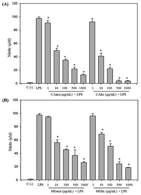
Fig. 1 Effect of CAacn, CAhx (Fig. 1A) and MAacn, MGhx (Fig. 1B) on NO production after 72 h of activated mouse peritoneal macrophages. Control (-) represent non activated peritoneal macrophages. LPS represents activated peritoneal macrophages with LPS without extracts treatments. NO production by LPS activated peritoneal macrophages was significantly (*p < 0.05) inhibited when different concentration (1, 10, 100, 500, 1000 μg/mL) of extracts were added. Data represent mean ± S.D. values from the three independent experiments.
Cell viability
Cell viability was evaluated using the mitochondrial-dependent reduction of 3-(4,5-dimethylthiazol-2-yl)-2,5-diphenyl tetrazolium bromide (MTT) to purple formazan. It was showed that the treatments with 1, 10, 100, 500 and 1000 μg/mL with the extracts did not alter the viability of mice peritoneal macrophages.
In vivo anti-inflammatory effects
In the carrageenan-induced hind paw oedema model (table 1), the non-polar (CAhx) and polar extracts (CAacn) of C. aromatisans at the dose of 200 mg/kg resulted in the maximum inhibition (92 and 96%, respectively) of paw oedema at 3 h after carrageenan injection. Similar results were obtained for the TPA-Induced mouse ear oedema model (table 2), in which both extracts of C. aromatisansshowed anti-inflammatory effects of up to 80% at the same dose. these effects were similar in both cases to the effects of the anti-inflammatory drug indomethacin. In contrast, the extracts obtained from M. grandiflora showed lower values of reduction of inflammation in the carrageenan-induced hind paw oedema model (table 2). In the TPA-induced mouse ear oedema model, no anti-inflammatory effect was observed for the non-polar or polar extract of M. grandiflora at the dose of 200 mg/kg.
Table 1 Effects of the CAacn, CAhx, MGacn and MGhx extracts on carrageenan-induced hind paw oedema in mice.
| Group | ||||||
|---|---|---|---|---|---|---|
| Dose (200 mg/kg, p.o.) | ||||||
| Control | Indomethacin 10 mg/kg | CAacn | CAhx | MGacn | MGhx | |
| Hind paw oedema at 3 h (x10-2 mm) ± S.D. | 44 ± 6.2 | 2.4 ± 1.6 | 1.8 ±4.7* | 3.6 ± 2.7* | 33.9 ± 2.4 | 23.3 ± 14* |
| Inflammation (%) | 100 | 4.5 | 3.4 | 6.7 | 76.3 | 41.6 |
Note: Values are expressed as the mean ± S.D. Statistically significant difference relative to the control group: *p<0.05 after one-way analysis of variance (ANOVA) with Bonferroni post hoc test. Untreated cells were used as a control in this evaluation.
Table 2 Effects of the CAacn, CAhx, MGacn and MGhx extracts on TPA-induced ear oedema in mice.
| Group | ||||||
|---|---|---|---|---|---|---|
| Dose (200 mg/kg, p.o.) | ||||||
| Control | Indomethacin 10 mg/kg | CAacn | CAhx | MGacn | MGhx | |
| Ear oedema at 3 h (x10-2 mm) ± S.D. | 6.1 ± 2.2 | 2 ± 1.0 | 2 ± 1.0 | 2.4 ± 0.5* | 7.2 ± 2.8 | 6.4 ± 2.6 |
| Inflammation (%) | 100 | 32.7 | 6.5 | 19.7 | 118 | 104.9 |
Note: Values are expressed as the mean ± S.D. Statistically significant difference relative to the control group: *p < 0.05 after one-way analysis of variance (ANOVA) with Bonferroni post hoc test. Untreated cells were used as a control in this evaluation.
Acute toxicity
In the acute toxicity test, the four groups treated with the extracts of C. aromatisansand M. grandiflora showed no signs of adverse effects, abnormal behaviour, toxicity or lethality at the dose of 200 mg/kg 72 h after administration.
GC-MS
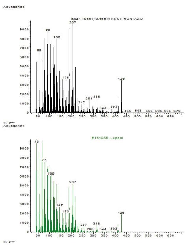
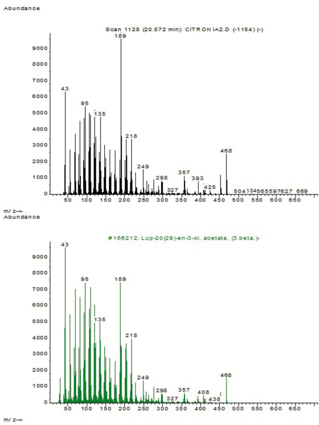
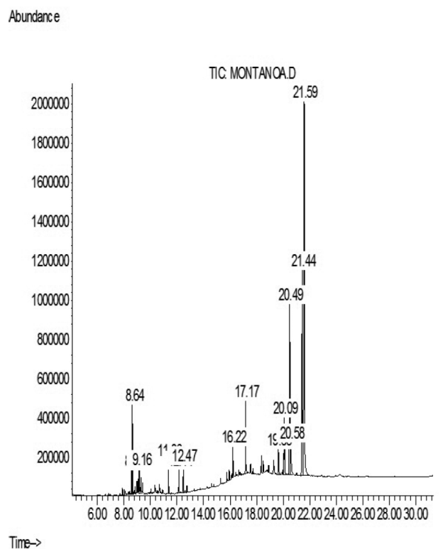
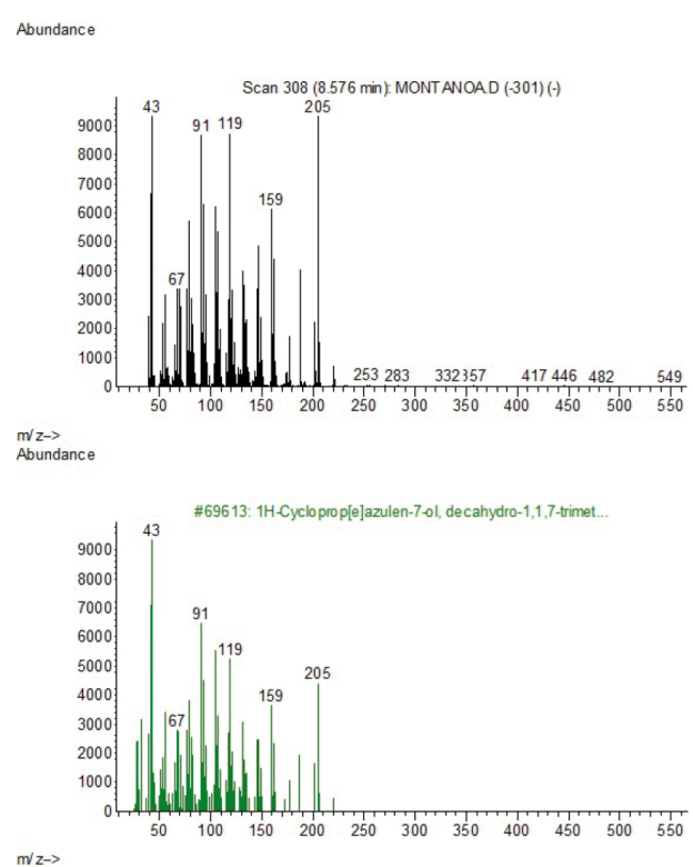
Gas chromatography-mass spectrometry analysis of the non-polar extracts of C. aromatisansand M. grandiflora showed that they were primarily composed of fatty acids and terpenoids (Figure S1-S22); C. aromatisans registered four major compounds at retention times (RT) of 7.51, 10.01, 10.94 and 20.57 min (Figure S1 in supplementary material) which included 2H-1-benzopyran-2-one, isolongifolen-5-one, methyl palmitate and Lup-20 (29)-en-3-ol, acetate, respectively (figure S2, S5, S7, S11). Other minor components were identified as ledol, 2-pentadecanone, stigmasterol, lupeol and β-sitosterol. M. grandiflora contained six main compounds at rt of 8.64, 17.17, 20.09, 20.49, 21.44 and 21.59 min (figures S14, S17-S19, S21,S22 in supplementary material) with mass fragmentation pattern similar to caryophyllene oxide, hentriacontane, 12-Oleanen-3-yl acetate, Lup-20 (29)-en-3-ol, acetate. Compounds with retention times at 21.44 and 21.58 min were not found in the NIST library (figures S21 and S22).
Discussion
Different models were used to evaluate the anti-inflammatory effects of C. aromatisansand M. grandiflora. the in vitro experiments were performed using the nitric oxide test. It is well known that during an inflammatory process, after signalling events, macrophages induce the expression of pro-inflammatory enzymes such as inducible nitric oxide synthase (iNOS). This enzyme is regulated by the secretion of pro-inflammatory cytokines and produces NO from L-arginine. Large amounts of NO can cause severe collateral damage in the tissue because NO can form peroxynitrite (ONOO-), a cytotoxic molecule related to inflammatory pathologies. For this reason, the regulation of the production of NO is an important target for inflammatory diseases (Guzik et al., 2003).
The in vivo anti-inflammatory effects of the extracts were measured using the carrageenan-induced hind paw oedema and TPA-induced ear oedema models. These two models are well-known indicators of acute inflammation, and both evaluations can be performed at the same time because they use the same animals and the same treatment doses. In addition, both models allow the rapid evaluation of the effect of an extract or compound with potential anti-inflammatory properties. When the polysaccharide carrageenan is injected into the hind paw, it produces oedema mediated by white blood cells (neutrophils, monocytes and lymphocytes), major inflammatory mediators such as NO, prostaglandin E2 (PGE2) and the cytokines TNF-α and IL-6 (Kim, et al., 2012). TPA is a topical oedematogenic agent and causes skin irritation and tumour promotion, because it stimulates PKC, which plays a critical role in signal transduction pathways and regulates cell growth and differentiation (Nishizuka, 1992). TPA affects the membrane, resulting in altered cell adhesion and the induction of arachidonic acid metabolism, specifically causing the release of prostaglandins, which are responsible for oedema (Weinstein et al., 1979). The results obtained in the present work, suggest that the different groups of anti-inflammatory metabolites of C. aromatisans are distributed between both polarities. Also, these results could indicate that the possible anti-inflammatory mechanism of MGhx (which showed an antiinflammatory effect only in the carrageenan model) does not involve the PKC pathway. Further in vitro experiments can elucidate the specific anti-inflammatory mechanism of MGhx. A literature search for C. aromatisans did not found any previous chemical study. A previous study of M. grandiflora revealed the presence of limonene, citronella, camphene, pinene and phellandrene in essential oils prepared from the inflorescences, stems and leaves (Pérez-Amador et al., 2006). The compound 6β-hydroxy-germacradien-8,12-olide (6-epi-desacetyllaurenobiolide) was obtained from the chloroform extract of M. grandiflora (Quijano et al., 1984). Different species of the Asteraceae family, including species of the genus Eupatorium, contain terpenoids, such as stigmasterol and taraxeryl acetate. Stigmasterol is able to reduce pro-inflammatory mediators such as NO and PGE2 in RAW 264.7 murine macrophages in vitro (Pandith et al., 2013). Taraxeryl acetate reduces inflammation in in vivo models such as hind paw oedema induced by carrageenan and mouse ear oedema induced by TPA (Pérez-García et al., 2005). In this preliminary evaluation of the non-polar extract of C. aromatisans, stigmasterol was found, but taraxeryl acetate was not. Stigmasterol has a known antiinflammatory effect, and other components of the non-polar extract may contribute to this effect in a synergistic manner. In the in vitro evaluation, it was observed that the extracts inhibited NO production in a concentration-dependent manner, and this activity might be responsible for their in vivo anti-inflammatory properties. However, considering that there exist different mediators that can be inhibited to produce the anti-inflammatory effect, further studies are necessary. The presence of terpenoids and fatty acids in the non-polar extracts could validate the use of this species in traditional medicine and the use of petrolatum to prepare the ointments. Because of its nonpolar composition, petrolatum would retain the non-polar compounds and essential oils from the leaves, and then it can be administered topically. Due to these interesting results, the isolation and characterisation of the bioactive compounds in each plant are recommended. Based on the present work, it can be concluded that the non-polar and polar extracts of C. aromatisans and the non-polar extract of M. grandiflora possess in vitro and in vivo anti-inflammatory activities, and both species seem to be useful to treat inflammatory symptoms.
Conclusion
The results obtained in the present study show fundaments to validate the traditional uses by Mayan communities that endorse the use of this species to treat inflammation. Additionally, C. aromatisans has shown a stronger in vivo and in vitro activity, and one of the possible mechanisms of the antiinflammatory response is the inhibition of the NO production.