


Servicios Personalizados
Revista
Articulo
 Inglés (pdf)
 Artículo en XML
 Referencias del artículo
 Enviar artículo por email
Indicadores
-
 Citado por SciELO
 -
 Accesos
Links relacionados
-
 Similares en
SciELO
Similares en
SciELO
Compartir

 Permalink
Polibotánica
versión impresa ISSN 1405-2768
Polibotánica no.31 México mar. 2011
Morphoanatomical characterization and antimicrobial activity of Tillandsia imperialis (bromeliaceae)
Caracterización morfoanatómica y actividad antimicrobiana de Tillandsia imperialis (Bromeliaceae)
J. Alejandro Vite-Posadas1, Alicia E. Brechú-Franco1, Guillermo Laguna-Hernández1, M. Gabriela Rojas-Bribiesca2 and H. Reyna Osuna-Fernández1
1 Laboratorio de Estructura y Fisiología de Plantas, Facultad de Ciencias, Universidad Nacional Autónoma de México, Ciudad de México;
2 Laboratorio de Microbiología, Centro de Investigación Biomédica del Sur (CIBIS), IMSS, Xochitepec, Morelos, México.
Recibido: 16 noviembre 2009.
Aceptado: 16 diciembre 2010.
Abstract
Tillandsia imperialis C.J. Morren ex Roezl is a bromeliad used in Mexican folk medicine mixed with other plant and animal parts as a good remedy for respiratory diseases, which are one of the main causes of morbility and mortality in the rural population of Mexico. The commercialization of medicinal plants for therapeutic use must comply with high standards for quality, safety and efficacy; structural, microbiological and pharmaceutical studies make a decisive contribution to this quality control. The aim of this study was to analyze the morpho-anatomical characteristics and histochemistry of bracts and leaves of Tillandsia imperialis and evaluate their antimicrobial activity to corroborate the effectiveness of this plant for traditional uses with experimental data. Crude extracts from the inflorescences and leaves were evaluated on Staphylococcus aureus (ATCC 29213), Streptococcus pyogenes (ATCC 08668), Escherichia coli (ATCC 25922), Streptococcus faecalis (ATCC 29212), Salmonella Typhi (ATCC 06539), Klebsiella pneumoniae (ATCC 13883) and Candida albicans (ATCC 10231). Results showed that T. imperialis possesses important morpho-anatomical characteristics useful for identification, including mesic and peltate trichomes, tetracyclic stomata and non-sclerotic hypodermis with spherical silica bodies. Microbiological research revealed specific antimicrobial activity against respiratory pathogenic microorganisms such as Staphylococcus aureus and Streptococcus pyogenes as well as six other bacteria. These results indicate the existence of antimicrobial compounds in the extracts and a correlation between the traditional uses of this plant and the experimental data.
Key words: antimicrobial activity, medicinal plants, structural and histochemical characteristics, Tillandsia imperialis.
Resumen
Tillandsia imperialis (Bromeliaceae) es una planta utilizada en la medicina tradicional mexicana para tratar ciertas afecciones respiratorias, las cuales son una de las principales causas de morbilidad y mortalidad en la población de nuestro país. No obstante, no se han realizado investigaciones que corroboren su uso en la medicina tradicional. Se realizó un estudio estructural e histoquímico de las brácteas y hojas de esta especie para aportar información útil para su correcta determinación farmacognóstica y se evaluó la actividad antimicrobiana de los extractos crudos obtenidos de las inflorescencias y hojas para corroborar su uso en la medicina tradicional. La actividad antimicrobiana de los extractos crudos se evaluó sobre Staphylococcus aureus (ATCC 29213), Streptococcus pyogenes (ATCC 08668), Escherichia coli (ATCC 25922), Streptococcus faecalis (ATCC 29212), Salmonella Typhi (ATCC 06539), Klebsiella pneumoniae (ATCC 13883) y Candida albicans (ATCC 10231). Las características morfoanatómicas útiles que se detectaron en T. imperialis son tricomas mésicos, peltados, estomas tetracíclicos e hipodermis no esclerótica con cuerpos esféricos de sílice. El estudio antimicrobiano reveló actividad contra microorganismos patógenos de vías respiratorias como Staphylococcus aureus y Streptococcus pyogenes lo cual corrobora y valida su uso en la medicina tradicional de México.
Palabras clave: actividad antimicrobiana, estructura e histoquímica, planta medicinal, Tillandsia imperialis.
NTRODUCTION
For many people around the world, traditional medicine is still used and it is an important medicinal resource (McGaw et al., 2000). Medicinal plants used in the indigenous cultures from developing countries are numerous and diverse (Nigenda et al., 2001). The traditional medicine in Mexico plays an important role in medical practice where approximately 40 million of people employ traditional remedies (García, 2002) which include a rich variety of medicinal plants. Some of them are used for the treatment of respiratory infections, one of the first causes of hospitalization in Mexico and responsible for fifty percent of deaths in developing countries (Salud Pública Méx., 2004; WHO, 1998). Phytotherapy as a part of Therapeutics requires the rational development of herbal medicinal products with guaranteed quality, safety and efficacy, along with accurate and assessable information as well as appropriate education in this field (Cañigueral, 2006). The confirmation of the authenticity of the botanical samples requires pharmacognostic studies. Few scientific investigations have analyzed the leaf, stem or root anatomy, which is useful to guarantee the authenticity of medicinal botanical materials and in turn contributes to their identification and characterization for the quality control of the products derived from these plants (Arambarri & Mandrile, 1999). It is necessary to make botanical tests for plant materials from markets to avoid the falsification or substitution in the use of any medicinal botanical material. Data acquired in scientific experimentation should guarantee and provide approaches for quality control, as recommended by the World Health Organization and the Mexican Health system.
In spite of the diversity of medicinal herbs, the scientific knowledge available is very limited as in the case of bromeliads. This group exhibits great diversity of habitats and represents the third most important botanical family of Mexican monocotyledons (Pulido-Esparza et al., 2004). Bromeliads have been used since ancient times and are currently being used by Mexicans as food, forage, ornamental and medicinal plants. Tillandsia imperialis Morren ex Roezl (Bromeliaceae) is a tank epiphytic bromeliad found in tropical mountane cloud forest, distributed throughout southeastern Mexico, Salvador and Honduras. In Mexico is known locally as "tencho", and has been widely exploited in florist trade. Infusions made from the leaves of T. imperialis are used in Mexican traditional medicine to treat respiratory ailments along with other plants including Gardenia jasminoides, Physalis pubescens, Juglans regia, Hibiscus rosa-sinensis, and Rosa gallica as well as Dasypus novemcinctus (an armadillo), (Chino & Jácquez, 1986).
Scientific studies supporting the medicinal use of this plant and verifing its pharmacological effectiveness are lacking. The aim of our work was to analyze comparative morpho-anatomical characteristics of this bromeliad and evaluate its biological activity, so corroborate the traditional uses of this plant with experimental data.
MATERIAL AND METHODS
Plant Material
Tillandsia imperialis C.J. Morren ex Roezl plants were bought within local markets of Mexico City between February and April 2004. The voucher specimen was deposited inside the herbarium of Universidad Autónoma Metropolitana Iztapalapa in Mexico (UAM-IZ), with the accession number 55702, (botanical identification) by Ph.D. Adolfo Espejo (UAM-IZ).
Color Distribution and Foliar Pigmentation
The color of bracts and leaves were compared in vivo with the help of a standard color handbook (Kornerup & Wanscher, 1963). Lamina and sheath leaf pigmentation was examined on foliar adaxial face as much as foliar abaxial face. For detailed analysis, each plant part (bract and leaf) was divided in three regions: apical, middle and basal.
Pigmentation type was determined using the Benzing and Friedman's protocol (1981).
Anatomical Characterization
Leaf paradermal and longitudinal fresh sections of T. imperialis were prepared by hand with single-edge razor blades. Other samples were fixed in FAA (Formalin, acetic acid, ethanol 100-50-500 mL in 1L), washed and dehydrated in increasing concentrations of ethanol solution (30°-50°-70°-96°-100°) and included in Paraplast MR. Sections were processed with the Johansens method (1940), and histochemical studies were carried out on some samples and were observed in a light microscope (Axiostar plus Carl Zeiss).
Preparation of Extracts
Inflorescences and leaves were dried at ambient temperature and powdered. Ten grams of the powder were extracted three times 8h each one sequentially with organic solvents: hexane, dichloromethane and methanol (J.K. Baker) using a Soxhlet apparatus. The crude extracts were then evaporated to dryness under vacuum conditions and the percentage yield for each extract was determined.
Microorganisms Evaluation
Test Microorganisms
Six standard microorganisms strains were used for testing antimicrobial activity: Staphylococcus aureus (ATCC 29213), Staphylococcus aureus OPS (clinical isolate; identified and obtained from the Laboratorio de Microbiología, Centro de Investigaciones Biomédicas del Sur, IMSS), Staphylococcus aureus AHD (clinical isolate), Streptococcus pyogenes (ATCC 08668), Escherichia coli (ATCC 25922), Streptococcusfaecalis (ATCC 29212), Salmonella Typhi (ATCC 06539), Klebsiella pneumoniae (ATCC 13883) y Candida albicans (ATCC 10231). All bacteria were maintained on Tryptic Soy agar (TSA Merck) at 37°C. Defibri-nated sheep blood (5%) was added to the medium for Streptococcus pyogenes. Yeast was maintained on Sabouraud 4% dextrose agar (SDA Merck).
Antimicrobial Assays
Antibacterial and antifungal activities were tested by two-fold dilution method (Rojas et al., 2001). Extracts were dissolved in 5% dimethylsulfoxide (DMSO; Merck) and added to melted agar culture medium in Petri dishes at the following final concentrations: 0.5, 1.0, 2.0, 4.0, 8.0 mg/mL. The antimicrobial assay was carried out on Muller-Hilton agar (MHA Merck); in the case of Streptococcus pyogenes sheep blood (5%) was added to Mueller-Hilton medium. Microbial suspensions with 0.5 McFarland standard turbidity equivalents were prepared by suspensions of the growth from Brain Heart Infusion (BHI BBL). Suspensions were further diluted 1:20 to obtain a concentration of 104 colony-forming units (CFU)/mL for the bacteria and yeast. The diluted inoculum was applied with a loop calibrated to deliver 0.002 mL, resulting in a spot covering a circle of 5-8 mm diameter. The plates were incubated for 24 h at 37°C. Gentamicin and Nystatin (5 and 10 Hg/mL respectively) and 5% DMSO were used as reference standards. Observations were performed in duplicate and results expressed as the lowest concentration of plant extract that produced a complete suppression of colony growth, the minimal inhibitory concentration (MIC).
RESULTS
Color and Foliar Pigmentation
Detailed results of bract and leaf pigmentation are shown in Fig 1.

The leaf color is not uniform; it varied considerably depending upon the surface and position along the axis. Reddish color (11E8, 11F8) abundantly dominated in both the adaxial and the abaxial foliar apical faces (Fig. 1A, 1D), green pigments (30D7, 30D8) were majorly found in the leaf's middle region (Fig. 1B. 1E). At the basal region (Fig. 1C, 1F) a pale yellow color was detected near to stem (1A2, 2A2). Cyanic color (10D8, 12E8, 14F5, 15F5) was localized between the middle and basal foliar region. Pigmentation pattern was linear and it was darker in the adaxial face (Fig. 1F). Apical bracts pigments included reddish color (9A8, 9C8) and pale pink (8A3), they were more brilliant at abaxial face (Fig. 1G) than the adaxial face (Fig. 1I). Yellowish white color (1A2) was found at the basal region (Fig. 1H, 1J) similar to the leaf basal region.
Anatomical characterization and histo-chemical test
The leaf presented a single epidermis with peltate trichomes located on both abaxial and adaxial foliar surfaces with a symmetric linear arrangement. The trichomes presented cell rings 4 + 8 + 32 mesic anatomy (Fig. 2A). The epidermal cells of abaxial surface showed straight thick inner periclinal walls whereas outer ones were thinner (Fig. 2B). Only abaxial epidermal cells presented a single siliceous spherical body in each cell (Fig. 2C). Tetracyclic stomata were distributed only in the apical abaxial leaf epidermis (Fig. 2D).
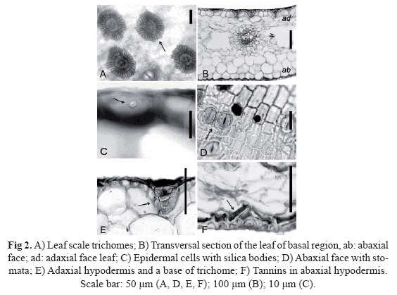
Under the epidermis was found a mono-layer of hypodermal cells (Fig 2E) with colorless thin-walled and spherical cells in the abaxial face; the adaxial hypodermal cells were rectangular or elongated. The hypodermal cells of the basal region of the leaf presented condensed tannin (red-brown colour), but only at the abaxial face (Fig. 2F). This microscopic features are useful for the characterization and botanical description of T. imperialis and will help in the right identification of the commercial herbal drug samples.
Antimicrobial Assays
The results of the antimicrobial activity of T. imperialis extracts are shown in Table 1. The highest yield was obtained with the methanolic extract in both leaves and inflorescence. Both hexane and dichloromethane extracts from inflorescence presented a high inhibitory activity. According to previous antimicrobial studies carried out at the CI-BIS laboratory (IMSS), crude plants extracts with MIC values between 2.5 and 8mg/mL (Navarro et al., 1996; Rojas et al., 2001; Salinas et al., 2009) have led to the isolation of strong antimicrobial compounds. Considering that in this study only crude extracts were employed, extracts with MIC values of 8mg/mL or below were considered active.

All extracts assayed (save the dichlorometh-ane extract from the leaves) exhibited antimicrobial properties against at least one of the tested microorganisms at concentrations of 8 mg/mL or below. None of the extracts showed antimicrobial activity against Candida albicans and Escherichia coli. Three extracts inhibited Staphylococcus aureus: the hexanic extract from the leaves (MIC 4mg/mL) and the hexanic and dichloromethanic extracts from inflorescence (MIC 0.5mg/mL) which showed a significant antibacterial activity by inhibiting the growth of bacteria at a relatively low MIC, so compounds of low polarity maybe are the responsible for the inhibitory response. The most active inhibitory value against S. aureus AHD (clinical isolate) was obtained with the hexanic extract from the inflorescence (MIC 8 mg/mL).
Streptococcus faecalis was inhibited by hexanic inflorescence extract (MIC 8mg/mL). Klebsiella pneumoniae and Salmonella typhi were inhibited by methanolic extracts from leaves and inflorescences (MIC 8mg/mL). In general, inflorescence extracts were more active than leaf extracts. These results indicate the existence of antimicrobial compounds in the extracts and show a correlation between the traditional uses of this plant and the experimental data.
DISCUSSION
According to the results, the color pattern of the leaves of T. imperialis is considered an ephemeral cyanic pigmentation (Benz-ing and Friedman, 1981). The cell rings (4 + 8 + 32) of the peltate trichomes with a symmetric linear arrangement, match up with the cell arrange in other genus like Catopsis, Glomeropitcairnia, Guzmania and Vriesea (Tomlinson, 1969). T. imperialis presents important morphoanatomical characteristics, some of which are common to the taxonomic family such as the mesic trichomes, tetracyclic stomata, non-sclerotic hypodermis and spherical silica bodies which are mainly distributed in the abaxial epidermis which contrast with the silica bodies deposited in the hipodermis in T. andicola (Prychid et al., 2004). The number andthe arrangement of the trichomes cells are specific characteristics for T. imperialis which are useful for identification and differentiation from other species. These microscopic characters are important for establishing an appropriate folair anatomical identification of T. imperialis.
The pharmacological research established the specific biological activity of the extracts of T. imperialis against respiratory microorganisms such as Staphylococcus aureus and Streptococcus pyogenes as well as suggested the presence of antimicrobial compounds in the extracts. These results correlate with the traditional uses of this plant. Nevertheless, phytochemical analyses of this species haven't been reported. In other species of bromeliads such as Nidularium and Tillandsia, flavonoids and triterpenoids (Delaporte et al., 2004) have been registered as major chemical components (Delaporte et al., 2004). Cabrera and Seldes, 1997 and Can-tillo-Ciau et al., 2001, identified cycloartane triterpenoids in extracts of T. usneoides and T. fasciculata. Adrián-Romero and Blunden (2001) detected high contents of glycinebetaine in other Tillandsia species, which have been reported to play a role in pathogen-resistance mechanisms/strategies in plants. Possibly this kind of compounds are responsible for the observed microbial inhibition in T. imperiallis, however, experimental research to identify active compounds in this bromeliad, responsible for the microbial inhibition observed, must be carried out, especially in the extracts that exhibited good antibacterial activity such as the ones effective against Staphylococcus aureus AHD (clinical isolate).
CONCLUSION
The microscopic characters which help out on an accurate foliar identification and differentiation for other species of T. imperialis are mesic trichomes, tetracyclic stomata, a non-sclerotic hypodermis, and spherical silica bodies. The pharmacological research reached out to determine the specific biological activity of the extracts of T. imperialis against respiratory microorganisms, demonstrated the existence of antimicrobial compounds in the extracts and show a positive correlation between the traditional uses of this plant and the scientific experimental data.
ACKNOWLEDGMENTS
All the authors are grateful to M.F.P. Ana Isabel Bieler Antolin from Laboratorio de Microcine, Fac. de Ciencias, UNAM. We also thank to Q.A. Veronica Muñoz Ocotero from Laboratorio de Fitoquímica, Facultad de Ciencias, UNAM, M.C. Aurora Zlotnik for her assistant in the anatomical technique. The authors thank Dr. Adolfo Espejo for the taxonomic identification of the plant and to Dr. Robert A. Bye Boetler for the English write revision.
LITERATURE CITED
Adrian-Romero, Maricela., G. Blunden, 2001. "Betaine distribution in the Bromeliaceae". Biochem. syst. ecol., 29: 305-311. [ Links ]
Arambarri, A.M., E.L. Mandrile, 1999. "Tillandsia L. (Bromeliaceae): Anatomy and ethnopharmacology". Acta Hort., 503: 133-140. [ Links ]
Benzing, D.H., W.E. Friedman, 1981. "Patterns of foliar pigmentation in Bromeliaceae and their adaptive significance". Selbyana, 5(3-4): 224-240. [ Links ]
Cabrera, M.G., A.M. Seldes, 1997. "Short side-chain cycloartanes from Tillandsia usneoides. Phytochemistry, 45(5): 1019-1021. [ Links ]
Cantillo-Ciau, Z., W. Brito-Loeza, L. Quijano. 2001. "Triterpenoids from Tillandsia fasciculata". J. Nat.Prod., 64: 953-955. [ Links ]
Cañigueral, S., 2006. "Las monografías de calidad seguridad y eficacia en el uso racional de los preparados a base de plantas medicinales". Fitoterapia 6(S1): 25-29. [ Links ]
Chino, V., R, Jácquez, 1986. "Contribución al conocimiento de la flora medicinal de Quimixtlán, Puebla". Tesis de licenciatura. ENEP-Iztacala, UNAM, Ciudad de México, 344 pp. [ Links ]
Delaporte, R., M. Sarragiotto, O. Takemura, G. Sánchez, B. Filho, C. Nakamur, 2004. "Evaluation on the antioede-matogenic, free radical scavenging and antimicrobial activities of aerial parts of Tillandsia streptocarpa Baker (Bromeliaceae)". J. Ethnopharmacol., 95: 229-233. [ Links ]
García, E.I, 2002. Catálogo de plantas medicinales de un mercado de la ciudad de Puebla. Gobierno del estado de Puebla. México, 211 pp. [ Links ]
Johansen, D.A, 1940. Plant microtechnique. McGraw-Hill, Book Company. Inc. New York and London. 523 pp. [ Links ]
Kornerup, A., J.H. Wanscher, 1963. Methuen Handbook of Colour. Ed. Eyre Methuen. London, 252 pp. [ Links ]
McGaw, L.J., A.K. Jãger, J. van Stauden, 2000. "Antibacterial, antihelmintic and anti-amoebic activity in South African medicinal plants". J. Ethnopharmacol., 72(1, 2): 247-263. [ Links ]
Navarro, V., M. Villarreal., G. Rojas., X. Lozoya. 1996. "Antimicrobial evaluation of some plants used in Mexican traditional medicine for the treatment of infectious disease". J. Ethnopharmacol., 53(3): 143-147. [ Links ]
Nigenda, G., G. Mora-Flores, S. Aldama-López, E. Orozco-Núñez, 2001. "La Práctica de la Medicina Tradicional en América Latina y el Caribe: el dilema entre la regulación y tolerancia". Salud Pública Méx., 43: 41-51. [ Links ]
Prychid, C., P. Rudall, M. Gregory, 2004. "Systematics and biology of silica bodies in monocotyledons". Bot. Rev., 69(4): 377-440. [ Links ]
Pulido-Esparza, V., A.R. López-Ferrari, A. Espejo-Serna, 2004. "Flora Bromeliológica del estado de Guerrero, México: Riqueza y distribución". Bol. Soc. Bot. Méx. 75: 55-104. [ Links ]
Rojas, G., J. Lévaro, J. Tortoriello, V. Navarro, 2001. "Antimicrobial evaluation of certain plants used in Mexican traditional medicine for the treatment of respiratory diseases". J. Ethnopharmacol., 74: 97-101. [ Links ]
Salinas, S.D.O., G.M. Arteaga-Nájera., I. León-Rivera., O. Dorado-Ramírez., M.G. Valladares-Cisneros., V.M. Navarro García, 2009. "Antimicrobial activity of medicinal plants from the Huautla sierra biosphere reserve in Morelos (México). Salud Pública de México. 2004, 46(5): 464-487. [ Links ]
Tomlinson, P.B, 1969. "Anatomy of the monocotyledons III. Commelinales-Zingiberales". Clarendon Press, Oxford. U.K. 446 pp. [ Links ]
WHO. "The World Health Report, 1998". Live in the 21st Century a vision for all world. Health Organization, Genova, France. [ Links ]

