SciELO - Scientific Electronic Library Online

 
vol.86 número4Diagnóstico prenatal y repercusiones perinatales de la persistencia de vena cava superior izquierda. Serie de casos y revisión bibliográficaMalformación arteriovenosa uterina congénita. Presentación de un caso con hemorragia uterina masiva índice de autoresíndice de materiabúsqueda de artículos
Home Pagelista alfabética de revistas  

Servicios Personalizados

Revista

Articulo

Indicadores

Links relacionados

  • No hay artículos similaresSimilares en SciELO

Compartir


Ginecología y obstetricia de México

versión impresa ISSN 0300-9041

Ginecol. obstet. Méx. vol.86 no.4 Ciudad de México abr. 2018

https://doi.org/10.24245/gom.v86i3 

Casos clínicos

Basal cell carcinoma of the vulva: A rare presentation of a common disease

Carcinoma de células basales de la vulva: una presentación rara de una enfermedad común

Gleycianne da Silva Oliveira Dumont Vieira1 

Ana María Gonçalves Rebelo2 

Guilherme Miranda Correia1 

Walberto Monteiro-Neiva-Eulálio Filho3 

Sabas Carlos Vieira4 

1 BSc. Differential Integrated School, FACID-DeVry, Tereslna, PI-BR Undergraduated in medicine.

2 MD. Attending Physician, Department of Pathology, São Marcos Hospital, Teresina, PI-BR.

3 BSc. University Federal of Piauí, Teresina, PI, Teresina, PI-BR.

4MD, Ph.D. Associate Professor, Department of specialized medicine, University Federal of Piauí, Teresina, PI-BR.


Abstract

Background

Basal cell carcinoma (BCC) is the most common skin malignancy, representing 75 % of non-melanoma skin cancers. BCC of the vulva is a very rare disease, accounting for only 2 to 3% of vulvar malignancies and less than 1% of all BCC. In the majority of cases occurs in postmenopausal women and usually presents as an asymptomatic lesion. However, itching, pain, bleeding and a sensation of a lump may occur.

Clinical case

We present the case of a 82-year old patient with a BCC located in the vulva. On physical examination, the patient had a lesion with elevated borders and central ulceration on the vulva, near the right side of the clitoris, measuring 2x2 cm, without any suspicious inguinal lymph nodes on physical examination and ultrasonography of the inguinal region. Excision of the lesion with safety margins was performed. She remains asymptomatic without signs of local recurrence or metastases 24 months after surgery.

Conclusion

El BCC por lo general ocurre en mujeres mayores y generalmente se presenta como una lesión asintomática. el examen físico y la biopsia son esenciales para el diagnóstico. La cirugía con márgenes libres es el tratamiento de elección. Aunque tiene un buen pronóstico, las recurrencias locales son comunes.

Keywords: Basal cell carcinoma; Vulva; Vulvar Neoplasms; Neoplasms

Resumen

Antecedentes:

El carcinoma de células basales es la neoplasia maligna más común de la piel, representa 75% de los cánceres de piel no melanoma. El carcinoma de células basales de la vulva es muy raro: representa de 2 a 3% de las neoplasias malignas de la vulva y menos de 1% de todos los carcinomas de células basales. En la mayoría de los casos ocurre en mujeres posmenopáusicas y casi siempre se manifiesta como una lesión asintomática, aunque puede causar picazón, dolor, sangrado y la sensación de un nudo.

Caso clínico:

Paciente de 82 años, con carcinoma de células basales de localización vulvar. En el examen físico se encontró una lesión con bordes elevados y ulceración central en la vulva, cerca del lado derecho del clítoris, de 2 x 2 cm, sin ganglios linfáticos inguinales sospechosos en el examen físico ni en la ecografía de la región inguinal. Se realizó la escisión de la lesión con márgenes de seguridad. Veinticuatro meses después de la cirugía la paciente permanece asintomática, sin signos de recurrencia local o metástasis.

Conclusión:

El carcinoma de células basales suele aparecer en mujeres mayores y generalmente se presenta como una lesión asintomática; el examen físico y la biopsia son esenciales para el diagnóstico. La cirugía con márgenes libres es el tratamiento de elección. Aunque tiene un buen pronóstico, las recurrencias locales son comunes.

Palabras clave: Carcinoma de células basales; vulva; neoplasma vulvar; neoplasmas

Background

Basal cell carcinoma is the most common skin malignancy, representing 75% of non-melanoma skin cancers. It occurs most frequently in men.1 Chronic sun exposure is the main related in ducer, which explains the increased incidence in individuals of more advanced age.2 It generally develops in people with light-colored skin and in locations that are exposed to sunlight. However, basal cell carcinoma may also affect regions that are relatively protected from sunlight such as the armpits, legs, groin and sites that are totally covered such as the perianal and genital regions.2 Vulvar location is extremely rare corresponding to approximately 2-3% of all vulvar carcinomas and less than 1% of all basal cell carcinomas.3

Case report

An 82-year old female hypertensive patient, with a surgical history of right leg amputation due to deep venous thrombosis four years ago, perineoplasty and cardiac catheterization sought medical care.

The patient reported the appearance of a vulvar mass one year previously, that increased in size and exhibited ulceration. On physical examination, the patient had a lesion with elevated borders and central ulceration on the vulva, near the right side of the clitoris, measuring 2 x 2 cm (Figure 1 ), without any suspicious inguinal lymph nodes on physical examination and ultrasonography of the inguinal region. Chest X-ray was normal. Complete blood count and serum biochemical tests showed no alterations. The patient chose to resect the lesion in a one-step approach, without previous biopsy. Excision of the lesion with safety margins was performed (Figures 2, 3). Microscopic analysis showed a malignant ulcerated epithelial tumor composed of atypical basaloid cells with peripheral palisading and occasional artifactual cleft between the tissue blocks and stroma. In the adjacent dermis, a mild nonspecific chronic inflammatory infiltration was observed, demonstrating that it was a basal cell carcinoma of solid ulcerated type, with free lateral and deep surgical margins (Figure 4).

Figure 1 Basal cell carcinoma of the vulva. 

Figure 2 Surgical specimen. 

Figure 3 Vulva after surgical resection with safety margins. 

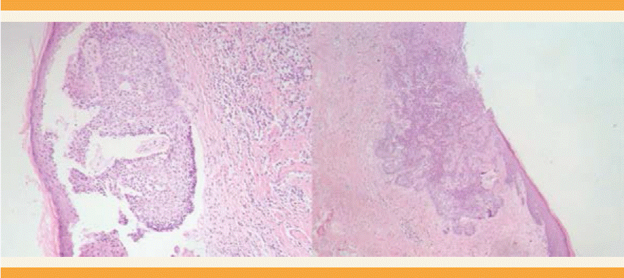
Figure 4 Microscopy: (100 x, hematoxylin and eosin) epithelial tumor composed of atypical basaloid cells with peripheral palisading and occasional artifactual cleft between the tissue blocks and stroma. In the adjacent dermis, a mild nonspecific chronic inflammatory infiltration. 

The patient remains asymptomatic without signs of local recurrence or metastases 24 months after surgery. She signed a consent term for permission to publish the case.

Discussion

Basal cell carcinoma is the most common malignancy in humans, particularly in individuals with light-colored skin. Exposure to ultraviolet radiation is the main risk factor associated with the origin of this cancer. There is evidence that these tumors have a higher occurrence in exposed areas, normally the skin of the head and neck.3,4 Other associated factors include exposure to chemical factors such as arsenic, alterations in immunologic vigilance, and some hereditary skin conditions, including Gorlin's syndrome and xeroderma pigmentosum.5 CBC on non-exposed skin, including the armpits, groin, buttocks, vulva, scrotum, penis, perianal and pubic skin is uncommon.3

Basal cell carcinoma of the vulva is a rare occurrence, corresponding to approximately 2-3% of all vulvar carcinomas and less than 1% of all basal cell carcinomas.2 It was first described by Temesvary in 1926. Since that time, few cases have been reported in the literature.1 Despite the frequent association between basal cell carcinoma and ultraviolet radiation and sunlight, it does not provide a plausible explanation for the vulvar lesions. Previous irradiation, syphilis and chronic irritation have been considered possible causes. Other factors may also be involved, such as genetic predisposition, traumas, chronic ulcers, burns, scars and states of systemic im-munosuppression.2 None of these factors was identified in the current case.

In the majority of cases, basal cell carcinoma of the vulva occurs in postmenopausal women and usually presents as an asymptomatic lesion. However, itching, pain, bleeding and a sensation of a lump may occur.2 There may be a delay in diagnosis. Patients may delay seeking medical attention, and consider the pruritus a simple irritation, or physicians may initially misdiagnose these lesions, classifying them as inflammatory or infectious dermatosis. Basal cell carcinoma has a harmless appearance in this region, therefore biopsy of all suspicious lesions is recommended.6 In the present case, the patient chose surgical resection due to the high index of clinical suspicion. She desired to undergo the surgical procedure in a one-step approach.

Basal cell carcinoma of the vulva is mainly located in the labia majora region, although some cases have already been described in the labia minora and clitoris. In general, the lesion is unilateral.2 This pathology shows a high capacity to infiltrate and cause local destruction with a recurrence rate of up to 20%, dependent upon histological type, tumor size and treatment chosen. The rate of metastasis is rare, estimated at around 0.0028%.7

The present clinical case corroborates results showing that the majority of lesions affect women over 70 years of age and behave in an indolent manner with a low propensity for lymphatic spread or distant metastases.8 A wide local excision with free margins is the treatment of choice. In patients who develop larger tumors and lymphatic dissemination, lymphadenectomy must be performed.9

When surgery is contraindicated other treatments may be used, such as curettage, electrocoagulation, cryotherapy, radiation, CO2 laser, photodynamic therapy or topical application of imiquimod.9

This malignancy has a favorable prognosis. Nevertheless, the rate of local recurrence is around 25%. Appropriate patient follow-up is mandatory with frequent physical examination and instructions about the symptoms and signs compatible with tumor recurrence.9

References

1. Reyes MV, Pascualini MF, Valente E, Kurpis M, Lascano AR. Carcinoma basocelular pigmentado de vulva. Arch. Argent. Dermatol. 2009; 59:115-117. [ Links ]

2. Mulvany NJ, Rayoo M, Allen DG. Basal cell carcinoma of the vulva: a case series. Pathology. 2012; 44(6): 528-33. [ Links ]

3. Montesdeoca GE, Grandal TG, Hervas IM, García ER, Pruñonosa JCM, Torras PBI. Carcinoma basocelular de vulva. Prog Obstet Ginecol 2014; 57: 169-71. DOI: 10.1016/j.pog.2013.09.002 [ Links ]

4. Chinem VP, Miot HA. Epidemiologia do carcinoma basocelular. An Bras Dermatol. 2011;86(2):292-305. http://dx.doi.org/10.1590/S0365-05962011000200013. [ Links ]

5. Mulayim N, Silver DF, Ocal IT, Babalola E. Vulvar basal cell carcinoma: two unusual presentations and review of the literature. Gynecologic Oncology 2002;85 (3): 532-7. doi:10.1006/gyno.2001.6582. [ Links ]

6. Gibson GE, Ahmed I. Perianal and genital basal cell carcinoma: A clinicopathologic review of 51 cases. J Am Acad Dermatol. 2001;45(1):68-71. DOI: 10.1067/mjd.2001.114588. [ Links ]

7. Estrada VM, Vidrio RMG, Ramos Garibay AR, Luna AE. Carcinoma basocelular ulcerado de vulva: presentación de un caso. Rev Cent Dermatol Pascua 2003; 12(3):123-6. [ Links ]

8. Benedet JL, Miller DM, Ehlen TG, Bertrand MA. Basal cell carcinoma of the vulva: clinical features and treatment results in 28 patients. Obstet Gynecol. 1997;90(5):765-8. [ Links ]

9. Ernesto G, Díaz JA, Torres SA, Rangel CM. Carcinoma de células basales de la vulva: reporte de un caso y revisión de la literatura. Rev. Asoc. Colomb. Dermatol. Cir. Dermatol. 2007; 15(4): 286-9. [ Links ]

This article should be cited as:

Gleycianne da Silva Oliveira Dumont Vieira, Ana Maria Gonçalves Rebelo, Guilherme Miranda Correia, Walberto Monteiro Neiva Eulálio Filho, Sabas Carlos Vieira. Carcinoma de células basales de la vulva: una presentación rara de una enfermedad común. Ginecol Obstet Mex. 2018 abril;86(4):267-271. DOI: https://doi.org/10.24245/gom.v86i3.

Received: November 2017; Accepted: February 2018

*Correspondence: Walberto Monteiro Neiva Eulálio Filho. walberto@outlook.com.

Creative Commons License This is an open-access article distributed under the terms of the Creative Commons Attribution License