


 nueva página del texto (beta)
 Inglés (pdf)
 Artículo en XML
 Referencias del artículo
 Enviar artículo por email
 Citado por SciELO
  Similares en
SciELO
Similares en
SciELO 
 Permalink
INTRODUCTION
Tissue engineering aims to establish, restore, or increase the function of tissues by means of the in vitro culturing of anchorage-dependent cells (either differentiated or undifferentiated) on scaffolds made of biomaterials. Growth factors are added to facilitate cell proliferation.
The in vitro tissues are then transplanted to a target organ [1] [2] with tissue injury. Such an injury is susceptible to infection by pathogenic microorganisms, which can lead to the loss or alteration of transplanted tissue, thus complicating tissue recovery and sometimes causing implant failure [1].
The biomaterials utilized in tissue engineering are biocompatible polymers, such as chitosan, that elicit robust cell proliferation [3]. The latter biopolymer is biocompatible, biodegradable, non-toxic, non-mutagenic, bioactive, cationic, antibacterial, and antifungal. It has been of great interest in developing drug-delivery scaffolds for tissue engineering and has been classified as “generally recognized as safe” (GRAS) by the US Food and Drug Administration (FDA) [4] [5]. Another material approved by the FDA to foster cell proliferation and adhesion is the poly(l-lactide-co-ɛcaprolactone) (PLCL) polymer, due to its biocompatibility and mechanical properties [6].
Chitosan can be combined with certain trace elements essential for biological, physiological, and enzymatic processes. Consequently, zinc (Zn), potassium (K), and magnesium (Mg) [7] are added to improve certain functions, including protection against bacteria or fungi, bone regeneration, the synthesis of proteins, the absorption of Fe3+, the protection of tooth enamel, and the production of skin collagen [8] [9] [10]. ZnO nanoparticles (NPs), on the other hand, promote cell proliferation, growth, and differentiation. Additionally, their nanometric size affords high reactivity and thus favors an antibacterial effect [11].
During a surgical intervention for organ or tissue implantation, the main agents of infection are bacteria, which are able to generate septicemia, impetigo, cellulitis, skin abscesses, and even patient death [12]. The combination of chitosan, PLCL, ZnO NPs, and trace elements has an exciting future in tissue engineering. Apart from biocompatibility, this complex has the capacity for cell proliferation as well as inhibition of infection by nosocomial pathogens (e.g., Staphylococcus aureus, Escherichia coli, Pseudomonas aeruginosa, and Serratia marcescens) in surgical incisions and other medical conditions (e.g., diabetic foot and venous ulceration) [13].
The aim of the present study was to develop an HQT hydrogel with chitosan physically cross-linked to PLCL, with the addition of ZnO NPs and trace amounts of K and Mg. The ZnO NPs were obtained by chemical coprecipitation. The HQT hydrogel and its components were characterized with X-ray diffraction (XRD), Fourier transformed infrared spectroscopy (FT-IR), and scanning electron microscopy (SEM).
The antibacterial behavior of the HQT hydrogel was assessed with S. aureus ATCC® 25923 and E. coli ATCC® 25922 by means of the Kirby-Bauer method, in accordance with the international standards recommended by the Antimicrobial Susceptibility Testing Subcommittee of the National Committee for Clinical Laboratory Standards (NCCLS) [14] and the Clinical & Laboratory Standards Institute (CLSI) [15].
MATERIALS AND METHODS
a) Synthesis of ZnO NPs
Nanoparticles were synthesized by the coprecipitation method. A 4.46 mmol solution of zinc acetate (Sigma Aldrich, 99%) was prepared in 42 mL of methanol (Meyer, 99%) by magnetic stirring at 600 rpm and 60 °C. A 7.22 mmol solution of potassium hydroxide (KOH) (Sigma Aldrich, 99%) was prepared in 23 ml of methanol as a precipitating agent. The resulting mixture was added dropwise to the zinc solution, followed by stirring at 600 rpm and 60 °C for 3 h. The white precipitate was washed with acetone and water, and then heated at 60 °C in an oven for 12 h.
b) Characterization of ZnO NPs and the hydrogel by XRD, FT-IR, and SEM
The ZnO nanoparticles were characterized by X-ray diffraction with a Bruker D8 Advance diffractometer, with a Cu kα source at 30 mA and 30 kV and a scanning speed of 1.2° steps/min (in the range of 10-80 2θ degrees). The size of the crystallites was determined by means of the Scherrer equation. FT-IR analysis of the samples was carried out in a PerkinElmer Frontier FT-IR spectrometer at a wavelength of 4000 to 400 cm-1. Scanning electron microscope analysis of the ZnO NPs and the hydrogel were performed on a JEOL JSM 6100 SEM at 20 kV, after coating the materials with gold for 40 s.
c) Preparation of the HQT hydrogel
The concentration of the elements contained in the hydrogel was previously defined by considering the bactericidal effect. A 2% (w/v) solution of chitosan (Sigma Aldrich, at a medium molecular weight and 85% deacetylated) was prepared in 1 M glacial acetic acid (Sigma Aldrich) and stirred at 600 rpm until dissolved. Subsequently, ZnO NPs, K, and Mg were each adjusted to a concentration of 300 mM. KCl2 and MgCl2 (Sigma Aldrich, 99%) were the precursors of K and Mg. PLCL (Sigma Aldrich) at 0.2 % (w/v) was added and the mixture was stirred at 600 rpm for 12 h. The hydrogel was heated in an oven at 40 °C for 48 h to promote the crosslinking reaction.
d) Antibacterial evaluation
The antibacterial activity of the HQT hydrogel was assessed with E. coli ATCC® 25922 and S. aureus ATCC® 25923 by means of the disk diffusion (Kirby Bauer) method in BD Mueller Hinton agar, following the CLSI guidelines. The turbidity of the bacterial suspension was adjusted to a 0.5 McFarland standard (1.5x10⁸ CFU/ml) and then incubated for 24 h at 37 ºC. Inhibition was shown as a clear circular zone and the diameter was measured in mm, indicating bacterial susceptibility to the sample under study. BD BBL Sensi-DiscTM antimicrobial sensitivity test discs (Becton Dickinson) of amoxicillin/clavulanic acid (30 µg) were used as the positive control for inhibition.
RESULTS AND DISCUSSION
a) Characterization by XRD, FT-IR, and SEM
ZnO NPs were characterized by SEM and XRD (Figure 1). The micrograph (Figure 1a) of the surface of the ZnO powder, viewed at 5000X, displays agglomerates of particles with irregular morphology. By analyzing the energy-dispersive X-ray spectroscopy (EDS, Fig. 1b) and elemental mapping (Figs. 1c, d), peaks corresponding to oxygen and zinc were identified on the surface of the material. Hence, the synthesis was successful. Moreover, the product had no contaminants. The diffractogram (Fig. 1c) exhibits peaks corresponding to the zincite phase (according to PDF #04-0032106) in space group P63mc, with lattice parameters a=3.25010Å and c=5.20710Å. Characteristic reflections were observed in the (100), (002), (101), (102), (110), (103), (112), and (202) lattice planes, similar to those found by Purwaningsih et al. 2016 [16]. The size of the crystallites was 30 nm, calculated with the Scherrer equation. No secondary phases were formed during synthesis.
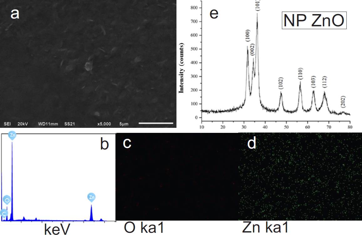
Figure 1 Characterization of ZnO NPs: a) Surface micrograph at 5000X; b) EDS spectrum; elemental mapping for c) oxygen and d) zinc; and e) the XRD pattern.
The hydrogel was morphologically characterized by SEM before and after adding the trace elements and the ZnO NPs (Figure 2). Before adding anything, chitosan displayed a smooth texture and large pores distributed all along the surface (Figure 2.1 a). The porosity of the hydrogel is a critical factor since it allows for the anchoring of cells and the loading of different drugs, elements, or biologically relevant substances. After adding the trace elements and the ZnO NPs (Figure 2.2 a), the surface had a distinct appearance. The particles were coated by a chitosan matrix. By comparing the results of EDS and elemental mapping (Figs. 2.1 b and 2.2 b), it was possible to identify carbon (C), nitrogen (N), and oxygen (O) (Figs. 2.1 c, d and e) in the hydrogel without trace elements. With the addition of these elements, a good distribution of Mg, K, and Zn existed on the surface of the hydrogel (Figs. 2.2 c, d and e). A chlorine (Cl) peak was also found for the inadequately washed hydrogels.
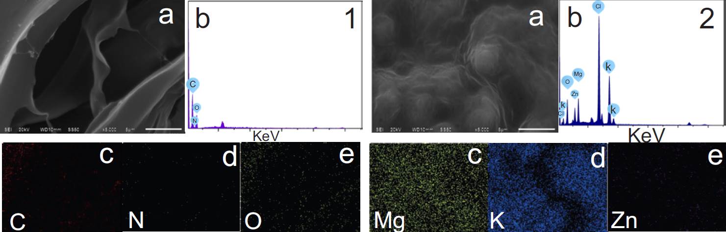
Figure 2 SEM spectra of the hydrogel. 1) Before adding trace elements, and 2) after adding them. Micrographs (a), EDS spectra (b), and elemental mapping (c, d, e, and f).
Correct washing eliminates the Cl residues derived from the precursors of the trace elements (KCl2 and MgCl2). During the microscopy analysis, no damaged or detached zones were detected on the hydrogel, which indicates that the physical crosslinking with PLCL was optimal.
The FT-IR spectra are shown for trace elements, PLCL, and the hydrogel (Figure 3). All samples were dissolved in 1 M acetic acid. The vibrational bands of ~3398-3300 cm-1 and 1637 cm-1 in spectra 1, 2, and 3 can be attributed to the OH group in the solvent [17]. The spectrum of chitosan (Figure 3.4) exhibits a band at ~3323 cm-1 for the OH/NH groups, at ~1644 cm-1 for C-N, at ~1392 cm-1 for C=O, and at ~1288 cm-1 for C-O-C (corresponding to amine 1), C-H vibrations, and C-C and C-O stretching (corresponding to amine 3), respectively [18] [19].
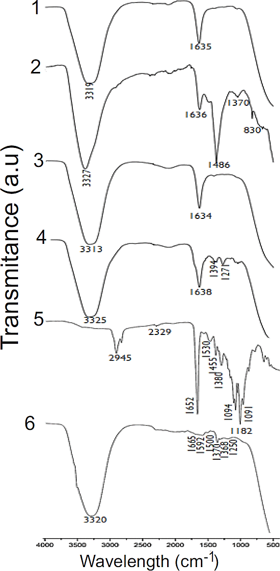
Figure 3 FT-IR spectra for the Mg precursor (1), the K precursor (2), ZnO NPs (3), chitosan (4), PLCL (5), and the HQT hydrogel (6).
The main contributions of PLCL are seen in Figure 3.5. The band at ~2980 cm-1 is assigned to alkyl stretching, at ~1750 cm-1 to carbonyl stretching, at ~1700 cm-1 to carbonyl ester stretching, and at ~1450 cm-1 to CH2 bending. The bands at ~1380 cm-1 were ascribed to C=O, at ~1189 cm-1 and ~1182 cm-1 to C-O-O and asymmetric CH2, and at ~1091 cm-1 to the stretching vibrations of C-O [20].
The FT-IR absorption bands of the HQT hydrogel (with all its components) are shown in Figure 3.6, observing a clear difference in the interaction of the functional groups. The bands at ~3300 cm-1 were designated as the NH2 and OH groups, at ~1750 cm-1 as the stretching of the carbonyl groups in PLCL, at ~1600 cm-1 as the CH2 and CH3 groups, and at ~1250 cm-1 as a stretching of the C-O-C present in chitosan. Finally, the band at ~1420 cm-1 may be due to the chitosan CH2 groups or PLCL-COO- groups [18] [19] [20].
b) Antibacterial activity
The antibacterial effect of the HQT hydrogel was evaluated with E. coli and S. aureus. The inhibition zones were more evident for S. aureus (25 mm for chitosan and 14 mm for ZnO NPs) than E. coli (14 mm for chitosan and 7 mm for ZnO NPs) (Figure 4). This result is attributed to the innate antimicrobial activity of chitosan and the oxidative stress caused by ZnO nanoparticles. Regarding the latter, reactive oxygen species (ROS) interact with the cell wall of bacteria and destroy the intracellular content, damaging proteins and lipids. Hence, positively charged nanoparticles bind to the negatively charged bacterial membrane, fomenting its disintegration or rupture [21]. Additionally, the size of ZnO NPs is effective against Gram-positive and Gram-negative bacteria because of the large surface area available to interact with the bacterial wall [22].
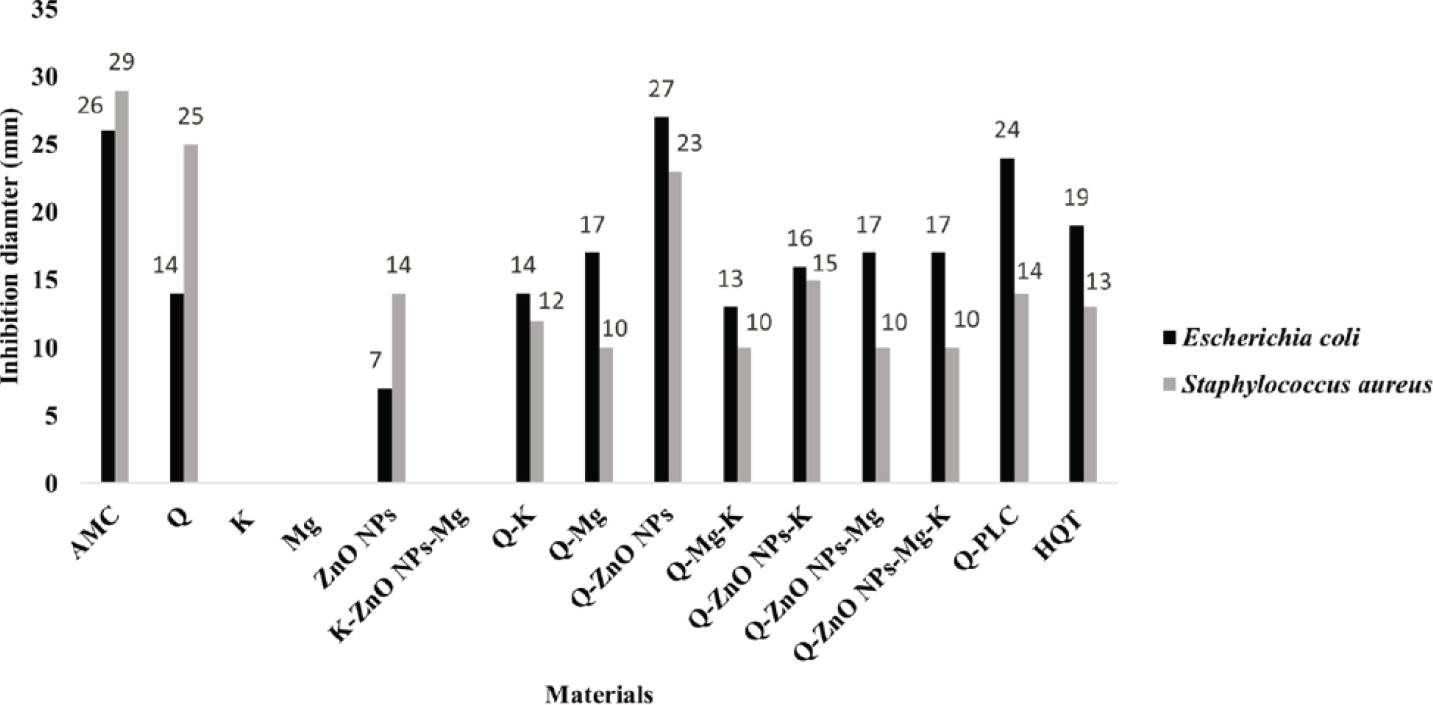
Figure 4 Inhibition zones (in mm) of E. coli and S. aureus exposed to the biomaterials herein tested.
For the biomaterials with trace elements, the inhibition of E. coli was more significant than that of S. aureus. The content of K and Mg affect the antibacterial activity of ZnO NPs, as can be appreciated by the lack of inhibition of both microorganisms. Concerning E. coli, the inhibition diameters resulting from K combined with Q (Q-K) materials were very similar to those found with chitosan alone (14 mm). The antibacterial effect of Q-K materials on S. aureus was reduced by about 50% (to 12 mm) (Figure 4).
K is required by microorganisms for the metabolism of carbohydrates, the activation of some enzymes, and bacterial osmoregulation [23]. Therefore, it is herein inferred that K was mainly used as a nutrient by the bacteria rather than a growth inhibitor. For E. coli, the inhibition diameter was greater with Q-Mg (17 mm) than chitosan alone (14 mm). Thus, Mg increased the antibacterial potential of chitosan. Mg and K did not show any antibacterial activity separately. They participate in vital functions as nutrients [23], which was probably their primary role. Q-PLCL generates a good antibacterial effect on E. coli, with an inhibition diameter of 24 mm. Finally, the HQT hydrogel significantly inhibited S. aureus (13mm) and E. coli (19mm), the latter being more susceptible.
The values of the inhibition diameters are observed in Figures 4 and 5. In both, the importance of the inhibitory activity of chitosan is evidenced by its greater inhibition diameter for all combinations. The concentration of 300 mM of the trace elements promoted bacterial inhibition in some of the materials. This concentration was determined in a previous study employing the same bacteria. The tests were conducted by means of the Kirby-Bauer method, exposing each bacteria to the biomaterials presently evaluated.

Figure 5 Results of the Kirby-Bauer method to examine the antibacterial effect of the HQT hydrogel with distinct combinations of trace elements and ZnO NPs. S. aureus: A, B, C, and D; E. coli: E, F, G, and H. 1) ZnO NPs; 2) Q; 3) Mg; 4) K; 5) K-ZnO NPs-Mg; 6) Q-K; 7) Q-ZnO NPs; 8) Q-Mg; 9) Q-ZnO NPs-Mg; 10) Q-Mg-K; 11) Q-ZnO NPs-K; 12) HQT; 13) Q-PLCL; and 14) Q.

Figure 6 Inhibition produced by the antibiotic disc (the positive control), containing amoxicillin/clavulanic acid (30 µg). A) S. aureus; B) E. coli.
E. coli and S. aureus are common pathogens responsible for tissue infection. Due to the structural and chemical differences of their cell walls, it was relevant to evaluate the inhibitory effect of the present biomaterials on both species [13]. According to the literature, chitosan, PLCL and ZnO NPs are non-toxic materials and have been used in various biomedical applications. The current results indicate that combining biomaterials on a chitosan hydrogel scaffold holds promise for combatting tissue infection caused by bacteria. Future research is necessary to examine the cytotoxicity of the hydrogel.
CONCLUSIONS
Zinc oxide nanoparticles of 30 nm were obtained in the zincite phase. It was possible to elaborate an adequately homogenized chitosan-based hydrogel physically cross-linked with PLCL and enriched with ZnO NPs as well as trace amounts of Mg and K. In vitro testing showed that this hydrogel has antibacterial activity. Since these biomaterials are organic, FDAapproved, and biocompatible with different biological systems, the resulting hydrogel represents a potentially useful antibiotic scaffold for tissue engineering.

