


Serviços Personalizados
Journal
Artigo
 Inglês (pdf)
 Artigo em XML
 Referências do artigo
 Enviar este artigo por email
Indicadores
-
 Citado por SciELO
 -
 Acessos
Links relacionados
-
 Similares em
SciELO
Similares em
SciELO
Compartilhar

 Permalink
Revista mexicana de ingeniería biomédica
versão On-line ISSN 2395-9126versão impressa ISSN 0188-9532
Rev. mex. ing. bioméd vol.34 no.3 México Dez. 2013
Artículos de investigación
Development of a neuromuscular junction model on surfaces modified by plasma polymerization
Desarrollo de un modelo de unión neuromuscular sobre superficies modificadas por polimerización por plasma
E. Zuñiga-Aguilar*, R. Godínez*, O. Ramírez-Fernández*, J. Morales*, R. Olayo**
* Universidad Autónoma Metropolitana, Departamento de Ing. Eléctrica, Distrito Federal, México.
** Universidad Autónoma Metropolitana, Departamento de Física, Distrito Federal, México.
Correspondence:
E. Zuñiga-Aguilar,
Departamento de Ingeniería Eléctrica.
Universidad Autónoma Metropolitana-Iztapalapa.
Apdo. Postal 55-534, Iztapalapa, México.
Correo electrónico: esza08@gmail.com
Fecha de recepción: 26 de Octubre de 2012
Fecha de aceptación: 25 de Septiembre de 2013
ABSTRACT
The aim of this work is to implement a biological model of neuromuscular junctions to study the mechanisms involved in intra and inter cellular processes using cell co-cultures. To optimize growth and development of the neuromuscular junction, cells were seeded on plasma polymerized pyrrole which has proven suitable for other types of cell cultures. The cell lines used were motor neuron NG108-15 and skeletal muscle C2C12. Cells were evaluated according to their morphology and electrophysiological characteristics. To observe the expression of specific proteins of the nerve synapse, immunocytochemical techniques were applied using dying antibodies. Proteins localized in nerve terminals were dyed and imaged by fluorescence microscopy. Images of cell co-cultures showed the formation of neuromuscular junctions. The preparation of neuromuscular junctions described in this work will allow the study of the mechanisms involved in their functions.
Keywords: cell co-culture, neuromuscular junction, plasma polymerization, polypyrrole.
RESUMEN
El objetivo de este trabajo es implementar un modelo biológico de unión neuromuscular para el estudio de los mecanismos involucrados en los procesos intra e intercelulares empleando co-cultivos celulares. Con el fin de optimizar el crecimiento y desarrollo de las uniones neuromusculares, las células se cultivaron sobre superficies de polipirrol obtenidas mediante polimerización por plasma que han mostrado ser adecuadas en otros tipos de cultivos celulares. Las líneas celulares que se emplearon fueron los modelos de motoneurona NG108-15 y muscular C2C12. Las células se evaluaron de acuerdo a su morfología y características electrofisiológicas. Para observar la expresión de proteínas clave de la sinapsis, se aplicaron técnicas inmunocitoquímicas utilizando anticuerpos específicos para la marcación de proteínas localizadas en las terminales nerviosas adquiriendo imágenes con microscopía de fluorescencia. Las imágenes de los co-cultivos celulares mostraron la formación de uniones neuromusculares. El método de preparación de uniones neuromusculares que se describe en este trabajo permitirá estudiar los mecanismos involucrados en sus funciones.
Palabras clave: co-cultivo celular, unión neuromuscular, polimerización por plasma, poli pirrol.
INTRODUCTION
The neuromuscular junction is a chemical synapse responsible for transmitting signals from the motor nerve terminal to the postsynaptic region of muscle fiber [1]. Studies in neuromuscular junctions are conducted to correlate structural aspects of synapse formation with its physiological role. These studies have led to the creation of new biological models of neruomuscular junctions used in pharmacology and anesthesiology. The neuromuscular junction is a target of many neurotoxins and models are needed to understand the mechanisms by which different diseases affect, directly or indirectly, the neuromuscular union site generating autoimmune and genetic disorders [2-5].
Many of the studies on neuromuscular junctions have been conducted using in vivo methods or by culturing tissue fragments for neuromuscular junction formation [6,7]. These cultures allow for adequate visibility of neuromuscular training and allow for structure-function correlation studies. In practice this is a laborious task. Generally, studies report the formation of neuromuscular junctions in tissue fragments but structure-function correlations are difficult to interpret due to the complexity of tissue and its complementary functions [8,9]. Skeletal muscle fibers are innervated by large myelinated nerve fibers and each nerve fiber branches many times and stimulates three to several hundred skeletal muscle fibers.
As a step towards the study of mechanisms of innervation at the level of unicellular interaction recently primary cell cultures in monolayers of cells extracted from skeletal muscle and spinal cord have been prepared to form neuromuscular junctions in culture [10-12]. The use of such cell cultures has some disadvantages, including the difficulty in isolating a single cell population, particularly of nervous origin, and the high risk of contamination of the cell culture [13, 14].
An alternative to cell isolation is the use of neuronal and muscle cell lines that can form neuromuscular junctions in culture. The use of cell lines helps to have control of the cellular environment, cell homogeneity and a better understanding of the cell type being studied [15,16].
The culture surface must display appropriate characteristics for cells to adhere and grow, allowing the cells to differentiate properly. There is still a need for research on developing surfaces for betrer cell adhesion. Recently, plasma modified surfacCs have been shown to be effective surfaces for cell culture and as biocompatible polymers [17, 18].
Because the study and development of new neuromuscular junction models remains a challenge, in this paper we present preliminary results of the development of a model for studying the formation of neuromuscular junctions on surfaces treated by plasma polymerization used for cell cultures of motor neuron and skrletal muscle cells. The plasma polymerization technique allows us to modify the surface of materials such that they can be used as growth surfaces of excitable cells, permitting the appropriate cell differentiation. The co-culture of muscle cells and motor neurons in a plasma polymerization-treated surface , allowed us to achieve the formation of neuromuscular junctions. The procedure ctescribed in this work will allow researchers to better characterize and understand the formation neuromuscular junctions between motor neurons and skeletal muscle.
METHODOLOGY
Cell Culture Protocol
The NG108-15 cell line in differentiated form is considered a neuron biological model; the cell line was purchased from ATCC No. HB-12317. Cells were seeded in culture dishes of 35 mm with 2 ml of DMEM (Cat. No. 12100-046 Dulbecco Gibco) supplemented with 10% FBS (Cat. No. 16000, Gibco), 1% L-glutamine (Cat. No. G-1517, Gibco), 1% antibiotic-antimycotic (Cat. No. 15240, Gibco), and 1x HAT Supplement which is a liquid mixture of sodium hypoxanthine (5 mM), aminopterin (20 ţM) and thymidine (0.8 mM) (Cat. No. 21060, Gibco). The culture dishes were placed in an incubator under optimum conditions of temperature and atmosphere (5% CO2 and 37 °C).
The C2C12 cell line is a biological model of skeletal muscle cell; cells were donated by Dr. Bulmaro Cisneros of the Genetics and Molecular Biology Laboratory, CINVESTAV. Cells were seeded in culture dishes of 35 mm with 2 ml of DMEM (Catelog No.: 12100-046, Gibco) supplemented with 10% FBS (Cat. No. 16000, Gibco), 1% L-glutamine (Cat. No. G- 1517, Gibco), 1% antibiotic-antimycotic (Cat. No. 15240, Gibco) and 1 mM sodium pyruvate (Cat. No. 21060, Sigma). The culture dishes were placed in the incubator at optimum temperature and atmosphere (5% CO2 and 37°C).
Plasma Polymerization
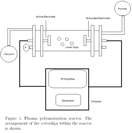
Glass microscope cover slips were coated by plasma polymerization. The polymerization was done in a glow discharge reactor under low pressure; pyrrole (98% Sigma-Aldrich) was used as monomer. The polymerization was conducted in a tubular glass reactor with stainless steel electrodes, using the configuration show in Figure 1. The corerslips were placed in the plasma reactor at a power of 30 watts (Dressler Cesar, RF Power Generator) for 10 min at a pressure of 3 x 10⁻² Torrs (Edwards Active Gauge Controller). Coated materials were placed and stored under UV light to ensure sterility in subsequent experiments.
Cell seeding on the polymerized surface
Once the C2C12 muscle cells in culture dishes reached confluence, the cells were trypsinized with 1 ml Trypsin + EDTA (Cat. No. 25200072, Gibco), and then seeded on the cover slips treated by plasma polymerization. The cover slips were placed inside untreated culture dishes. Cells were seeded using the micro culture technique, thus ensuring that only the cells that grow on the treated surface were evaluated. An aliquote of 400 μl with 300 x 103 cells/ml was placed on the substrate with DMEM (Cat. No.12100-046, Gibco) supplemented with 10% FBS (Cat. No. 16000, Gibco), 1% L-Glutamine (Cat No. G-1517, Gibco) and 1% antibiotic-antimycotic (Cat. No. 15240, Gibco). The dishes were placed in an incubator at optimal temperature and atmosphere (5% CO2 and 37°C).
Cell Differentiation Protocol
Once the C2C12 cells reached 60% of confluence on the cover slips, the medium was changed to 2 ml of DMEM differentiation medium (Cat. No. 12100-046 Gibco), supplemented with 5% horse serum (Cat. No. 26050-088, Gibco) and 1% antibiotic-antimycotic (Cat. No. 15240, Gibco). Cells were maintained for 5 days with this medium and kept in the incubator at optimum temperature and atmosphere (5% CO2 and 37°C).
Electrophysiological Recordings
Motor neuron and muscle cells were immersed in mammalian extracellular solution containing: 4.5 mM KCl, 151.5 mM NaCl, 1 mM MgCl2, 2 mM CaCl2, and 5 mM HEPES. Glass pipettes (World Precision Instruments, FIL glass 4IN 1BBL NO) were used as electrodes; the pipettes were stretched with a horizontal puller (Sutter Instruments, Model P-97) and had resistances of 2-3 MΩ. The micropipettes were filled with intracellular solution (140 mM KCl, 10 mM NaCl, 1 mM MgCl2, 1 mM CaCl2, 11 mM EGTA, 10 mM HEPES). An Axopatch 200A amplifier (AxoIntruments) was used; it was set in the voltage-clamp configuration to measure ionic currents, and in the current attachment configuration for the measurement of changes in the potential of the membrane. Patch clamp was used in the experiments with a holding potential of -80mV. Rectangular depolarizing pulses of 10 mV were applied to evoke voltage-dependent ionic currents of Na+, K+.
In electrical current clamp experiments, a DAGAN amplifier was used. Cells were immersed in mammalian extracellular solution .Stretched borosilicate glass pipettes with resistances of 60 MΩ or more were prepared. The pipettes were filled with 3M KOOCCH3 (95%) + 3M KCl (5%). From the resting potential, the membrane potential was changed by the injection of rectangular current pulses depolarizing and hyperpolarizing it. The membrane resistance and the induced action potentials were measured.
The voltage and current stimuli and the synchronous acquisition signals were digitized through a Digidata 1200 converter card. The signals were processed and analyzed using Clampfit software. Experiments were performed at room temperature (24 ř C).
Application of immunocytochemistry
In co-culture and the simple culture of NG108-15 the cells were fixed to the cover slips for 15 minutes with a solution of 4% paraformaldehyde (Cat. No. 158127, Sigma). The samples were then washed 3 times with PBS (Cat. No. 003002) with a 10 min rest between each wash. The membranes in the samples were made permeable by washing with TRITON (Cat. No. T8787, Sigma) in PBS for 10 min, followed by 3 washes with PBS with a 10 min rest between each wash. Samples were blocked with 1% donkey serum (Cat. No. D9663, Sigma) diluted in PBS for 30 min. Primary antibodies were applied to each cover slip with neuronal cells; the first primary antibody was applied was Anti-Vesicular Acetylcholine Transporter antibody mouse (Cat. No. AB68984) with a 1:800 dilution and FITC rabbit as secondary (Cat. No. F0205, DAKO) diluted 1:400. The second primary antibody was Synaptophysin (Cat. No. M0776, DAKO) made in mouse with a 1:800 dilution. Texas Red (Cat. No. G-862, Gibco) made in horse with 1:200 dilution was used as secondary antibody. To the co-culture, nicotinic receptors alpha-3 was used to localize the motor neurons. All samples were mounted in glycerol (Cat. No. 15514-011, Gibco) and checked for presence of antibodies by observing them by fluorescence microscopy.
RESULTS
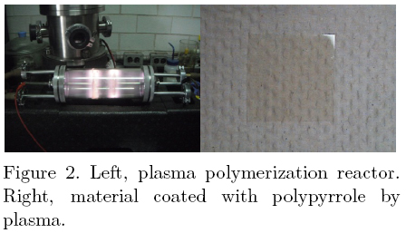
The plasma polymerization technique generates a thin film of synthetic material on the surface of glass substrates, which can be used as culture surfaces for C2C12 muscle and NG08-15 mot o r neuron cells.
Figure 2 shows the plasma reactor (during the polymerization. Glass cover slips are placed inside the reactor and the surface is coated by a thin film of plasma polymerized pyrrole.
The FT-IR absorption spectrum of plasma polymerized pyrrole is shown in Figure 3. All characteristic peaks of the plasma polymerized pyrrole functional groups can be seen [19]. The peaks at 3357 cm–1 and 1627 cm–1 correspond to the amines in the polypyrrole structure. The wide peak in the range of 500-1000 cm–1 contains many absorptions corresponding to alkenes from broken pyrrole rings. The width of the peak indicates the complex absorption in the polymer because of branching, crosslinking and interactions among the pyrrole groups closely packed in the film.

Figure 4 shows the microculture of cells on the cover slips. The microculture technique allows us to delimit the cell growth area to the cover slips which are coated wit h polypyrrole prepared by plasma polymerizat ion. C2C12 muscle cells and NG108-15 motor neurons grown on the plasma polymerized pyrrole surface did not show any morphological alteration or cell death.
In order to study the electrical functionality of the motor neurons and the muscle cells, electrophysiological tests were performed. Figure 5 (a) presents action potentials induced by depolarizing current injection. From a membrane potential of -50 mV, cells were stimulated with hyper and depolarizing; currents; the first action potential was recorded when the voltage threshold was surpassed. To test that the electrical properties of motor nerve cells are not affected by the presence of the polymer, currents of Na+ and K+ were measured with the voltage clamp method as Figure 5 (b) shows. Note the appearance of negative fast currents (Na–) created by the opening; of these channels, followed by the delayed positive currents (K+) caused by the activation of voltage-dependent channels. Figure 5 (c) shows the action potentials of C2C12 muscle cells, recorded by setting the resting potential at -75 mV and then applying a train of depolarizing current pulses from 0.1 nA to 1.0 nA.
Immunocytochemical testing was done to reveal if protein synthesis remained present in the motor neurons The synaptophysin antibody was used to reveal the presence of neuron synapses in the cells seeded on the polymerized material since this protein is found only in the membrane of synaptic vesicles.
Figure 6 shows the neuron cell growth on the polymer surface and the same cell group after the synaptophysin antibody treatment; to enhance the visual contrast the micrograph was colored with imageJ software (Figure 6c).
Another way to observe the formation of synapses formation is to detect any released protein with neurotransmitter function in the synapse. In the case of NG108-15 motor neurons the presence of acetylcholine would indicate synapse formation. Figure 7 shows the neuron cells on the polymerized material and the fluorescence images of the same cells after the antibody treatment (V-Chat-FITC) directed at the vesicular acetylcholine transporter. The results indicate the presence of acetylcholine in the nerve terminals of the seeded motor neurons.
Once the adhesion, differentiation and functionality of the neuron cells was tested, and the adhesion and differentiation of C2C12 muscle cells was achieved, the next step is to perform the cell co-culture. Motor neuron NG108-15 cells were differentiated and then tripziniced and reseeded in the polymerized material with the muscle cells. NG108-15 adhered and began issuing ramifications to the C2C12 muscle cells 24 hours after being; planted. Figure 8 shows micrographs of the two kinds of cells separately and the co-culture under the same growth environment.
After 48 hours of culture, the nerve prolongations of the motor neurens extended to the surface of C2C12 muscle cell, forming the junctions. Figure 9 shows a bipolar motor neuron that extents its ramifications to reach the surface of a muscle cell and genwates two neuromuscular junction.
DISCUSSION
A critical part of the cell culture technique is the growth surface. There is abundant literature on this subject. In recent years, it has been shown that surfaces treated with polypyrrole have advantages over conventional cell growth surfaces, being biocompatible and enhancing cell proliferation. In this work we demonstrate the use of a polypyrrole surface prepared by plasma polymerization for the co-culture of motor neurons and skeletal muscle cells to study the formation of neuromuscular junctions.
Plasma polymerization of pyrrole monomers allowed us to generate very thin films of polymerized material on the surface of glass cover slips. The surfaces thus prepared were demonstrated to be adequate for cell growth. The infrared characterization of the surfaces shows the presence of amine groups which have been associated with good cellutar adhesion [17, 18].
The funrtionality of the cells was established by the electrical functional testing. Action potentials of motor neurons NG108-15 and muscle cells C2C12 were obtained. In the former, evoked ionic currents of Na+ and K+ were observed.
Immunocytochemical techniques allowed us to observe the properties of protein synthesis in motor neuron cells NG10815. The presence of proteins characteristic of this type of cells, such as Synaptophysin, located on the surface of the cell membrane of nerve cells, and vesicular acetylcholine transporter, a protein found in nerve terminals, was observed. We confirmed that the protein synthesis is maintained in the presence of the polymer surface. Additionally, we found that the plasma polymer surfaces were adequate for co-culture of neuron and muscle cells. Marking nicotinic receptors alpha-3 of motor neurons demonstrated the presence of these receptors in the cell membrane, which was a proof of the presence of this cell type in the cell co-culture.
The technique of co-cultured cells allows us to make the formation of neuromuscular junctions on polypyrrole surfaces prepared by plasma polymerization. Neuromuscular junctions are evident in optical microscopy images which show the characteristic morphology of neuromuscular junctions reported in the literature.
We, therefore, conclude that the surface modification of materials with polypyrrole synthesized by plasma allowed the muscle cell and motor neuron cell growth, did not alter the electrophysiological properties in both cells and allowed protein synthesis of motor neuron cells. This makes it an attractive material for use in cell cultures and co-cultures of motor neurons and muscle cells to generate neuromuscular junctions and as in vitro model to study cellular communication processes.
ACKNOWLEDGMENT
We thank to CONACYT and ICyT-DF (Instituto de Ciencia y Tecnología del Distrito Federal) through the project PIUTE 10-63, ICyTDF 276/2010 for the support given to this work.
REFERENCES
1. Engel A.G. (1999). "Anatomy and molecular architecture of the neuromuscular junction". En: Myasthenia Gravis and Myasthenic Syndromes. Engel AG (eds). Contemporary Neurology Series. Oxford University Press, New York. [ Links ]
2. Vincent A. (2008). "Autoantibodies in neuromuscular transmission disorders". [Journal Article], Ann Indian Acad Neurol; Vol.11(3):140-145. [ Links ]
3. Vrolix K., Fraussen J., Molenaar P.C., Losen M., Somers V., Stinissen P., De Baets M.H., Martínez-Martínez P. (2010). "The auto-antigen repertoire in myasthenia gravis". Autoimmunity, Vol.43(5-6):380-400. [ Links ]
4. Tzartos S.J., Langeberg L., Hochschwender S., Swanson L.W., Linstrom J. (1986). "Characteristics of monoclonal antibodies to denatured Torpedo and to native calf acetylcholine receptor: species, subunit and region specificities". J Neuroinmunol, Vol. 10:235-253. [ Links ]
5. Marx A., Wilisch A., Schultz A., Gattenlöhner S., Nenninger R., Müller-Hermelink H.K. (1997). "Pathogenesis of myasthenia gravis". Virchows Arch Vol.430:355-364. [ Links ]
6. Takahashi T., Nakajima Y., Hirosawa K., Nakajima S., and Onodera K. (1987). "Structure and Physiology of Developing Neuromuscular Synapses in Culture". Journal Neuroscience Vol. 7 (2):473-481. [ Links ]
7. Shimada Y., Fischman D. A., and Moscona A. A. (1969), "The development of nerve-muscle junctions in monolayer cultures of embryonic spinal cord and skeletal muscle cells". The Journal of Cell Biology Vol. 43 (2): 382-387. [ Links ]
8. Wilson, GG, Karlin, A. (2001). "Acetylcholine receptor channel structure in the resting, open and desensitized states probed with the substituted-cysteine-accesibility method". Proc. Natl. Acad. Sci. USA. Vol 98(3):1241-1248. [ Links ]
9. Corringer P.-J. et al. (2000). "Nicotinic receptors at the amino acid level". Ann. Rev. Pharmacol. Toxicol. Vol.40:431-458. [ Links ]
10. Courtney J., Steinbach J.H. (1981). "Age changes in neuromuscular junction morphology and acetylcholine receptor distribution on rat skeletal muscle fibres", J. Physiology Vol. 320: 435-447. [ Links ]
11. Matthews-Bellinger J., Salpeter M. (1983). "Fine structural distribution of acetylcholine receptors at developing mouse neuromuscular junctions". The Journal of Neuroscience Vol. 3(3):644-657. [ Links ]
12. Coutinho de Barcelos C., Assunção Braga A., Da Silva Braga F., Braga G. , Antoniassi S., Oshima Y., Rodrigues L. (2008). "In Vitro and In Vivo Neuromuscular Effects of Atracurium and Rocuronium in Rats Treated with Carbamazepine for Seven Days". Rev Bras Anestesiol Vol. 58(2):137-151. [ Links ]
13. Michikawa M, Kobayashi T, Tsukagoshi H. (1991). "Early events of chemical transmission of newly formed neuromuscular junctions in monolayers of human muscle cell co cultered with fetal rat spinal cord explants". Brain Res Vol.538: 79-85. [ Links ]
14. Wyskovsky W, Hohenegger M, Plank B, Hellamnn G, Klein S, Suko J.(1990). "Activation and inhibition of the calcium-release channel of isolated skeletal muscle heavy sarcoplasmic reticulum". Eur J Biochem Vol. 194: 549-559. [ Links ]
15. Jiang J., Choi R., Siow N., Lee H., Wan D. and Tsim K. (2003). "Muscle Induces Neuronal Expression of Acetylcholinesterase in Neuron-Muscle Co-culture". The Journal of Biological Chemistry Vol. 278:45435-45444 [ Links ]
16. Das M., Rumsey J.W., Gregory C. A., Bhargava N., Kang J.F., Molnar P., Riedel L., Guo X. and Hickman J. J.(2007). "Embryonic motoneuron-skeletal muscle co-culture in a defined system". Neuroscience Vol.146:481-488. [ Links ]
17. Pérez-Tejada E., Gómez Quiroz L. E., Morales J., Gutiérrez M. C., Olayo M. G., Cruz G. J., Olayo R. (2007). "Cultivo de Hepatocitos sobre vidrio modificado con un polímero de pirrol sintetizado por plasma". Superficies y Vacio Vol. 21 (3): 10-14. [ Links ]
18. Olayo R., Ríos C., Salgado-Ceballos H., Morales J. (2008). "Tissue spinal cord response in rats after implants of polypirrole and polyethylene glycol obtained by plasma" J. Mater Sci: Mater Med. Vol. 19:817-826. [ Links ]
19. Cruz G.J., Morales J., Olayo R. (1999). "Films obtained by plasma polymerization of pyrrole" Thin Solid Films, Vol. 342:119-126. [ Links ]

