


Services on Demand
Journal
Article
 English (pdf)
 Article in xml format
 Article references
 Send this article by e-mail
Indicators
-
 Cited by SciELO
 -
 Access statistics
Related links
-
 Similars in
SciELO
Similars in
SciELO
Share

 Permalink
Revista mexicana de ingeniería biomédica
On-line version ISSN 2395-9126Print version ISSN 0188-9532
Rev. mex. ing. bioméd vol.32 n.1 México Jul. 2011
Artículo de investigación original
Magnetic characterization of solid food to gastric emptying studies by mechanical-magnetogastrography assessment
Córdova-Fraga T*, Rodriguez D*, De la Roca-Chiapas JM*, Sosa M*, Hernández MA**, Vargas M*, Solorio SE**, Bernal JJ*
* Departamento de Ingeniería Física-DCI, Universidad de Guanajuato Campus León.
** Unidad Médica de Alta Especialidad IMSS, Clínica T-1 León, Guanajuato.
Correspondence:
Teodoro Córdova-Fraga,
E-mail: theo@fisica.ugto.mx.
Tel. 52 (477) 7885100.
Received article: 26/may/2010.
Accepted article: 14/february/2011.
ABSTRACT
Mechanical-Magnetogastrography (M-MGG) is a technique that has been used to measure gastric emptying in healthy subjects and patients with gastrointestinal pathologies. This has allowed implementing a non-invasive technology, free of ionizing-radiation and it may be used in diagnostic tests in clinical medicine. The characterization of a phantom which has a magnetic behavior similar to that observed in gastric emptying studies carried out in persons is presented. A fluxgate magnetometer was used to record the signal in a magnetic unshielded room. Phantom included nine identical deposits prepared with magnetite, Fe3O4 and flour. The behavior of the experiment is identical to studies previously reported in evaluations of healthy people and patients.
Key Words: Mechanical-magnetogastrography, gastric emptying, magnetic stimulator.
RESUMEN
La magnetogastrografía mecánica (M-MGG) es una técnica que ha sido usada para medir el vaciado gástrico en pacientes sanos y pacientes con patologías gastrointestinales. Esto ha permitido la implementación de una tecnología no invasiva, libre de radiación ionizante que puede ser utilizada para pruebas clínicas en medicina. En este artículo, la caracterización de un «phantom» que tiene un comportamiento magnético similar a los estudios realizados para el proceso de vaciado gástrico es presentada. Un magnetómetro de flujo magnético fue utilizado para registrar la señal en un cuarto sin aislamiento magnético. El «phantom» incluyó depósitos idénticos preparados con magnetita, Fe3O4 y flúor. El comportamiento de los experimentos obtenidos es idéntico a estudios previamente reportados en las evaluaciones de personas sanas y pacientes con alguna patología.
Palabras clave: Magnetogastrografía mecánica, vaciado gástrico, estimulador magnético.
INTRODUCTION
Biomagnetic gastrointestinal (GI) system assessment began in the late fifties and early sixties1,2. However, these biomagnetic devices and techniques began to expand their applications until the early seventies of the past century. Cohen in 1969 used the magnetometer to measure the direct current fields of many parts of the body, and established the first orders of magnitude depending on the biomagnetic source registered3-5. In the seventies, further studies of the GI system were published6-8. This technique, applied to GI system studies, has been growing since then to date9-15. In this regard, a significant progress has been made in the techniques for gastric emptying assessment, including theoretical and experimental developments to incorporate new magnetometers that work at room temperature, with high sensitivity devices to record magnetic fields around nT, so that, with appropriate quantities of oral contrast agent, the biomagnetic signal intensity is increased in that order.
Particularly, in previous studies9-12 it has evaluated the gastric emptying half-time to predict the behavior of ingested food and associate it with the behavior of the drain on health12 and the disease10, it has been used as food semisolid and solid15 and it has been gotten similar results to scintigraphy, it was also used the same type of test food in order to make studies comparable. Which one was done in order to create a technological development that is a diagnostic option with a lot of advantages because of it is not invasive, low cost, and it lacks of ionization radiation, such in all studies were assumed they were performed close to use of scintigraphy and a decaying behavior has been tested the sensors have a good signal to noise ratio, which measure the different magnetic intensities, nevertheless, it has not been done, so far it is known, or there are not laboratory experimental evidence showing that what is being measured is raised at the theoretical level, hence the importance of a study of phantoms which may be fitted with gastric emptying and that results in an average time of gastric emptying similar to that found in previous studies.
Recently in our laboratory, M-MGG technique has been used in different studies, to evaluate the gastric segment peristaltic activity, the colon transit time in different phases of menstrual cycle and motility evaluation in patients with functional dyspepsia10 and healthy subjects11-14 to reproducibility evaluation. Several studies has been done in order to assess the gastric emptying through magnetic tracer diluted in semi-solid contrast media, in both healthy subjects and gastro paresis patients6,7,14. The authors analyzed the magnetic signalof magnetictracers mixed in a solid food (yogurt) to measure gastric emptying using M-MGG in male healthy subjects (defined as subject without GI symptoms or known GI disease). As long as, others researches have reported in preliminary studies, motility, nutritional value, taste perception and possible side effects of solid food contrast agent15.
The aim of this study is to evaluate the magnetic properties of solid test meal by using an experimental phantom performed for this purpose (containers with food test). The theoretical importance of this investigation was to verify the gastric-emptying rate of solid foods, similar to those reported by other authors, through a controlled experiment.
It is pretend that the food prepared and characterized in our laboratory could be used for further studies to gastric emptying time assessment using M-MMG technique, which represents an alternative simple and novel similar to other diagnosis methods employed in clinical medicine evaluation.
MATERIAL AND METHODS
A magnetic stimulator constructed and characterized in our laboratory was used in this study, this device whose results have been previously published11,12, was implemented with two identical coils assembled in a Helmholtz coils array, these two coils are plugged in series with a capacitor bank and fed with electrical line at 220 v. This configuration produces a nearly homogeneous magnetic field with intensity around 32 mT in the central region of the system16. This magnetic flux density is generated with a discharge of a capacitance bank, in 17 ms, with a capacitance value of 46 mF (Figure 1). It is enough strong to magnetize the particles of magnetite contained in the food, and then, it produces magnetic field in order of nT, which is recorded with a fluxgate magnetometer sensor.

Phantoms were prepared in nine identical plastic cylindrical deposits, with 5.6 ± 0.1 cm diameter and 3.0 ± 0.1 cm height. These containers were prepared with 1 g of magnetite (Fe3O4 ) in 9 g of flour, the weight container of magnetite was 22.5 ± 0.1 g. The nine containers were placed in different configurations into magnetization system. The settings used are shown in Figure 2, the containers were formed into three columns, standing just above the fluxgate magnetometers. The columns were placed, with respect to the sensors, in four different positions: The first assay consisted in to place the columns forming a triangle between the two sensors. In the second assay, the columns are placed above one sensor. The third configuration was the same as previous one, except that the columns were placed on the other sensor. Finally, the columns were placed forming a straight line between the two sensors.
Once each configuration was established, the capacitors were charged at 100 %, in 60 s and then they were discharged in 17 ms, this guarantees the current flow through coils to generate a magnetic pulse around 32 mT.
Recordings were made during one minute at a sampling frequency of 30 Hz, so 1,800 samples were measured, then they were averaged and this magnetic field value was considerate for the corresponding sample in each case. After the first measurement, a container was removed in order to reduce the amount of ferromagnetic material in the phantom and the process was repeated, that is, loading, unloading and measurement of the magnetic field strength, as has been described previously. It is important to emphasize that this characterization was performed in a non-magnetic shielding room.
The function used in the gastric emptying, in order to adjust the experimental data, has been reported previously10 and is described as an exponential decay function

Where t½ = π0 In (2) is the gastric emptying half time.
Additional to characterization, measurements of gastric emptying test food with M-MGGtechnique were made in people who agree to take part in the study. Values were obtained in both healthy subjects and diabetics with gastroparesis syndrome for 100 minutes, in intervals of 10 minutes.
RESULTS
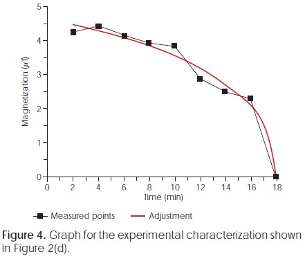
The four experimental configurations used in this study were similar, so they presented identical behavior. Results of the magnetic variations corresponding to the first two experiments are shown in Figures 3 and 4. The graphics show the magnetization value measured by M-MGG versus time as the containers are removed from the phantom. Continues curve represents the exponential data fit according to equation (1). It is showed that the removal of material only affects the magnitude of the overall signal and simulates the emptying of ingested material in the process of intake, processing and disposal of food in the stomach.

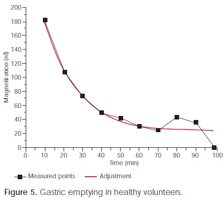
In order to show the gastric emptying evaluated in human subjects, Figure 5 exhibits the M-MGG evaluation in a healthy volunteerwho ingestedan oral solid food similar to that described previously.. As long as, Figure 6 shows the results in a patient with gastroparesis disorder.

DISCUSSION
Magnetic material placed in a phantom had a decaying behavior similar to oral magnetic contrast in the gastric emptying in subject and both exhibit an exponential decay fitting modeling by equation (1). The magnetic intensity decreases exponentially due to a decreasing into the food volume. Our findings provide enough information to continue using this method to infer gastric emptying half-time11,12.
Experimental results of meal magnetization show that the change is consistent with previously reported values11,12,15, because of the magnetic intensity begins to drop as biomagnetic material was removed. Also, our results show that solid food test is useful for measuring gastric emptying in healthy subjects15. Also, Figure 5 shows a good agreement with theoretical model of gastric emptying in healthy subject.
CONCLUSIONS
Althoughanalysisof GI diseaseadjusted models was not our study purpose, based on our results we conclude that magnetization decreased almost linearly during the first 50 minutes of starting the analysis, with a subsequent fluctuation unclear, in patients with gastroparesis syndrome. To verify this protocol could be interesting to reduce the acquisition sampling interval to times shorter than 5 minutes, thus obtaining a greater number of points for the adjustment.
Finally, this solid food test has similar characteristics to conventional meal test employed in scintigraphy, whose technique is considered gold standard method to assess gastric emptying in certain GI disorder. Also, among advantages of M-MGG is the fact that it is a non-ionizing radiation technique; it is a non-invasive so it could be implemented in our hospitals as a routinely GI alternative diagnosis technique, and it can be used in the monitoring of dyspepsia syndrome and gastroparesis due to diabetes mellitus disease.
ACKNOWLEDGMENTS
Authors want to thank CONACYT and DAIP for financial support under grant numbers J50182 and 000017/10.
REFERENCES
1. Wenger MA, Engel BT, Clemens TL and Cullen TD. Stomach motility in man as recorder by the magnetometer method. Gastroenterology 1961; 41: 479-85. [ Links ]
2. Wenger MA, Henderson EB, Dinnin JS. Magnetometer method for recording gastric motility. Science 1957; 125: 192-195. [ Links ]
3. Cohen D. Ferromagnetic contamination in the lungs and other organs of the human body. Science 1973; 180: 743748. [ Links ]
4. Cohen D. Detection and analysis of magnetic fields produced by bioelectric currents in humans. Journal of Applied Physics 1969; 40: 1046-1048. [ Links ]
5. Cohen D, Edelsack EA, Zimmerman JE. Magnetocardiogram taken inside a shielded room with a superconducting point-contact magnetometer. Appl Phys Lett 1970; 16: 278-280. [ Links ]
6. Benmair Y, Dreyfuss F, Fischel B, Frei EH, Gilat T. Study of gastric emptying using a ferromagnetic tracer. Gastroenterology 1977; 73: 1041-1045. [ Links ]
7. Benmair Y, Fischel B, Frei EH, Gilat T. Evaluation of a magnetic method for the measurement of small intestine transit time. American Journal of Gastroenterology 1977; 68: 470-475. [ Links ]
8. Frei EH, Benmayr Y, Yerashlmi S, Dreyfuss F. Measurements of the emptying of the stomach with a magnetic tracer. IEEE Trans Mag 1970; 6: 348-349. [ Links ]
9. Córdova-Fraga T, Carneiro AA, De Araujo DB, Oliveira RB, Sosa M, Baffa O. Spatiotemporal evaluation of human colon motility using three-axis fluxgates and magnetic markers. Med Biol Eng Comput 2005; 43: 712-715. [ Links ]
10. De la Roca-Chiapas JM, Córdova-Fraga T, Zarate A, Solis Ortiz S, Bernal JJ, Vargas M, Sosa M. Magnetogastrography in patients with functional dyspepsia. Biomagnetism: Interdisciplinary Research and Exploration Proceedings of the 16th International Conference on Biomagnetism, Hokkaido University Press (Japan). 2008: 15-17. [ Links ]
11. Córdova FT, Gutiérrez JG, Sosa AM, Vargas LF, Bernal AJJ. Gastric activity studies using a magnetic tracer. Physiol Meas 2004; 25(5): 261-270. [ Links ]
12. De la Roca CJM, Hernández SE, Solís MS, Sosa AM, Córdova FT. Magnetogastrography (MGG) reproducibility assessments of gastric emptying on healthy subjects. Physiol Meas 2007; 28(2): 175-183. [ Links ]
13. Carneiro AA, Baffa O, Oliveira RB. Study of stomach motility using the relaxation of magnetic tracers. Phys Med Biol 1999; 44(7): 1691-1697. [ Links ]
14. Forsman M. Intragastric movement assessment by measurement magnetic field decay of magnetized tracers particles in a solid meal. Med Biol Eng Comput 2000; 38(2): 169-174. [ Links ]
15. Reynaga-Ornelas MG, De la Roca-Chiapas JM, Cordova-Fraga T, Bernal JJ, Sosa M. Solid test meal to measure the gastric emptying with magnetogastrography. AIP Conference Proceedings, 2008; 1032: 246-248. [ Links ]
16. Walter Greiner. Classical Electromagnetics. Springer-Verlag (USA), 1996. [ Links ]
Nota
Este artículo también puede ser consultado en versión completa en: http://www.medigraphic.com/ingenieriabiomedica/

