


 nova página do texto(beta)
 Inglês (pdf)
 Artigo em XML
 Referências do artigo
 Enviar este artigo por email
 Citado por SciELO
  Similares em
SciELO
Similares em
SciELO 
 Permalink
Placozoans are the simplest free-living animals and probably the most primitive Eumetazoan. With only six cell types organized as an irregular ciliated flat disc (Smith et al., 2014) their body plan is comprised of marginal versus interior and top versus bottom regions and no symmetry axes are evident. These organisms posses san upper and lower epithelium -with no basal lamina or extracellular matrix- that enclose a loose network of interconnected branched cells called fiber cells. Discovered by F. E. Shultz in the 19th century, Trichoplax adhaerens is the only described species of the phylum Placozoa (Schulze, 1883). Its simplicity and taxonomic basal position make Placozoans unique orga nisms to study early metazoan evolution. A recent phylogenomic study that compared the relationships of early diverging metazoan lineages, unambiguously placed Porifera as the sister group of all Metazoans; Ctenophora emerged as the second branch of Eumetazoa and Placozoa resulted the basal taxon of the clade Placozoa+Cnidarian+Bilateria (Simion et al., 2017).
Placozoans have been found in tropical and subtropical waters around the world (Eitel et al., 2013) and despite their identical morphology, molecular studies imply the existence of many species and different lineages occurring sympatrically (Voigt et al., 2004). In one extensive study about the biodiversity found in the Gulf of Mexico, the phylum Placozoa is listed as part of the species of several phyla found (Felder & Camp, 2009); however, no additional data, formal description, or scientific publication was done.
Here, we describe findings dozens of Placozoans adhered to the glass walls of a seawater aquarium after performing several partial changes of water obtained from the Tuxpan reef in June and July 2015. The Tuxpan reef is an emerged platform located 12 km from the coast line in the Southwest of the Gulf of Mexico.This littoral region has estuarine waters with mangrove swamps a Tuxpan harbour is situated at the mouth of the Tuxpan. River. It is known that the ocean currents in the Gulf of Mexico transports water along the coastal line from the Southeast (Campeche Bank) to the Southwest (Zavala-Hidalgo et al., 2003). Parallel to this coastal line, several reefs form an ecological corridor (Ortiz-Lozano et al., 2013), which are inhabited by species that arise from the Caribbean Sea. Therefore, the Placozoans we found may be a native population but derived from the Caribbean zone, probably related with the H1-4 haplotypes (Grell & López-Ochoterena, 1987; Voigt et al., 2004). The aquarium that harbors the Placozoans is three years old and has colonies of corals, sponges, and sea grass beds that were obtained from the same reef. No synthetic water, exotic species, or commercial food for corals were introduced in the aquarium. The setup, with a volume of 400 L, has a sump filter with live rock obtained from the reef zone and the temperature is maintained at 26-28 °C. Three organisms were fixed in 4% glutaraldehyde in seawater and registered as Voucher specimens in the National Collection of Phylum Porifera, UNAM (catalog number CNPGG-1488, CNPGG-1489, and CNPGG-1490).
Most Placozoans were around 600 mm in diameter although some smaller or larger specimens were also found (ranging from 200 to 1500 mm). They showed an irregular “hairy plate” form (“tricho-plax”) and gliding movement (Figuras 1 a-b ). The characteristic birefringent vesicles or “shiny spheres” of this taxon were clearly observed on the upper side (Figura 1 c ), except in the outer margin where upper and lower epithelium apposed (Figura 1 e ). Images also show yellow bodies scattered below the upper epithelium containing vacuoles of digested algae (Figura 1 c ). The Trichoplax moved constantly with amoeboid movements but without a particular direction; at intervals, the margins folded such that the lower epithelium formed transient grooves (Figura 1 a , arrows). We observed that sometimes one groove was extended from side to side and the Placozoan slides on its own lower epithelium (Figura 1 f ). Occasionally, the edges rose and enclosed the upper epithelium for a few minutes (Figura 1 g-k ).
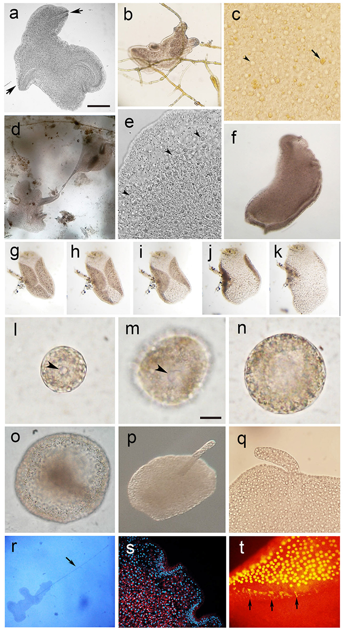
Figures 1a-t Placozoans confined in marine aquariums. a) Living Trichoplax showing the characteristic epithelial grooves and irregular shape. b) Gliding Trichoplax adhered to Feldmannia sp. algae. c) The characteristic yellow vacu oles of digested algae (arrow) and the shiny spheres (arrow head). d) Asexual reproduction by binary fission. e) Detail of the upper marginal region showing the characteristic shiny spheres (arrow heads) and radially aligned fiber cells. f) Bent Trichoplax with apposed lower epithelium. g) Time-lapse image of Tri choplax bending the epithelial borders up ward. h) Downward movement of the marginal epithelia. i) With seconds, Trichoplax extends the marginal epithelia. j)Trichoplax returns to extended position. k) Extended Trichoplax starts the gliding movement. l) Small-size open-sphere swarmer. m) Medium size open-sphere swarmer, arrow heads point to the opening. n) Cup shape swarmer. o) Spherical form of Trichoplax. p) Specimen with upper rod-like protuberance. q) Close-up showing the upper protuberance. r) Trichoplax with upper filament (arrow). s) Fluorescence image of Mito Tracker and DAPI-stained Trichoplax. t) Lyso Tracker stained lipophilic vesicles (shiny spheres); arrows point to small vesicles stained in marginal epithelium. Scale bars: a = 100 mm; m = 50 mm, scale apply to Figura l and n.
We observed two of the four modes of reproduction reported in the literature: binary fission (Figura 1 d ) and flagellated, hollow swarmers (Figuras 1 l-n ) (Thiemann & Ruthmann, 1991). By scraping the glass walls of the aquariums, spinning swarmers were obtained mostly adhered to algae. On glass slides introduced in the aquarium and examined one week later, we observed one spherical form of Trichoplax that slowly rotated, maintaining a strong adhesion to the substrate without signs of degeneration (Figura 1 o ). This spherical Trichoplax appears to be of the type previously reported as “hollow sphere with interior compartment” (Thiemann & Ruthmann, 1990). Interestingly, on other glass slides we captured some Trichoplax with a long protuberance in the upper side (Figuras 1p-q). This structure lasts for several hours but disappears after 24h. It seems to be the same slender threadlike extension of around 1 mm long that was often observed in those Trichoplax living in areas where the water flow is stronger (Figura 1 r ). This filamentous structure swings with the stream of water and maintains a constant size for hours; it is probably used to sense the currents of water. To our knowledge, this structure has never been reported before. Finally, we assayed fluorescent dyes to stain specific cellular structures, such as the nucleus (DAPI), mitochondrion (Mito Tracker, Molecular Probes) (Figura 1 s ), and acidic organelles (Lyso Tracker, Molecular Probes) (Figura 1 t ) to confirm the staining patterns reported in other studies (Smith et al, 2014).
Placozoans grew and multiplied well with these aquarium condictions and grazed upon Feldmannia sp. algae that grew on the glass walls (Figura 1 b ). They shared a micro-ecosystem with an assemblage of small organisms such as Vorticella, ciliates, heliozoans, nematodes, metamorphosing sponges, and cyanobacteria. Ciliates and nematodes made contact randomly with Placozoans but they did not elicit any specific response or reaction.
We have been able to maintain a healthy ecosystem in the marine aquarium with the appropriate conditions to obtain a constant supply of Trichoplax, making it possible to perform other experiments, for exam ple, molecular studies to determine the haplotype of this strain.

