


 nueva página del texto (beta)
 Inglés (pdf)
 Artículo en XML
 Referencias del artículo
 Enviar artículo por email
 Citado por SciELO
  Similares en
SciELO
Similares en
SciELO 
 Permalink
Introduction
There are different herbicides that can be used to increase the production, quality, and control of the growth of different weeds of commercial crops (Bolognesi, 2003; González et al., 2011). Although these herbicides offer great advantages, some tend to cause adverse effects on crop production and can also be genetically dangerous, that is to say, genotoxic (Aksakal, 2013; Valencia-Quintana et al., 2013), so it produces mutations in DNA and can cause hereditary diseases and cancer. Atrazine and dicamba, either individually or in combination, are two of the most used herbicides to control the growth of weeds on maize and other crops (Golla et al., 2011). Although some authors have reported inconclusive results regarding the genotoxicity of dicamba, their results indicate that dicamba has mutagenic effects on legume cells. In addition, it has been shown to cause sister chromatid exchanges in Chinese hamster ovary cells and increased micronucleus frequency in fish blood cells (Cenkci et al., 2010; González et al., 2009; Lee et al., 1983; Ruiz et al., 2014; Ruiz et al., 2018).
The Environmental Protection Agency (US-EPA, 2009) decided to reassess the risks of atrazine to the health and the environment; additionally, Srivastava & Mishra (2009) reported its effect in altering the mitotic index, increasing the number of chromosomal aberrations in plants. Genotoxic activity of atrazine was clearly demonstrated in different bioassays using earthworm, quail, micro-algae, fishes, anurans, rodents, human lymphocytes, HepG2 cells, and plants (Adeyemi et al., 2015; Campos-Pereira et al., 2012; García-Villada & Reboud, 2007; Gonçalves et al., 2017; Nwani et al., 2011; Powell et al., 2011; Song et al., 2009). However, information on its genotoxic effects in aquatic organisms is rather scarce (Cavas, 2011). On the other hand, there are other studies reporting that this compound has no genotoxic activity (Brusick, 1994; International Agency for Research on Cancer [IARC], 1999; Sánchez et al., 2018; Zeljezic et al., 2006). Valencia-Quintana et al. (2013) reported that marvel (atrazine/dicamba combination) did not show any DNA damage in meristematic cells of Vicia faba root at a 62.5 μg/ml-31.25 μg/ml concentration, but it occurred from the 125 μg/ml-62.5 μg/ml concentrations.
In order to assess the genotoxic activity of herbicides, multiple bioassays have been used (Alvarez-Moya et al., 2011; Alvarez-Moya et al., 2014; Valencia-Quintana et al., 2013; Zúñiga-González et al., 2016). The comet assay is a bioassay that consists of performing simultaneous electrophoresis of individual cells. The result is cells with tails that look like stellar comets when viewed under a microscope, hence, its name “comet test”. The length of the tail is directly proportional to the genetic damage. Comet assay is a reliable test to detect different types of DNA primary damage (Tice et al., 2000). Additionally, it has been proven to be highly sensitive and reproducible (Singh et al., 1988). This test detects different types of DNA damage such as in single and double-strand breaks, covalent cross-links between DNA/DNA, DNA/protein, or base deamination by oxidative damage occurring in apurinic or apirimidinic sites (Azqueta & Collins, 2013; Brendler-Schwaab et al., 2005). It is also worth mentioning that the comet assay has been widely used for assessing the genotoxic activity of a variety of pesticides (Alvarez-Moya et al., 2014; Garaj-Vrhovac & Zeljezic, 2002). Due to the paucity of data on the genotoxicity of the commercial formula marvel and the controversial genotoxicity of dicamba and atrazine, in this study, the genotoxicity of marvel at different concentrations was assessed. The selection of concentrations was based on previous studies which used high marvel concentrations and reported positives results, so we explored the lower concentration to detect the initial genotoxicity point of marvel. The alkaline comet assay was used in human and mice lymphocytes, as well as in fish erythrocytes, all of them potentially exposed organisms that can contribute to evaluate the genotoxic effect in species of various ecosystems, including humans (De Lapuente et al., 2015; Vischetti et al., 2020).
Materials and Methods
Chemicals used
Marvel commercial combination according to Marvel-Syngenta data sheet was obtained from Syngenta Agro, Mexico, D.F.: atrazine (3,6-dichloro-2-methoxybenzoic) (22.23%), dicamba (6-Chloro-N2-ethyl-N4isopropyl-1, 3, 5-triazine-2, 4-diamine) (11.45%), inert compounds (66.32%). Water was used as a solvent to prepare the marvel concentrations (0.5 μg/ml-0.26 μg/ml, 5 μg/ml-2.64 μg/ml, 50 μg/ml-26.4 μg/ml, 500 μg/ml-264 μg/ml atrazina-dicamba) and the positive control (N74 nitrosodiethylamine [NDEA, CAS No. 55-18-5] at a 5 mM).
Human study
Ethical approval was conferred from the institutional ethics committee for experimental treatments (DBCYM/184/2020). Written informed consent for participation in the study was obtained from each of the participants. Furthermore, all research was conducted in accordance with the ethical standards on human experimentation in the Helsinki Declaration of 1975.
Obtaining samples and cells preparation
Human. Blood cells were chosen for this study because it has been shown that this type of cells offers advantages for monitoring the effects of genotoxic agents (Faust et al., 2004). In this sense, 10 human participants over 18 years of age (six males and four females) responded to a questionnaire to rule out exposure to genotoxic agents. Individuals on medical treatment, smokers, drug users, and residents of the polluted area of Guadalajara, Jalisco, México, were excluded from the study. In Table 1, individual factors that can induce genetic damage are shown. As negative controls, blood samples from young people with the best indicators mentioned in the previous questionnaire were used. For the preparation of human lymphocytes, peripheral blood samples were obtained by finger puncture from participants. Each blood sample (100 μl) was placed in a test tube containing 4 ml of phosphate-buffered saline (PBS) (160 mM NaCl, 8 mM Na2HPO4, 4 mM NaH2PO4, and 50 mM EDTA, pH 7) and immediately centrifuged at 3000 rpm for 5 min. The supernatant was removed, and the pellet was suspended in phosphate buffer and immediately stored at 4 °C until used. Both positive and negative control groups for monitoring were used as recommended by Møller et al. (2020).
Table 1 Individual factors of human blood donors that can influence DNA integrity.
| Individual factor | Bioassay used | Reference |
| Ionizing radiation | Micronuclei | Balasen & Ali 1991; Erexon et al.,1991; Zúñiga-González et al., 2016. |
| Smoking | Comet assay |
Sardas, Aygün, Gamli, Ünal, Ünal, Berk & Karakaya, 1998;
Lebailly et al., 1998. |
| Age | Micronuclei | Maluf, 2004. |
| Diet | Cancer test |
Maluf, 2004; Pinto et al., 2015. |
| Occupational and environmental exposition to genotoxic agents |
Demarini, 2013. Alvarado-Hernández, Montero-Montoya, Serrano-García, Arellano-Aguilar, Jasso-Pineda & Yáñez-Estrada, 2013; Pitarque et al., 2002. |
|
| Exercise | Comet assay |
Maluf, 2004; Pinto et al., 2015. |
Source: Author’s own elaboration.
Mice. Ten mice (Balb/c strain) of four to six weeks of age were provided by the Molecular and Cellular Department of CUCBA and were kept at 12-h light/dark cycle, temperature of 25 ºC-30 ºC, humidity rvange of 55%-15%, and with access to food and water ad libitum. Animal use was in accordance with the Guide for the Care and Use of Laboratory Animals (Norma Oficial Mexicana, 1999). All procedures were approved by the institutional ethics committee. The inhalation of ethyl ether, as an anesthetic process, was carried out by placing the animal in a closed cube for 5 min; after that, cardiac puncture was performed, and 100 μl blood samples were obtained from each animal. Samples were maintained following the procedure mentioned in human samples. Mice were euthanized by CO2 asphyxiation.
Fishes (Oreochromis niloticus). Ten fishes were provided by the Banco Nacional Genómico and acclimatized in aquariums of 5000 L of tap water with air recirculation, under a natural photoperiod with the following physicochemical conditions: salinity 0, temperature 20 °C ± 1 °C, pH 7.3 ± 0.2, and dissolved oxygen 8.1 mg/L ± 0.5 mg/L. The fishes were fed with fish roe every other day. Animal use was in accordance with the Guide for the Care and Use of Laboratory Animals (Norma Oficial Mexicana, 1999. Blood was collected by branchial puncture from specimens 15 cm-23 cm in length. Hence, 100 μl blood sample was obtained from each animal and maintained following the procedure mentioned in human samples.
It is pertinent to mention that Oreochromis niloticus erythrocytes and human and mouse lymphocytes have been used as biomonitors of genetic damage induced by pesticides (Alvarez-Moya et al., 2022).
Comet assay
The comet assay was carried out using total blood of human, mice, and tilapia, following the procedures described by Singh et al. (1988) and Alvarez-Moya et al. (2001). Each blood sample previously obtained was divided into four parts. Then, each part was mixed (v/v) with a different final concentration of the marvel (0.76 μl/1 ml, 7.68 μl/1 ml, 76.8 μl/1 ml, and 768 μl/1 ml) for 6 h at 25 °C. Untreated cells, resuspended in 500 μl of PBS, were used as negative controls; on the contrary, cells treated with a final concentration of NDEA at 5 mM were used as positive controls. After that, all samples were washed three times with PBS and centrifuged at 3000 rpm for 10 min. Later, the pellet was resuspended in 100 μl of PBS and subsequently placed on agarose gels. This procedure was performed in duplicate slides for each sample of the three species used. For the nuclear lysis, all slides were immersed in a lysis buffer (2.5 M NaCl, 100 mM Na2 EDTA, 10 mM Tris-HCl, 1% sodium lauryl sarcosine, 1% Triton X-100, and 10% DMSO, pH 10) for 2 h at 4 °C and then washed with distilled water. Furthermore, the slides were placed in a horizontal electrophoresis system (Bio-Rad, model A6) containing electrophoresis buffer (NaOH 300 mM, EDTA 1 mM, pH 13) for 45 min at 4 °C to allow the DNA to unfold. Afterwards, the electrophoresis was carried out for 20 min at 1.0 V/cm with an amperage of ~300 mA (Labconco, model 4333280). The same electrophoretic unit and power supply were used throughout the study. After the electrophoresis was carried out, all slides were gently washed with distilled water to remove the alkaline solution. Then, they were all immersed in a neutralization buffer (0.4 M Tris base, pH 7.5) for 5 min. The gels were stained with ethidium bromide (100 μl at 20 μg/ml) for 3 min and then washed three times using distilled water to remove possible bromide residues. The slides were analyzed using a fluorescence microscope (Zeiss, model Axioskop 40) with a 515-560 nm excitation filter. Comets were observed at 40X, and their tail length was determined using the comet assay II software (Zeeiz Sinoptic Mikro S.A De C.V. México, 2012), which automatically indicates the start and the end of the comet length (Ma et al., 1994). Approximately, 200 comet cells per studied subject were analyzed.
Statistical analysis
In all experimental groups, mean and standard deviation of the comets’ tail length were obtained, but only the significant differences between experimental groups are shown. Then, it was analyzed by one-way analysis of variance (Anova) using the CoStat program (Ma et al., 1994). All experimental groups were compared with their corresponding negative and positive control using the Tukey test. Two slides for each experimental treatment, including the negative and positive controls, were evaluated. A value of p ≤ 0.05 indicated significance.
Results
Figure 1 is for the sole purpose of showing images of electrophoresed cells (single cell electrophoresis) from humans, mice, and tilapia. It can be seen how the negative controls have a lower migration than the experimental groups. The mean length of the tail of 100 cells is the value that is taken as a reference to establish comparisons: 100 cells negative control, 100 cells positive control, and so on. The representation of these values is shown in the bars of Figure 2.
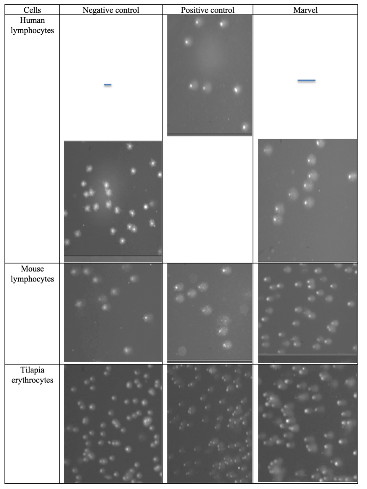
Source: Author’s own elaboration.
Figure 1 Comets from human and mice lymphocytes and tilapia erythrocytes exposed to positive control and marvel herbicide. The blue line indicates the tail length parameter, and all cells are at the same magnification scale (10X). The nuclear size is not considered.

Source: Author’s own elaboration.
Figure 2 Genetic damage induced by different concentrations of marvel herbicide in human (a, b) and mice (e, f) lymphocytes and fish erythrocytes (c, d) using tail length and tail moment.
There are two parameters used in the comet test: tail length and tail moment, both parameters were used to evaluate genetic damage in the three species studied. The genotoxic activity of different concentrations of marvel in human lymphocytes is shown in Figure 2a and 2b. All concentrations showed significant genotoxic activity when compared to the negative control (p < 0.05); however, no significant difference was observed between the different marvel concentrations and the positive control.
The genotoxicity of the different concentrations of marvel in tilapia erythrocytes are shown in Figure 2c and 2d. All concentrations showed significant genotoxic activity when compared to both negative (p < 0.05) and positive (p < 0.05) controls. Only the concentration at 0.76 μl/1 ml showed a significant difference (p < 0.05) in respect to the other concentrations and the positive control. A significant difference was not observed between the concentrations at 7.68 μl/1 ml, 76.8 μl/1 ml, and 768 μl/1 ml. Similar to that observed in blood cells of human and fish, the genotoxicity of the marvel herbicide in mice lymphocytes was significant when compared to the negative control (p <0.05) in both tail length and tail moment (Figure 2e and 2f). Using tail moment and tail length, no significant difference was observed between the positive control and the marvel concentrations (Figure 2e and 2f). In tilapia erythrocytes and mice lymphocytes, migration averages increased starting at the 0.76 μl/1 ml concentration; however, the average tail length remained similar in the rest of the concentrations.
Discussion
Studies in various cell types have shown that atrazine, a compound of the marvel herbicide, causes DNA damage (Clements et al., 1997; Dos Santos & Martínez, 2014; Ribas et al., 1995); however, its genotoxicity is still under discussion (IARC, 1999; Kligerman et al., 2000) since other authors report absence of genotoxicity (Brusick 1994; IARC, 1999; Zeljezic et al., 2006). On the other hand, dicamba, another compound of the marvel herbicide, has also proved to be genotoxic (Asita & Matebesi, 2010; Cenkci et al., 2010; Filkowski et al., 2003; González et al., 2007; González et al., 2011; Perocco et al., 1990). Additionally, even though the genotoxic potential of marvel (atrazine-dicamba combination) has been reported in sweet corn (Reynoso et al., 2015) and Vicia faba (Sánchez et al., 2018), it is very important to determine the genotoxic effect of this commercial combination and its different concentrations in organisms, which will contribute to evaluate the genotoxic effect in different species and ecosystems. Moreover, it is very important to evaluate the genotoxicity of marvel in exposed people because the existence of genetic damage can alter the state of health (Rasgele et al., 2014).
In this study, the commercial form of marvel herbicide showed to be highly genotoxic, just as previously reported on evaluations of its individual compounds, atrazine and dicamba, in different species (Campos-Pereira et al., 2012; Gonçalves et al., 2017; Huang et al., 2015; Larramendy et al., 2010; Perocco et al., 1990; Sharma & Vig 2012; Song et al., 2009; Srivastava & Mishra, 2009; Ruiz et al., 2014).
These results indicate that marvel has considerable genotoxic activity at a concentration of 0.76 μl/1 ml in human and mouse lymphocytes, while in fish erythrocytes (0.76 μl/1 ml) the genotoxic activity is poorly detectable. Both parameters, tail length and tail moment, showed similar results. Schaaf (2015) designed a numerical scale to evaluate the characteristics of substances in relation to human health and damage to the environment, which is called “environmental impact assessment” (VIA). On this scale, atrazine reached a VIA of 310 with low ecotoxicological impact, high genotoxic impact, and high irritative capacity, being highly persistent in soils and waters. Dicamba, with a VIA of 180, showed moderate toxicological impact, low carcinogenicity, and genotoxicity, but it is highly irritating and low persistent in water and soil.
Our results confirm the genotoxicity of marvel, and considering the model of Schaaf (2015), this herbicide will probably show more prominent IVA than atrazine and dicamba. At the ecotoxicological level, it will be moderately toxic with some effect on bees, high genotoxicity, high irritability, presence in sediments, and soil, but with little bioaccumulation. In a previous study, our working group reported that marvel induced damage in sweet corn kernels at concentrations that went from 384 μg/1 ml to 768 μg/1 ml (Reynoso et al., 2015); however, other work groups reported genotoxic effects only starting at 187.5 μg/1 ml and up to 6000 μg/1 ml concentrations (Sánchez et al., 2018). These results strongly differ with the absence of marvel genotoxicity reported by Zeljezic et al. (2006).
This study showed that marvel is genotoxic at very low concentrations, and it could be associated with changes in DNA methylation induced by dicamba (0.26 μg/1 ml-0.79 μg/1 ml) (Arslan 2020; Yildirim et al., 2014) and atrazine (0.126 μg/1 ml and 0.005 μg/1 ml) (Cavas, 2011; Sharma & Vig, 2012). Data of the genotoxicity-concentration relationship of the commercial formula marvel and its active ingredients are shown in Table 2. To ease the comparisons, different reported concentrations at ppm were converted into concentration values at μg/1 ml.
Marvel was equally genotoxic in all the cells studied, therefore, continuous exposure to this herbicide will represent a risk to all organisms, even in very low concentrations. The possible genotoxic effect of adjuvant substances (secondary diluent, solvent, dispersant, antifreeze) is not ruled out, which in the case of marvel constitute 65.32% of its weight, and they are not properly described (Marvel-Syngenta data sheet); it appears that this is a frequent situation Argüello-Rangel et al., 2015). The absence atrazine genotoxicity and presence of genotoxicity in a commercial atrazine-based herbicide Zeljezic et al., 2006) are attributed to adjuvant substances present in the commercial herbicide and suggest the need to evaluate the genotoxicity of these chemical compounds (Solomon et al., 2013). In addition, there are only few reports regarding the genotoxic evaluation of the chemically pure dicamba-atrazine combination, as shown by the empty spaces in Table 2.
Table 2 Genotoxicity of the commercial herbicide marvel and its active ingredients atrazine and dicamba studied individually or in combination. Commercial herbicides based on atrazine or dicamba and their adjuvants are also included.
| Authors | Organism or cell studied |
Marvel (Atrazina- dicamba- adjuvants) |
Atrazine | Dicamba | Atrazine-dicamba combination chemically pure |
Atrazine-based herbicide and adjuvants |
Dicamba-based herbicide and adjuvants |
| Arslan (2020) |
Triticum Aestivum (Randomly Amplified Polymorphic) DNA methylation change |
0.076+ 0.152+ 0.228+ |
|||||
| Sharma & Vig (2012) |
Allium cepa Micronuclei |
0.126+ | |||||
| Our data | Erythrocytes of Oreochromis niloticus and human and mice lymphocytes comet assay |
0.76+ 7.68+ 76.8+ 768+ |
0.5 5.04 50.40 504.0 |
0.26 2.64 26.40 264.00 |
|||
| Reynoso et al. (2015) |
Zea mayz L. comet assay |
384-768+ 768-1536+ |
252-504 504-1008 |
132-264 264-528 |
|||
| Sánchez- Alarcón et al. (2018) |
Vicia faba Micronuclei |
93.7- 187.5+ 375+ 1400+ 3000+ 4500+ 6000+ |
62.5- 125+ 250+ 1000+ 2000+ 3000+ 4000+ |
31.25- 62.5+ 125+ 400+ 1000+ 1500+ 2000+ |
|||
| Cavas (2011) |
Carassius auratus L. Micronucleicomet assay |
0.005+ 0.010+ 0.015+ |
0.005+ 0.010+ 0.015+ (Gesaprim herbicide) |
||||
| Zeljezic et al. (2006) | Human lymphocytes comet assay |
0.047- 0.47- 4.7- |
0.047+ 0.47+ 4.7+ |
||||
| Yildirim et al. (2014) |
Phaseoulus vulgaris (changes in methylation) |
0.26+ 0.52+ 0.79+ |
|||||
| Sorensen et al. (2005) | CHO cell comet assay |
10- | |||||
| Filkoski et al. (2003) |
Arabidopsis tatiana recombination markers |
120 i | |||||
| Lee et al. (1983) |
Drosophila melanograster |
252 504 |
0.396 i 132 i 264 i |
||||
| Mohammed & Ma (1999) |
Tradescantia Micronuclei stamen hair mutation bioassay |
12 600+ | |||||
| González et al. (2011) | CHO-K1 Mn | 12.6 + 100.8 + |
+ means Positive results; - means Negative results; i means Inconclusive
Note.In order to establish an accurate comparison, the data reported previously were converted to μg and adjusted to μg/1 ml. Our experimental results from this study are included in the table in bold letters.
Source: Author’s own elaboration.
Globally there is no clear consensus regarding the carcinogenicity or another indicator of ambient impact of dicamba or atrazine individually or in combination, neither is there agreement about the marvel commercial formulation, so further information regarding this issue is needed.
Conclussion
Results obtained in this study suggest that marvel is highly genotoxic even at concentrations as low as 0.76 μg/1 ml, and perhaps even lower. These concentrations are the lowest reported for marvel. It may be possible that the differences reported in regards of marvel genotoxicity and its active ingredients are due to the marked differences between the concentrations studied, the chemical adjuvants present in the commercial formulations, and methodological variations between this and other studies. It is necessary to evaluate the genotoxicity of the chemically dicamba-atrazine combination, without adjuvants, and to make comparisons with commercial formulations based on dicamba or atrazine.
Conflict of interest
The authors declare that they have no conflict of interest.

