


 nova página do texto(beta)
 Inglês (pdf)
 Artigo em XML
 Referências do artigo
 Enviar este artigo por email
 Citado por SciELO
  Similares em
SciELO
Similares em
SciELO 
 Permalink
Introduction
Danon syndrome was first described by Danon MJ in 1981. Its alterations are framed in a triad consisting of dilated cardiomyopathy, myopathy and mental retardation. Initially, it was believed that it was expressed only in males. However, it is now known that it can affect both men and women. Most cases and severe forms of the condition have been found in men. Mutations in the LAMP2 gene, found in the Xq24 locus, have been known to cause the disease. This gene is responsible for the production of a protein named lysosomal associated membrane protein-2 (LAMP-2). It plays an important role in the mechanism of autophagy mediated by chaperones, mainly in skeletal and cardiac muscle cells, which explains its clinical manifestations.
To date, there is no clarity about the incidence, prevalence and prognosis of this disease. What is known is that its cardiac manifestations, both electrical and structural, play a leading role in the severity the phenotype. It is likely that in addition to its low incidence, Danon disease is also underdiagnosed, as it is easily confused with more prevalent conditions with similar phenotypes and genetic studies are scarce in countries like Colombia. From the point of view of treatment, the measures that have been adopted are focused on reducing and mitigating these alterations.
Here we present a case of a 29-year-old male patient who underwent multiple diagnostic and therapeutic interventions without being able to accurately document his pathology until adulthood. Only immunohistochemistry and posterior genetic testing done by CES Cardiology and Genoma CES was the diagnosis of Danon disease confirmed.
Case presentation
A 29-year-old male patient, without specific ethnic distinction, with no family history of known heart disease, was diagnosed with hypertrophic cardiomyopathy in the first year of life. His condition was extensively studied and thought to be Pompe disease, which could not be corroborated. His psychomotor development was normal during childhood. In his preadolescence stage, he began to notice progressive loss of muscle mass associated with elevated Creatine Kinase (CK) and elevation of liver enzymes. The patient received treatment for polymyositis until 2011 without a response, which was suspended when no progress was observed in skeletal muscle biopsy. Concomitantly, he presented functional impairment and multiple syncopes, and at the age of 15, a bicameral pacemaker was implanted. 5 years later a ventricular septum of more than 30 mm was documented. Therefore, the decision was made to change the device for an implantable cardioverter defibrillator (ICD) as a primary prevention strategy for sudden death.
He was first evaluated by the CES Cardiology in July 2014 during the reprogramming of his pacemaker. At this point he was found with a marked loss of muscle mass (42 kg weight) as well as elevated liver enzymes and total CK. A computerized axial tomography (CT) of the heart (Figure 1) and echocardiogram (Figure 2) were performed in August 2014, where a 44 mm interventricular septum with a left atrium of 31 mm and an area of 16 cm2 was described. Left Ventricle ejection fraction was of 70% without ventricular dilatation but with severe hypertrophy (calculated mass 1295.86 g, mass index 900 g/m2, relative thickness 1.94). Until that moment, the patient had never presented discharges from the device. However, the patient presented an emergency two months later for inappropriate ICD discharges generated by atrial fibrillation (AF) with rapid ventricular response, despite the maximum doses of metoprolol and verapamil.

Figure 1: Computerized axial tomography of the heart, in which there is evidence of the thickening (septal hypertrophy) myocardium.

Figure 2: Transthoracic echocardiogram with different visualizations of the great cardiac hypertrophy.
Given the patient’s clinical history and the phenotypic manifestation, Danon disease was considered as a diagnostic option. He was programmed for electrical isolation of pulmonary veins due to his history of AF in order to maintain the sinus rhythm as long as possible and to avoid inappropriate discharges due to the rapid ventricular response associated with AF. During this procedure, an endomyocardial biopsy was performed. Here, reports indicated focal hypertrophy of fibers with slight degenerative changes, sarcoplasmic autophagic vacuoles with lamellar membranes, cellular detritus and fine granular material in lysosomes, with no evidence of amyloid deposition or inflammation and presence of cytoplasmic inclusions. These findings are histologically compatible with Danon disease. Figure 3 shows electron microscopy images obtained from the endomyocardial biopsy of the patient and their corresponding interpretation.

Figure 3: Electronic microscopy images obtained from the endomyocardial biopsy of the patient. They show autophagic vacuoles (B) in the sarcoplasm occupied by lamellar membranes (A) and lipofuscin pigment (C). These findings are compatible with Danon disease.
Whole exome trio sequencing was used to determine mutations in the genome of the patient, as well as the mother and father. Genomic DNA was extracted from peripheral blood samples obtained. Several quality criteria were considered before library preparation, such as DNA free of RNA and proteins, with a ratio between 1.8 and 2.0 of A260/A280. Libraries were created using the x Gen Whole Exome Panel kit, Integrated DNA Technologies (IDT). Exome sequencing was performed on the Illumina HiSeq 2500 platform. Average sequencing coverage was of 120X. The reads were aligned with the human reference Hg19 genome by means of the BWA-MEM algorithm with predetermined parameters. The variants were determined with GATK Lite, Samtools and Free Bayes, generating a consensus of variants for classification. The annotation and variant information uses databases such as genomic Super Dups, NHLBI Exome Sequencing Project (ESP6500), 1000Genomes Phase 3, dbSNP 138, dnNSFP, Exome Agregation Consortium and ClinVar. Population frequencies were determined by the information of the Colombian population of 1000Genomes. The classification of variants was determined by the classification of specific genetic change with clinical evidence reported in ClinVar and Clinvitae. Additional information on the variants is obtained from OMIM, Pubmed, GeneCards and NIH Genetics Home Reference.
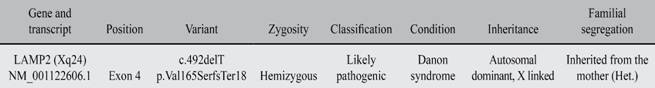
The whole exome trio sequencing allowed us to find a mutation with a likely pathogenic classification, which correspond to the reported phenotype of the patient. The specific mutation corresponds to a hemizygous single nucleotide deletion in the LAMP2 gene, resulting in a change in reading frame (c.492delT p.Val165SerfsTer18). The LAMP2 gene encodes a protein associated with lysosomal membrane type 2. Lysosomes are components that digest and recycle cellular material. This protein is important in the normal functioning of muscles, including cardiac muscle. Mutations in LAMP2 had been previously associated with Danon disease and several cardiomyopathies. This gene is linked to the X chromosome, which led us to suspect the origin of the mutation belonging to the mother. The trio analysis revealed that indeed, the mother was a heterozygous carrier of the single nucleotide deletion. The father did not have mutations in LAMP2. The changes and other specific mutation information can be found in Table I. To complete the family studies, transthoracic echocardiograms were performed on the two siblings of the patient, both of which are males, as well as both of the parents. All family members tested had normal results for their respective ages and no alterations were noted.
Discussion
Danon disease is a dominant X-linked hereditary syndrome,1 with no ethnic distinction and no clearly established epidemiology to date. This entity affects both men and women, although the condition is more prevalent and severe in males. It was initially described by Danon MJ in 1981, who described two 16-year-old men with mental retardation, cardiomegaly, and proximal skeletal myopathy, in whom they demonstrated accumulation of glycogen in their muscles without alterations in the enzymes responsible for their metabolism. Today, there are several cases reported in the world that expose a wide spectrum of alterations that compromise from the cardiovascular, muscular and neurological systems to specific organs such as liver and the spleen. Therefore, the degree of involvement and prognosis of the disease is variable, which is evident in the heterogeneity of the investigations carried out so far.2
This conditions primarily caused by a mutation in the LAMP2 gene, located on chromosome Xq24, which encodes a lysosomal membrane-associated protein type 2. Until now, published literature only mentioned mutations in this gene as causing the disease. Histological tests show severe vacuolization and degeneration of cardiomyocytes, including myofibrillar disruption and lipofuscin accumulation, changes that may explain the phenotype as the disease manifests itself.3,4
Although the exact pathophysiological mechanism of the disease is still not understood, the presence of cardiomyopathy has been defined as a classical triad in 100% of cases, dilated or hypertrophic. Myopathy is observed in 90% of the cases, generally mild and silent, and mental retardation in 70% of the cases. With respect to cardiac involvement, for men, cardiomyopathy is progressive and of concentric hypertrophic type, preserves the ejection fraction and at least, at initial stages, cavitary dimensions are normal. However, concentric hypertrophy over time may evolve to eccentric hypertrophy. In women, dilated cardiomyopathy is the predominant variant and usually does not have manifestations as severe as in men. In addition, literature shows that women who are carriers of the mutation may experience onset of symptoms later in life and not as quickly as in men. A previously reported case having performed genetic studies on parents of patients affected by the disease observed that mothers may experience cardiomyopathies later in life, even without demonstrating alteration of the cardiac anatomy or function when initially tested.5,6 This may explain why the mother of our patient showed no signs of a pathological phenotype in the echocardiograms. In addition, cardiac tissue exhibits severe fibrosis, which favors the development of supraventricular and ventricular arrhythmias. Among the most common electrophysiological alterations, reports to date showed that atrioventricular conduction abnormalities occur in 86% to 100% of patients, as well as Wolff - Parkinson - White pre-excitation phenomena. However, a recent study with 7 patients diagnosed with Danon disease, found that AF was the most common arrhythmia in this type of patients and no accessory pathways were documented,7 event that was evident in our patient, in whom AF was documented for rapid ventricular response. Also, the alterations of the ventricular myocardium behave as a good substrate for the generation of ventricular arrhythmias and, consequently, as an independent risk factor for sudden death.3,4
However, the disease also compromises other systems and organs other than cardiovascular. Among the most described include: skeletal myopathy, usually mild but may present with muscle cramps; neuropathy; Intellectual disability with consequent learning problems; Liver disease manifested as hepatomegaly, elevation of hepatocellular enzymes such as aspartate aminotransferase (AST) and alanine aminotransferase (ALT) and mild portal fibrosis.3,4 It should be noted that visual, retinal, gastrointestinal, pulmonary, neurological and psychiatric manifestations also present in different proportions.8 It is worth mentioning that in the case of this patient, the myopathy observed was significantly marked and manifested at early age. Usually the clinical onset of the disease is during adolescence. It appears earlier and more aggressively in men than in women. In many of the series reported in the medical literature, men have an early death and heart transplantation is necessary to improve their survival.9 As for the diagnosis, a complete and detailed medical history, with special emphasis on cardiovascular family history, death and unexplained diseases is essential, as well as a rigorous physical examination. As an initial and easily accessible measure, an echocardiogram or a CT scan of the heart can be requested evidencing the significant cardiac hypertrophy that they present. Cardiac biopsy with an adequate interpretation is the method that had been used to date for the diagnosis. However, the gold standard is a genetic analysis.10,11
Whole exome trio sequencing allowed us to determine the inheritance pattern of the mutation. The mother, which was a heterozygous carrier of the LAMP2 deletion, did not show pathological evidence in the echocardiogram. As mentioned previously, the carrier status also increases the probability of developing cardiovascular problems in the future. Although both siblings of the patients were not studied via whole exome sequencing, they are most likely carriers of the unaffected copy of the X chromosome inherited from the mother, as both are males in their late teens. Echocardiograms also showed unaffected cardiac tissue.
The treatment is focused on the type of manifestations that the patient presents, as no definitive intervention exists that corrects or avoids the progression of the disease. Because cardiac function is the main predictor of mortality, in the vast majority of cases, to achieve greater survival, cardiac transplantation before the age of 25 was required. For now, this is considered the most effective intervention. Likewise, according to the type of arrhythmia that is present, implantation of intracardiac devices such as pacemakers or defibrillators may serve as primary or secondary prevention of sudden death.10,11
It is worth noting that the possibility of performing genetic studies due to the suspicion of diseases such as this is fundamental to arrive at a clearer and more accurate diagnosis and thus, to be able to impact in an early manner in these patients.
Conclusions
Danon disease is a dominant X-linked hereditary syndrome, with no ethnic distinction and no clearly established epidemiology to date. This entity affects both men and women, being more frequent, in presentation as in severity, in males. The exact pathophysiological mechanism of the disease is still not understood, although the presence of cardiomyopathy has been defined as a classical triad in 100% of cases, dilated or hypertrophic, myopathy in 90%, generally mild and silent, and mental retardation in 70%. This entity is primarily caused by a mutation in the LAMP2 gene, located on chromosome Xq24, which encodes a lysosomal membrane-associated protein type 2, which functions as a membrane receptor in chaperone-mediated autophagy. This activity is important for the proper functioning of skeletal and cardiac muscle cells. Histologically, cardiomyocytes show severe vacuolization and degeneration, including myofibrillar disruption and lipofuscin accumulation, changes that may explain the phenotype as the disease manifests itself. As of now, heart transplant seems to be the only alternative. We believe that genetic screening as a first tier test in childhood for patients with symptoms of myopathy or specifically myocardiopathy could be beneficial for early detection and diagnosis.

