


 nueva página del texto (beta)
 Inglés (pdf)
 Artículo en XML
 Referencias del artículo
 Enviar artículo por email
 Citado por SciELO
  Similares en
SciELO
Similares en
SciELO 
 Permalink
Introduction
Among the edible fungi cultivated worldwide with higher production are Agaricus bisporus (J.E. Lange) Imbach (Agaricaceae) and Pleurotus ostreatus (Jacq.) P. Kumm. (Pleurotaceae; both families belonging to Agaricales, Agaricomycetidae, Agaricomycetes, Agaricomycotina, Basidiomycota, Fungi; Index Fungorum, 2019) (Royse, 2014), which present diseases and pests such as green mold caused by species of Trichoderma Pers. (Hypocreaceae) (Oei, 1991; Park et al., 2004), fire mold by Neurospora sp. (Sordariaceae) and brown spot of Pseudomonas tolaassi Paine, as well as some by viruses (Kim et al., 1995; 2000).
The disease generated by fungi of the genus Trichoderma is difficult to distinguish, because in the culture of mushrooms of the genus Pleurotus (Fr.) P. Kumm. (Pleurotaceae), both mycelia have similar characteristics, so the infection is detectable after approximately two weeks only, as it turns green due to the strong sporulation (Danesh et al., 2000), causing the inhibition of mycelial growth of Pleurotus because they compete for space and nutrients. The strategy of Trichoderma species is the secretion of hydrolytic enzymes, such as chitinases, (-glucanases and cellulases, which lyse the cell walls of the Pleurotus mycelium (Goltapeh and Danesh, 2000), causing the fruiting bodies not to form (Beyer et al., 2000).
The first report of Trichoderma infection in P. ostreatus culture occurred in North America (Sharma et al., 1996), then in Italy (Woo et al., 2004), South Korea (Park et al., 2006), Romania (Kredics et al., 2006), Hungary (Hatvani et al., 2007) and Spain (Gea, 2009). The species of Trichoderma are asexual and they inhabit the soil (Kredics et al., 2009). The main Trichoderma species that cause green mold disease in mushroom cultures of the genus Pleurotus are T. pleuroticola S.H. Yu & M.S. Park (Hatvani et al., 2007), T. harzianum Rifai (Sobieralski et al., 2012) and T. pleuroti S.H. Yu & M.S. Park (as 'pleurotum') (Hypocreaceae, Hypocreales, Hypocreomycetidae, Sordariomycetes, Pezizomycotina, Ascomycota, Fungi; Index Fungorum, 2019) (Marik et al., 2017). It has been estimated that Trichoderma together with mushroom virus X (MVX is a relatively new disease that affects the production of the commercial mushrooms) has caused a 25% loss in the production of cultivated edible mushrooms (Soković and van Griensven, 2006; Sobieralski et al., 2012).
Trichoderma pleuroti and T. pleuroticola have been found in the substrate of mushroom cultivation of the genus Pleurotus in different countries; however, the second species has also been found in soil and wood, as well as associated with P. ostreatus that grows in natural environments in countries such as Canada, United States of America, Iran, New Zealand and some European ones (Komon-Zelazowska et al., 2007; Kredics et al., 2009). This represents a higher risk of infection for the mushroom cultivation, since the sources of infection by T. pleuroticola can be diverse (Kredics et al., 2009). Trichoderma pleuroti could be specialized to an ecological niche related to substrates of mushroom cultivation of the genus Pleurotus (Komon-Zelazowska et al., 2007). One way to control the disease caused by Trichoderma has been to use disinfectants (chlorine, calcium hydroxide) and/or fungicides; the latter could generate resistant strains that require more of these chemical compounds to control the problem. In this sense it is very important to look for alternatives of biological control of the disease of the green mold, knowing the Trichoderma species involved in the cultures of P. ostreatus.
It is worth mentioning that the fungi of the genus Pycnoporus P. Karst. (Polyporaceae, Polyporales, Incertae sedis, Agaricomycetes, Agaricomycotina, Basidiomycota, Fungi; Index Fungorum, 2019), although not considered edible due to their corky consistency, have medicinal and biological properties, such as antimicrobial activity according to reports in China, Japan and the rest of Asia (although less frequent) (Calonge, 2011). In Mexico it has been used to combat acne, warts, diarrhea, dysentery and tinea capitis (cutaneous mycosis) (Martínez-Alfaro et al., 1983; Guzmán 1994; 2003; 2008). Currently, there are no reports on the use of Pycnoporus species against fungi of the genus Trichoderma, but there are a few that indicate activity against other organisms of the same kingdom. Recently, it was informed that the culture broth of P. cinnabarinus (Jacq.) P. Karst. had a bacteriostatic and bactericidal effect against Escherichia coli Escherich and Staphylococcus aureus Rosenbach, respectively, suggesting that cinnabarin and cinnabaric acid are the compounds produced by the fungus with antimicrobial activity (Díaz-Godínez et al., 2016). Al-Fatimi et al. (2013) reported that the methanolic extract of fruiting bodies of Pycnoporus sanguineus (L.) Murrill showed considerable antifungal activity against the human pathogens Candida albicans (C.P. Robin) Berkhout, C. krusei (Castell.) Berkhout, Microsporum gypseum (E. Bodin) Guiart & Grigoraki, Trichophyton mentagrophytes (C.P. Robin) Sabour. and against C. maltosa Komag., Nakase & Katsuya, which is not a pathogenic fungus for humans. In another study, aqueous and methanolic extracts of the mycelium of P. sanguineus showed antifungal activity against different species of wood-degrading fungi; the methanolic extract had greater inhibition, particularly against Lentinus sp., Microporus affinis (Blume & T. Nees) Kuntze, M. xanthopus (Fr.) Kuntze and Trametes menziesii (Berk.) Ryvarden, but presented a lesser effect against Schizophyllum commune Fr. (Teoh, 2011). Based on the above, it was proposed to use extracts of fruiting bodies of Pycnoporus sp. to try to inhibit the growth of Trichoderma strains isolated from cultures of Pleurotus ostreatus.
Materials and Methods
Isolation of Trichoderma strains
Isolates of Trichoderma spp. were obtained by collecting spores of the mycelium that showed typical characteristics of the disease of green mold on the substrate of the culture of Pleurotus ostreatus. Sampling was carried out in eight production modules located in the municipalities of Tlaquitenango and Cuernavaca, Morelos, Mexico. Dextrose potato agar (PDA, Difco Laboratories) was used at the beginning of the isolation process of the strains of Trichoderma spp. (Chen et al., 1999). The inoculated plates were incubated at 25 °C (Incubator, LabTech LIB-150M, Jalisco, Mexico) in the dark, until mycelial growth was observed. To assure the process, the hyphal tip isolation was carried out, which consisted in taking with a sterile needle a portion of the sporulated mycelium, followed by inoculation in Petri dishes with water-agar, and incubation. After 24 h, the tip of a hypha was cut with a dissecting needle under a stereoscopic microscope (Motic SMZ 161, Mexico City, Mexico), placed on PDA, and then incubated at 25 °C in darkness (Zehr, 1978).
Molecular identification and morphological characteristics of Trichoderma strains
Mycelia were obtained by growing each Trichoderma strain isolated in potato dextrose broth by means of the inclined tube technique. For DNA extraction from previously dried mycelium, the PrepMan® Ultra Sample Preparation Reagent KIT (Thermo Fisher Scientific, Waltham, USA) was used following the manufacturer's instructions. The Translation Elongation Factor 1-alpha was amplificated by PCR using the primers Tef 728M-f (5’-CAT YGA GAA GTT CGA GAA GG-3’) and Ef2r (5’-GGA RGT ACC AGT SAT CAT GTT-3’), which give a product of 400 bp. The 50 μl reaction mixture contained 1 (mole of both primers, 2.0 U of Taq DNA polymerase (Promega), 0.1 μl of 4 mM dNTP, 5 μl of 10X buffer and 100 ng of DNA. The parameters for PCR amplification were 94 °C for 2 min; 35 cycles of 94 °C for 40 s, 66 °C for 55 s, 72 °C for 2 min and final extension 72 °C for 8 min, were cleaned with ExoSAP-IT (USB Corp., Cleveland, USA). The sequencing was performed in Macrogen, Maryland, USA. The rDNA sequences were compared with the data already published in GenBank database using the tool BLAST (NCBI, 2019). The morphological characteristics of each of the strains were also determined using an optical microscope (Motic BA310, Mexico City, Mexico) by means of preparations with 3% KOH, and conidia, phialides and conidiophores were measured. The keys used for their identification were those described by Samuels and Prakash (2015).
Mycelial growth rate of Trichoderma strains
The mycelial growth rate (Vc) of two strains of Trichoderma isolated (Tr1 and Tr5, each one as representative of the two determined species, see Table 1) as well as of T. aggressivum Samuels & W. Gams (Hypocreaceae, Hypocreales, Hypocreomycetidae, Sordariomycetes, Pezizomycotina, Ascomycota, Fungi; Index Fungorum, 2019) (CPM-113, donated by CREGENHC (Centro de Recursos Genéticos de Hongos Comestibles del Colegio de Postgraduados) which was used as a positive control) in PDA with and without photoperiod was measured. Each of the strains was inoculated (4 mm plug) in Petri dishes with PDA, incubated at 25 °C. One experiment was performed in complete darkness and another with photoperiod (12 h light/12 h dark). Each experiment was performed in triplicate and the mean ( SD of the Vc was reported in mm/h (Téllez-Téllez et al., 2003). An ANOVA and Tukey test using the STATISTICA (StatSoft Inc., 2004) were also performed.
Table 1: Molecular identification and morphological characteristics of strains of Trichoderma Pers. (Hypocreaceae) isolated from the culture substrate of Pleurotus ostreatus (Jacq.) P. Kumm.
| Molecular identification | Morphological characteristics | |||
| Strain | Identity (%) GenBank | NCBI GenBank accession number | Species | The values are long x wide (minor value) average (higher value) |
| Tr1 | 97 | HM142382.1 | T. pleuroti S.H. Yu & M.S. Park | Green color Gliocladium Corda type conidiophore Conidia: ellipsoidal shape, (2.9-)3.0-3.2(-3.6) ( (1.9-)2.0-2.1(-2.5) μm. Phialides, (11.5-)11.9-12.0(-12.2) ( (2.5-)2.6-2.7(-2.8), in 45° position of the base of the conidiophore |
| Tr2 | 95 | HM142382.1 | T. cf. pleuroti S.H. Yu & M.S. Park | Green color Gliocladium Corda type conidiophore Conidia: ellipsoidal shape, (2.9-)3.0-3.1(-3.3) ( (1.9-)2.0-2.2(-2.5) μm. Phialides, (7.0-)7.5-7.8(-8.0) ( (1.9-)2.0-2.7(-3.0) μm. |
| Tr3 | 99 | KJ665413.1 | T. atrobrunneum F.B. Rocha, P. Chaverri & Jaklitsch | Green color Pyramidal type conidiophore Conidia subglobose shape, (2.8-)3.2-3.5(-4.0) ( (2.5-)2.8-3.0(-3.6) μm Phialides, (5.0-)5.2-5.5(-6.0) ( (2.6-)2.9-3.0(-3.2) μm |
| Tr4 | 99 | KJ665413.1 | T. atrobrunneum F.B. Rocha, P. Chaverri & Jaklitsch | Green color Pyramidal type conidiophore Conidia subglobose shape, (2.8-)3.2-3.5(-4.0) ( (2.5-)2.8-3.0(-3.6) μm Phialides, (5.0-)5.2-5.5(-6.0) ( (2.6-)2.9-3.0(-3.2) μm |
| Tr5 | 99 | KJ665413.1 | T. atrobrunneum F.B. Rocha, P. Chaverri & Jaklitsch | Green color Pyramidal type conidiophore Conidia subglobose shape, (2.8-)3.2-3.5(-4.0) ( (2.5-)2.8-3.0(-3.6) μm Phialides, (5.0-)5.2-5.5(-6.0) ( (2.6-) 2.9-3.0(-3.2) μm |
| Tr6 | 96 | HM142382.1 | T. cf. Pleuroti S.H. Yu & M.S. Park | Green color Gliocladium Corda type conidiophore Conidia: ellipsoidal shape, (2.3-) 2.5-3.0(- 3.3) ( (1.5-) 1.8-2.0(-2.4) μm. Phialides, (5.5-)6.0-6.8(-7.0) ( (2.7-) 3.0-3.5(-3.8) μm. |
Inhibition of the growth of Trichoderma strains
An extract with acetone was obtained from the dry fruiting body of Pycnoporus sp. (strain HEMIM-51 donated by Herbario Micológico de Morelos); the mixture was stirred at 125 rpm for 12 h, then the solvent was removed by a rotary evaporator (Janke & Kunkel GMBH-IKA Labortechnik RV 06 System, Staufen, Germany). Inhibition tests were performed in both Petri dishes with agar and on lignocellulosic substrate. In the first case, different concentrations of the extract (1.2, 2.4 and 3.6% v/v) were added to the PDA and the strains isolated from Trichoderma (Tr1 and Tr5) and T. aggressivum were inoculated separately. The strain of P. ostreatus (HEMIM-50) was also used to determine the effect of the extract on its growth. All the cultures were incubated at 25 °C and every 12 h the Vc was measured until total invasion of the plate. The percentage of inhibition was calculated with the following formula:
For tests on lignocellulosic substrate, 10 g of wheat straw (75% humidity) were placed in cylindrical glass containers with a nominal volume of 250 ml, then sterilized (90 min/120 °C). To each experimental unit the extract of Pycnoporus sp. was added at 3.6% (v/w) considering the weight of straw. A parallel experiment was carried out without the addition of the extract as control. Consequently, the Trichoderma strains isolated (Tr1 and Tr5) were inoculated separately and incubated at 25 ºC during 10 days. Mycelial invasion and sporulation were observed in comparison with the control.
Results
In the different production modules of P. ostreatus, the characteristic symptoms of Trichoderma disease were observed in the substrates. Initially, there was presence of compact white mycelium that is confused with the Pleurotus mycelium and which gradually turns green, due to the sporulation. The isolation of Trichoderma strains was achieved by obtaining spores only in six of the eight samples. The six Trichoderma strains isolated were called Tr1, Tr2, Tr3, Tr4, Tr5 and Tr6. Through the molecular identification, the strains Tr1, Tr2 and Tr6 showed an identity of 97, 95 and 96%, respectively with T. pleuroti, reason why the first strain was considered as T. pleuroti and the last two as T. cf. pleuroti. Three strains showed an identity of 99% with T. atrobrunneum F.B. Rocha, P. Chaverri & Jaklitsch (Hypocreaceae, Hypocreales, Hypocreomycetidae, Sordariomycetes, Pezizomycotina, Ascomycota, Fungi; Index Fungorum, 2019) (Tr3, Tr4 and Tr5). In general, the strains presented typical morphological characteristics of each species (Table 1).
It was decided to work with Tr1 and Tr5, since the first presented the highest percentage of similarity with T. pleuroti, and the second was used as a representative strain of those identified as T. atrobrunneum, considering the same strain as Tr3 and Tr4. The culture in photoperiod or in darkness caused changes in the pigmentation of the mycelium. In general those with photoperiod showed a distinctive yellow coloration of young conidia; however, in the dark, they presented the typical green pigmentation (Fig. 1). The Vc of Tr1 and Tr5 were 0.52 and 0.55 mm/h in photoperiod, respectively, and in darkness 0.32 and 40 mm/h, for the same strains. For T. aggressivum with photoperiod was 0.57 and in the dark 0.49 mm/h.

Figure 1: Mycelial growth of strains of Trichoderma Pers. (Hypocreaceae) in PDA. A. Tr1 (darkness); B. Tr1 (photoperiod); C. Tr5 (darkness); D. Tr5 (photoperiod) and Trichoderma aggressivum Samuels & W. Gams in E. darkness and in F. photoperiod.
Figure 2 shows the inhibition of mycelial growth of Trichoderma and P. ostreatus assessed in Petri dishes with the extract of Pycnoporus sp. A clear effect of the extract on the Trichoderma strains was observed, while the mycelial growth of Pleurotus was minimally affected (8%) by the highest concentration of the extract, and no effect was observed with the other two concentrations.

Figure 2: Inhibition of mycelial growth of strains of Trichoderma Pers. (Hypocreaceae) and Pleurotus ostreatus (Jacq.) P. Kumm. on agar in Petri dishes added with the extract of Pycnoporus sp.
In general, the effect of the extract on Trichoderma strains was concentration dependent; Tr5 and T. aggressivum were the most affected, with 1.2% of the extract retarded mycelial growth in 48 and 33%, respectively. With concentrations of 2.4 and 3.6% the inhibition was 61-74% without significant difference (p<0.05). Strain Tr1 was also affected by the extract, observing inhibition of 21, 27 and 38% with concentrations of 1.2, 2.4 and 3.6%, respectively.
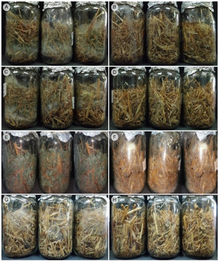
Strains Tr1 and Tr5 grown in wheat straw had abundant mycelium after 10 days of culture (Figs. 3A, C), while the addition of the extract of Pycnoporus sp. (3.6 %) completely inhibited their growth (Figs. 3B, D). The strain of T. aggressivum was less affected by the extract; at 10 days of incubation, the sporulation was delayed by the extract, since the typical green pigmentation could be observed in the absence of the extract (Fig. 3E) and only mycelium in the presence of the extract (Fig. 3F). Pleurotus ostreatus showed abundant mycelium (Fig. 3G); however, when the extract was added, the mycelial invasion was delayed by ten days (Fig. 3H).
Discussion
The disease caused by Trichoderma strains has been reported in several parts of the world, among them Europe, Asia, North America and Latin America; many studies have been realized to identify the species causing this problem, specifically for the cultivation of P. ostreatus. Choi et al. (2003) reported isolates of Trichoderma spp. obtained from P. ostreatus and P. eryngii (DC.) Quél. cultures using rice straw, cotton waste and sawdust as substrates. The strains found were Trichoderma cf. virens (J.H. Mill., Giddens & A.A. Foster) Arx (70.8%), T. longibrachiatum Rifai (16.7%) and T. harzianum (12.5%). Park et al. (2004) performed the molecular and morphological analysis of two isolates of Trichoderma from cultures of oyster mushroom (P. ostreatus) in Korea. These were proposed as new species with the names T. koreanum S.-Y. Oh, M.S. Park & Y.W. Lim and T. pleuroti, K1 and K2, respectively. Later, Park et al. (2006) renamed these two species as T. pleurotum and T. pleuroticola. It should be mentioned that T. pleuroticola is associated with the cultures of all mushrooms of the genus Pleurotus, while T. pleurotum has been found only in P. ostreatus cultures, both wild and cultivated, as well as in soil and wood in countries such as Canada, United States of America, Iran, New Zealand and some European ones (Kredics et al., 2009). Komon-Zelazowska et al. (2007) informed that the origin of infection by T. pleurotum (T. pleuroti) was through the substrate, so it must have an adequate management of straws used in the cultivation of mushrooms to reduce the frequency of the appearance of the disease. Trichoderma harzianum has been observed on decaying wood and associated with mushrooms in North America and Europe (Chaverri et al., 2015) and T. aggressivum in mushroom cultivation in Europe, Iran and South Korea Komon-Zelazowska et al. (2007).
Sobal et al. (2016) conducted a study in Mexico where they isolated 24 strains of Trichoderma from substrates of commercial cultivation of edible mushrooms in the states of Puebla, Tlaxcala, Morelos, State of Mexico and Veracruz, identifying T. agressivum f. agressivum, T. koningii Oudem, T. harzianum, T. atroviride Bissett, T. citrinoviride Bissett and T. pleuroti. In our work, T. pleuroti and T. atrobrunneum were found in the state of Morelos, and it is important to mention that the latter was not reported by Sobal et al. (2016), so this contributes to the knowledge of the species that cause diseases in the cultivation of mushrooms of the genus Pleurotus in the central region of Mexico.
It was communicated that T. pleuroticola showed a Vc similar to that of T. aggressivum while T. pleurotum was slower (Komon-Zelazowska et al., 2007). In this work, it was observed that in general T. aggressivum presented higher Vc compared with T. pleuroti. Chaverri et al. (2015) documented that T. atrobrunneum grown on PDA after 96 h at 25 ºC, presented abundant aerial and cottony or woolly mycelium, the conidia were abundant within 48 to 72 h in wide concentric rings, which coincides with the characteristics presented by the Tr5 strain.
Qiu et al. (2017) said that the high temperature favored a greater production of conidia in less time for T. asperellum Samuels, Lieckf. & Nirenberg, together with the hyphae of P. ostreatus. In this work three strains of T. atrobrunneum were isolated, Tr3 and Tr4 from the northern area of the municipality of Cuernavaca, Morelos, where the temperature ranges from 15-31 ºC, and Tr5 from the municipality of Tlaquitenango, where the temperature is 19-35 ºC. It is suggested that the Tr5 strain grew faster and sporulated (presented the green coloration) in less time, in comparison with Tr3 and Tr4 (data not shown), given the temperature of the place of origin that reaches up to 35 ºC, although more tests that can confirm the effect of temperature should be performed.
Given that the disease of green mold causes great losses in the production of mushrooms of the genus Pleurotus, many works have been carried out with the purpose of establishing strategies for its control, including the use of commercial fungicides and natural compounds, as well as the modification of the temperature and the pH of the cultures. However, this has had little success since it affects both the guest and the host (Woo et al., 2004; Wang et al., 2016). Hatvani et al. (2012) reported that certain natural compounds of phenolic type such as thymol, ferulic acid, (+)-menthol, and (-)-menthol had an inhibitory effect on the growth of T. pleurotum, T. pleuriticola and T. harzianum; however, these also inhibited the growth of the edible mushrooms.
Reyes (2013) tested an ethanolic extract of Ruta graveolens L. (4 g/L) in T. agressivum (CPM-113), obtaining an inhibition of 53% at 24 h, and 44% after 48 h, and they found that the fungus adapted to the presence of the compound over time, with abundant mycelium and change in the color of the spores. This phenomenon was not observed in any of the strains of Trichoderma studied in this work; only delays were obtained in the mycelial growth rate and as a consequence also in sporulation, the latter happened in both plate and on wheat straw. After ten days of incubation on wheat straw, the strain T. agressivum (CPM-113) did not show the green coloration with the extract, while without the addition of the extract the sporulation appeared, which indicates that although it does not completely inhibit this strain, its growth and sporulation were slower. Tr1 and Tr5 were completely inhibited at ten days of incubation on wheat straw by concentration of 3.6% (v/w) of the extract, and the strain of P. ostreatus at the same conditions showed a mycelial invasion delay of five days. It is suggested that the concentration of the fruiting body extract of Pycnoporus sp. at 3.6% could be effective for the control of green mold disease in mushroom cultures of the genus Pleurotus. It should be mentioned that the effect of the extract may also depend on the species, since each of them will have phenotypic and genotype characteristics that allow it to grow in adverse conditions. Marik et al. (2017) evaluated the aggressiveness of T. aggressivum (91 and 61%, respectively) and T. pleuroti (98 and 100%, respectively) against A. bisporus and P. ostreatus. In this work, T. pleuroti was the most resistant strain since the extract only decreased its growth by 38%. The extract of Pycnoporus sp. showed effectiveness, so it could be an alternative to control the green mold disease. It could be applied as a spray on the culture to prevent the invasion of Trichoderma spp., incorporating the extract during the incubation phase of Pleurotus, where the temperature increases due to the metabolic heat due to the mycelial growth of both fungi.
Conclusions
The extract of the fruiting body of Pycnoporus sp. was able to reduce the growth of the strains of T. pleuroti, T. atrobrunneum and T. aggressivum, although the effect varied depending on the species. The Pycnoporus extract is suggested as an alternative for the control of green mold disease since the effect on the growth of P. ostreatus strain was minimal. The mushrooms are a source of obtaining metabolites that have antifungal activity, and they are a potential alternative to counteract the effects of Trichoderma in the cultivation of Pleurotus.

