


 texto en
  Inglés (pdf)
 Artículo en XML
 Referencias del artículo
 Enviar artículo por email
 Citado por SciELO
  Similares en
SciELO
Similares en
SciELO 
 Permalink
Introducción
Sea turtles can harbor a variety of microorganisms that normally behave as saprophytes and do not cause any disease (Köhler 2006, Santoro et al. 2006). However, when the immune system weakens as a product of stress generated by natural and anthropogenic causes, individuals are more susceptible to pathogens such as bacteria, viruses, parasites, and fungi that cause diseases and even mortality. Among the main routes of entry of pathogens into sea turtles are injuries caused by traumas (Dobbs 2001), which affect the dermal tissues (Glazebrook and Campbell 1990).
Dermatitis is a problem frequently reported in captive-raised sea turtles (Glazebrook and Campbell 1990), but wildlife turtle reports are scarce (Maas 2013, Mazzarella et al. 2020). Commonly, in reptiles this disease is a consequence of abrasive skin lesions in animals that are in environmental conditions that favor bacterial infection (Boylan et al. 2017). Moreover, traumatic skin lesions represent the opportunity for pathogenic bacteria to invade tissues and reach the bloodstream, resulting in fatal septicemia (Orós et al. 2005).
Ulcerative and necrotizing dermatitis (UND) is a cutaneous disease characterized by blisters that often rupture, forming erosions and ulcers (Yager and Wilcock 1994). The open wounds created by these ruptured vesicles then provide a venue for secondary complications, such as infections (Maas 2013). The predisposition to UND is due to several factors, including water pollution (fecal pollution), epibionts, warm water temperatures, overpopulation, states of immunosuppression, and wounds from fighting in the breeding areas (Barragán 2002, Santoro et al. 2007, Duignan et al. 2020).
Abnormalities affecting the skin are an important aspect of aquatic wildlife health monitoring because the skin can be reflective of injurious external factors, such as water quality, and a host of systematic factors, such as diet and the function of the immune system and other organs, as well as reflecting the presence of diseases (Mazzarella et al. 2020). Therefore, in this study we report the first findings of UND in free-ranging olive ridley turtles (Lepidochelys olivacea) in La Escobilla, Oaxaca. This information adds new insights about this disease in the southern coastal region of Mexico.
Materials and methods
La Escobilla Sanctuary is in the Santa Maria Tonameca municipality (15°43ʹ37.56ʺ N, 96°44ʹ49.23ʺ W), on the southwest Mexican Pacific coast. The beach is approximately 25 km long, and the turtles nest along an 8-km-long strip at its western end. To facilitate the night counts of nesting females during the arribadas, the Centro Mexicano de la Tortuga has divided the nesting area into 160 permanent stations, marked every 50 m with a fixed concrete post (CONANP 2009).
While conducting a research project focused on assessing blood parameters in nesting olive ridley turtles (L. olivacea) at La Escobilla, we found several skin injury conditions on the necks of nesting turtles during the arribada events (September, October, and November) of the 2021 nesting season. We selected 6 of the permanent stations (approximately 200 m) from which to perform the counting and examination of the turtles present in that area to register the prevalence of these injuries. Because of the high prevalence of dermatitis in the nesting turtles, we used a simple skin lesion classification system consisting of 5 levels: level 1, discoloration; level 2, thickening; level 3, ulceration; level 4, necrosis; and level 5, “mix” (a combination of injuries). In the absence of a reference to the degree of skin lesions in sea turtles, we proposed this arbitrary criterion as a reference for our region. Lesion size was recorded with a rigid L-shaped forensic scale (Critical Path model), measuring the longest axis. Digital images of the lesions were obtained using an iPhone (XR model, 12 MP resolution).
Samples from lesions were taken with sterile swabs from 12 turtles with gross pathological changes (discoloration, thickening, ulceration, and necrosis) suggestive of UND. The swabs were then placed in Stuart transport medium and kept refrigerated at 4 °C until processing 5 h later in the laboratory (Vega-Manríquez et al. 2018). To identify bacteria associated with lesions, samples were seeded by streaking directly onto blood agar, Mannitol salt, Sabouraud, and MacConkey media and incubated for 72 h in microaerobiosis at 37 °C (Vega-Manríquez et al. 2018). The isolated colonies were analyzed by Gram stain; Gram-negative and Gram-positive bacteria were assessed for oxidase and catalase, respectively.
Antimicrobial resistance profiles were studied using the standard procedures (Blasi et al. 2020) with the 18 most commonly used commercial antibiotics (10 μg each, Table 1). Interpretation of results was conducted by referring to the Clinical and Laboratory Standards Institute ranges (CLSI 2018). Although fieldwork was carried out under permit SGPA/DGVS/03919/21, from the Secretaria del Medio Ambiente y Recursos Naturales (Mexico), for handling and extraction of blood, it is necessary to clarify that this permit did not include taking skin samples.
Table 1. Inhibitory growth response to antibiotics by bacteria sampled from olive ridley neck lesions. I = individual, R = resistant, S = sensitive, XX = moderate growth, X = little growth.
| Bacteria/fungus | Antibiotics | I1 | I2 | I3 | I4 | I5 | I6 | I7 | I8 |
| Pseudomonasp. | Amikacin | R | R | S | R | R | R | ||
| Ampicillin | R | R | R | R | R | R | |||
| Carbenicillin | R | R | R | R | R | R | |||
| Cephalothin | R | R | R | R | R | R | |||
| Cefotaxime | R | R | S | R | R | R | |||
| Ciprofloxacin | S | S | S | S | S | S | |||
| Clindamycin | R | R | R | R | R | R | |||
| Gentamycin | R | R | S | R | S | R | |||
| Netilmicin | R | R | S | R | R | R | |||
| Nitrofurantoin | R | R | R | R | R | R | |||
| Norfloxacin | R | R | S | R | R | R | |||
| Trimethoprim/sulpha | R | R | R | R | R | R | |||
| Staphylococcussp. | Ampicillin | R | R | R | R | R | R | R | R |
| Cephalothin | R | R | R | R | R | R | R | S | |
| Cefotaxime | R | R | R | S | R | R | R | R | |
| Ciprofloxacin | S | S | S | S | S | S | S | S | |
| Clindamycin | R | R | R | R | R | R | R | R | |
| Dicloxacillin | R | R | R | R | R | R | R | R | |
| Erythromycin | R | R | R | R | R | R | R | S | |
| Gentamycin | R | R | R | R | R | R | R | R | |
| Penicillin | R | R | R | R | R | R | R | R | |
| Tetracycline | R | R | R | R | R | R | R | R | |
| Trimethoprim/sulpha | R | R | R | R | S | R | R | R | |
| Vancomycin | R | R | R | R | R | R | R | S | |
| Candidasp. | XX | X | X | XX | XX | XX | X | X |
Results
During 3 arribada events, 525 turtles were examined in a stretch of approximately 200 m. Of these, 278 turtles had injuries on the back of the neck (52.9% prevalence). The severity of injuries varied (Fig. 1): 41.4% (n= 115) of injuries were level-1 injuries; 38.8% (n= 108), level-2 injuries; 12.6% (n= 35), level-3 injuries; 4.7% (n= 13), level-4 injuries; and 2.5% (n= 7), level-5 injuries. We did not find any epibionts or any residual marks suggestive of prior epibiont attachment on the affected turtles examined. The size of the injuries varied between the distinct levels. The average length by width was 6.5 × 3.5 cm for level-1 injuries, 10.0 × 3.5 cm for level-2 injuries, 3.0 × 3.0 cm for level-3 injuries, and 2.5 × 4.0 cm for level-4 injuries. Level-5 injuries varied in length.
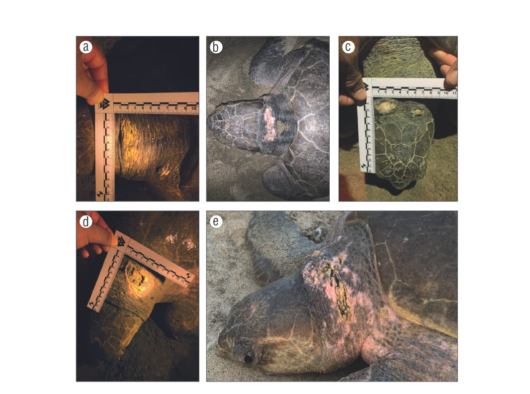
Figure 1. Skin injuries with gross pathological changes suggestive of ulcerative and necrotizing dermatitis on the necks of nesting turtles in La Escobilla, Oaxaca, Mexico. (a) Level 1 (discoloration), (b) level 2 (thickening), (c) level 3 (ulceration), (d) level 4 (necrosis), and (e) level 5 (mix).
A total of 2 bacteria (Pseudomonassp. andStaphylococcussp.) and 1 fungus (Candidasp.) were identified in 8 turtles with UND. Among the bacterial isolates,Staphylococcussp. was present in all samples and resistant to 4 antibiotics (ampicillin, clindamycin, dicloxacillin, and penicillin), whereasPseudomonassp. was present in 6 of 8 samples and resistant to 5 antibiotics. Furthermore, the bacteria were sensitive to other antibiotics (Table 1); however, ciprofloxacin was the only antibiotic with high inhibition ofStaphylococcussp. andPseudomonassp.
Discussion
In this study, we report the first findings of UND inL. olivaceain a high-density nesting beach. Dermatitis has been documented in captive sea turtles (Wiles and Rand 1987, Glazebrook and Campbell 1990, Vega-Martínez et al. 2018) and in some free-ranging sea turtles (Santoro et al. 2007, Gamez-Vivaldo et al. 2009, Mazzarella et al. 2020). Particularly in Mexico, there is an isolated dermatitis report forL. olivaceain Colima (Gamez-Vivaldo et al. 2009).
Skin lesions in free-ranging turtles could be produced by epibionts such asOzobranchusspp. (Santoro et al. 2007), net entanglement (Gamez-Vivaldo et al. 2009), and fights among individuals in the breeding areas (Booth and Peters 1972, Peralta and Luna 2016), which likely open cutaneous portals of infections with the subsequent colonization of skin lesions by bacteria (Glazebrook and Campbell 1990).
The injuries observed on the necks of the turtles in La Escobilla Sanctuary may be due to 3 explanations. First, injuries could be related to mating behavior during the reproductive season. During this process, a male mounts a female, plastron to carapace, with the male using claws to grasp the female carapace (Booth and Peters 1972). Particularly during the arribada phenomenon, massive groups of individuals occur in front of the nesting beach, a situation that generates fighting among individuals in the breeding areas. A high density of individuals (arribadas) can incite bite traumas, and typical places of aggression are neck, head, and tail (Barragán 2002). Evidence of claw marks and bites has been observed on female carapaces during the nesting season (Booth and Peters 1972, Peralta and Luna 2016), and we do not rule out the possibility of bites in the female neck region, which may be associated with secondary infection of the skin (Boylan et al. 2017). Second, warm water temperatures and daily rain events on the Pacific coast of southern Mexico may contribute to favorable conditions for bacterial growth and poor water quality during the sea turtle nesting season. Some bacterial species and other microbes may be carried out to the sea by sources of pollution such as effluents and runoff (Santoro et al. 2006). However, routine bacterial monitoring of beaches and coastal waters have not been conducted in this region. As the turtles exhibited dermatitis while nesting, it is possible that they could have also encountered poor water quality at their foraging grounds or during migration (Mazzarella et al. 2020). Third, the reason only some animals get sick may be related to alterations of their immune system (Vega-Manríquez et al. 2018). Biotic and abiotic factors such as water temperature, the presence of microorganisms, and contamination can influence the immunity of animals under stress (Zimmerman et al. 2010); therefore, both the high number of turtles during an arribada and the process of reproduction could be stressful factors that can immunosuppress their systems. In addition, it has been hypothesized than exposure to algal toxins in a red tide may have sublethal effects on health and immune function (Perrault et al. 2017, 2020). Herrera-Galindo et al. (2015) reported several dead sea turtles on the coast of Oaxaca, Mexico, and after having performed the necropsies, they found salps andPyrodinium bahamensein the stomach contents, which are related to red tides.
To our surprise, both bacterial isolates showed high resistance to most antibiotics tested, except ciprofloxacin, which generated the greatest inhibition ofStaphylococcussp. andPseudomonassp. We recommend initiating a continuous monitoring program to follow the occurrence of dermatitis inL. olivaceain subsequent years to better document prevalence and follow progression of skin injuries in more individuals. Furthermore, the addition of hematology and blood chemistry would provide more information about the health status of affected turtles beyond that evident from external examination and reproductive metrics.

