


 text new page (beta)
 English (pdf)
 Article in xml format
 Article references
 Send this article by e-mail
 Cited by SciELO
  Similars in
SciELO
Similars in
SciELO 
 Permalink
INTRODUCTION
The greatest threat to corals today is accelerated global climate change (GCC). GCC is mainly defined as the increase in the average temperature of the planet, due to the accelerated increase in atmospheric CO2, as a result of increasing anthropogenic activity since the post-industrial era (Hughes et al. 2017). Among other extreme changes caused by GCC, the increase in seawater temperature is the one that most affects the mutualistic relationship between corals and their endosymbiont dinoflagellates of the Symbiodiniaceae family (sensuLaJeunesse et al. 2018). Extreme and prolonged increases in seawater temperature cause the loss of the endosymbiotic relationship and, consequently, the bleaching of scleractinian corals. Bleaching can cause mass mortality in most species, resulting in reef degradation and worldwide environmental crisis (Eakin et al. 2019). Therefore, one of the main concerns is to understand the capacity of corals to acclimatize to environmental conditions (phenotypic plasticity) and to cope with GCC (Palumbi et al. 2014, Barshis 2015). Laboratory and field experiments that help elucidate the acclimatization capacity of scleractinian corals are thus of great relevance.
The acclimatization capacity of Pocillopora corals is of particular interest, largely because their phenotypic plasticity is extremely high. Nevertheless, the effects of high plasticity on the main cellular stress response pathways have been scarcely studied. Considering all the above, we examined the messenger RNA (mRNA) expression profiles for the morphospecies Pocillopora cf. capitata, Pocillopora cf. damicornis, and Pocillopora cf. verrucosa, which are the most abundant morphospecies at Carrizales Reef, off the coast of Colima, Mexico. Using quantitative PCR (Mayfield et al. 2013a, 2013b) and Pocillopora transcriptome sequencing (Mayfield et al. 2014a, 2014b) techniques, we evaluated the expression levels of carbonic anhydrase (CA) mRNA, copper,zinc-superoxide dismutase (CuZnSOD), green fluorescent protein-like chromoprotein (GFP-like cp), and 70-kDa heat shock protein (HSP70). In addition, we evaluated the ratios of nucleic acids and proteins, the content of pigments and mycosporine-like amino acids, and the densities of endosymbionts in coral tissues because these indicators have proven useful for assessing coral physiology and health (Hinrichs et al. 2013). To date, our results are the first to reveal the plasticity of gene expression in Pocillopora morphospecies from Carrizales Reef. We conclude that these results can be used as baseline elements for the study of acclimatization in the different Pocillopora morphospecies on the Pacific coast of Mexico.
MATERIALS AND METHODS
Study site and sample collection
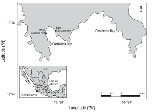
Carrizales Reef is located in Carrizales Bay (19°05´42” N, 104°26’21” W), just southwest of the coast of Manzanillo, Colima, Mexico (Fig. 1). On 13 February 2017, an expedition to Carrizales Reef was carried out using autonomous diving equipment, during which a total of 36 fragments of the corals P. cf. capitata, P. cf. damicornis, and P. cf. verrucosa were collected from 12 colonies on the east and west margins of the reef (n = 6). To reduce the effects of environmental variation, colonies that were sympatrically distributed at 3 m depth were selected. Each colony was identified by the typical morphological characteristics (following Veron 2000). However, since colony morphology is generally incongruous with genetics (Pinzón and LaJeunesse 2011), there is a possibility that the colonies belong to homogeneous genetic groups.
After the expedition, biological samples were divided into 3 groups of 12 fragments, which were immediately processed in the field and protected from light during transportation. Two of the groups were separately preserved in plastic vials, one with 10 mL of 10% formaldehyde and the other with 10 mL of cold RNAlater solution. Afterwards, samples in formaldehyde were refrigerated and samples in RNAlater were frozen at -80 °C until laboratory analysis. The remaining group of samples were initially transferred into microtubes with 2 mL of 100% methanol, subsequently stored at 4 °C for 24 h in total darkness, and lastly analyzed in the laboratory.
In addition, 6 samples of 1.0 L of seawater were collected at 1.0 m depth at each reef sampling site (east and west), at approximately 1-h intervals throughout the sampling duration (~4 h). Chlorophyll a (Chla) concentration in these samples was determined with a Jenway 6500 spectrophotometer (Chelmsford, Essex, UK), according to the methodologies of Strickland and Parsons (1972) and Grasshoff et al. (1999). The results are shown as Chla concentration per volume of seawater (mg·m-3). Owing to the technical difficulties in continuously recording light and temperature data during the expedition on 13 February, a second expedition was conducted on 27 May 2017 to obtain a new log. To do this, Onset HOBO Pendant UA-002-08 sensors (Onset Computer Corporation; Bourne, MA, USA) were used. Prior bi-monthly monitoring showed that seasonal variability in the reef occurs every 4 months (Muñiz-Aguiano et al. 2017). So even though these data do not correspond to the conditions on 13 February, we believe the values obtained on 27 May do allow for a reliable comparison of the spatial variation between the east and west sides of Carrizales Reef.
Laboratory analysis
The density of endosymbiont dinoflagellates was quantified from the fragments in formaldehyde by employing a modification to the protocol proposed by Zamoum and Furla (2012). Briefly, coral tissue was removed from the skeleton by incubation in 10 mL of 4 M NaOH at 65.0 °C for 1 h. The homogenate was then kept overnight at 37.5 °C and Symbiodiniaceae cell density was later quantified from a 10-μL aliquot using a Neubauer hemocytometer (n = 8 replicates).
Pigment concentration was determined from the fragments that were preserved in 100% methanol at 4.0 °C. After storing for 24 h, the homogenate was sonicated for 15 s and then centrifuged at 1,500 × g for 5 min at 4 °C; the resulting extracts were protected from the light and immediately used to measure absorbance by scanning wavelengths from 300 to 750 nm every 1 nm. Duplicate measurements were performed in a Spectronic Genesys 5 spectrophotometer (Thermo Fisher Scientific; Waltham, MA, USA) using 96-well microplates. Pigment content was calculated from the extinction coefficients for the β-carotene (145.0 L·g-1·cm-1) and perinidine (132.5 L·g-1·cm-1) carotenoids (Jeffrey and Haxo 1968) and for the diadinoxanthin (223.0 L·g-1·cm-1) and dinoxanthin (135.0 L·g-1·cm-1) xanthophylls (Vernon 1960), and total chlorophyll concentration (Chla and Chlc) was calculated according to the equations by Jeffrey and Humphrey (1975), with the recommended turbidity correction.
Nucleic acids and total proteins were extracted from the fragments in RNAlater after thawing. First, RNAlater excess was removed with a Kimwipes tissue (Kimberly-Clark; Roswell, GA, EUA). Then, samples were ground in a mortar, and approximately 100 mg of tissue and skeleton were transferred into 1.5-mL microcentrifuge tubes with 1.0 mL of TRIzol reagent (Life Technologies); samples were then homogenized using a mechanical disruptor (FastPrep24, MP Biomedicals; Santa Ana, CA, USA). The extractions were made following the specifications of the manufacturer with the following modifications. Briefly, up to 2 μL of 6M HCl were added to the homogenate to prevent the neutralization reaction that occurs between the calcium carbonate in the skeleton and the acidic TRIzol (Anderson et al. 2016). RNA was precipitated by adding isopropanol and incubating overnight at -20 °C, and the solution was then centrifuged to precipitate the RNA, which was eluted in 50 μL of ribonuclease-free water (RNase). DNA was recovered from the rest of the phenol solution following the recommendations by Mayfield et al. (2012). Briefly, a back extraction buffer (4M guanidine thiocyanate, 50 mM sodium citrate, and 1M Tris free base) was used. Equal volumes of back extraction buffer and phenol solution were mixed, and the mixture was subsequently centrifuged to obtain an aqueous phase, which was transferred into a new tube. DNA was precipitated and eluted in 200 μL deoxyribonuclease-free water (DNase), with an additional 10-min incubation at 55 °C. Lastly, proteins were precipitated from the remaining phenol solution by modifying the time of incubation and final solubilization (40 min at 50 °C in 1% SDS). The quantity and quality of nucleic acids and proteins were analyzed in a Nanodrop spectrophotometer and the samples were stored at -80 °C.
For the gene expression analysis, all RNA samples were thawed and purified by treatment with DNase I enzyme (without RNase) following the specifications of the manufacturer (Invitrogen, Thermo Fisher Scientific). With the purified RNA, 300 ng were reverse transcribed to the first strand of complementary DNA (cDNA) using the high-capacity reverse transcription kit and following the recommendations of the manufacturer (Applied Biosystems, Life Technologies; CA, USA). Aliquots of 20 ng·μL-1 of cDNA were prepared and then stored at -20 °C.
Relative mRNA expression was done by amplifying target genes (CA, CuZnSOD, GFP-like cp, and HSP70) and an internal control fragment (18 ribosomal RNA) to calibrate the complementary DNA template (Table 1). Eight independent real-time quantitative PCR (q-PCR) assays were performed, in a total volume of 20.0 μL with 6.3 μL of 2X SYBR Green Master Mix (Applied Biosystems), 0.6 μL of each primer for the target gene and the internal control gene (10 mmol·μL-1), 5.0 μL of the cDNA sample (20 ng·μL-1), and 6.9 μL of RNase/DNase-free water. Q-PCR assays were carried out in a StepOnePlus thermocycler (Applied Biosystems) with the following thermocycling conditions: (1) “hot start” at 95 °C for 10 min, (2) amplification of 40 cycles at 95 °C for 15 s followed by 60 s at 60 °C, and (3) fusion at 95 °C for 15 s followed by 60 s at 60 °C and 15 s at 95 °C. Results are given in terms of relative mRNA expression as calculated with the 2-ΔΔCt method (Schmittgen and Livak 2008).
Table 1 Parameters for the primers used in the real-time quantitative PCR assays.
Primer name |
Biological process |
Sequence 5′-3′ |
Tm (ºC) |
Source |
CA |
Metabolism/transportation |
F: AGGATGATGAGGAGGATGAGG |
60 |
Mayfield et al. (2013b) |
F: ATAGCAGGGAGGGGTGGTAA |
||||
CuZnSOD |
Oxidativestress |
F: GCTGTTTGCGTTTTATTGCCC |
60 |
Mayfield et al. (2014a) |
F: TCTTGCATGTGTCTCCCAC |
||||
GFP-like cp |
Light absorption |
F: AGGCAAACAAACGGGGACATT |
60 |
Mayfield et al. (2014b) |
F: GGCACTCCCTCCATCTTCA |
||||
HSP70 |
Stress response |
F: ATCCAGGCAGCGGTCTTGT |
60 |
Mayfield et al. (2014b) |
F: TCGAGCAGCAGGATATCACTGA |
||||
18S |
Internal control |
F: GCTAAATCCAAAATCCAACAGAG |
60 |
This study |
F: TTGAATGTCTTTCATTCTAGTATTCAA |
Tm: melting temperature.
F: forward primer.
R: Reverse primer.
Statistical analysis
Statistical analyses were performed using R software. All data were analyzed with the Shapiro-Wilk normality test and the Bartlett test to determine homoscedasticity (95% confidence intervals). For the environmental parameters, an unpaired 2-sample t-test was used. For biological parameters, 2-way analysis of variance (ANOVA) was used, followed by multiple comparisons (Tukey’s test) to assess significant differences (P < 0.05). In some cases, the bidirectional non-parametric Friedman test and the paired sign test were used. All data were presented as mean values ± standard error of the mean (n = 6).
Results
The expression of CA in P. cf. capitata was higher in the western (P < 0.05) than in the eastern reef colonies (Fig. 2). The GFP-like cp expression was higher in the P. cf. verrucosa colonies from the west side (P < 0.05) than in the colonies from the east side (Fig. 3). On the other hand, the CuZnSOD expression in P. cf. capitata was also higher in the western colonies (P < 0.05) compared to eastern reef colonies. Contrary to CA and GFP-like cp expressions, the expression of the CuZnSOD enzyme was lower in the P. cf. verrucosa colonies (P < 0.05) in relation to the P. cf. capitata colonies (Fig. 4). Finally, the HSP70 expression did not show significant differences between the east and the west; nevertheless, all mean values of the HSP70 expression were consistently higher in western corals (Fig. 5).
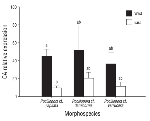
Figure 2 Expression levels of carbonic anhydrase (CA) mRNA in Pocillopora morphospecies on the east and west sides of Carrizales reef. Values are the mean ± standard error of the mean (n = 6). Letters denote significant differences between the data (P < 0.05).
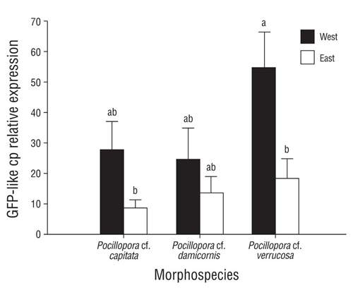
Figure 3 Expression levels of green fluorescent protein-like chromoprotein(GFP-likecp) mRNAin Pocillopora morphospecies on the east and west sides of Carrizales reef. Values are the mean ± standard error of the mean (n = 6). Letters denote significant differences between the data (P < 0.05).
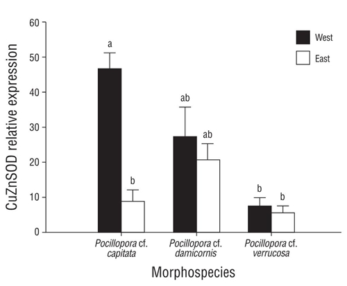
Figure 4 Expression levels of copper,zinc-superoxide dismutase (CuZnSOD) mRNA in Pocillopora morphospecies on the east and west sides of Carrizales reef. Values are the mean ± standard error of the mean (n = 6). Letters denote significant differences between the data (P < 0.05).
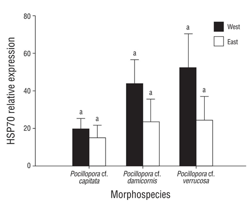
Figure 5 Expression levels of heat shock protein (HSP70) mRNA in Pocillopora morphospecies on the east and west sides of Carrizales reef. Values are the mean ± standard error of the mean (n = 6). Letters denote significant differences between the data (P < 0.05).
In the joint analysis of Chla and Chlc, carotenoids, xanthophylls, endosymbiont density, mycosporine-like amino acids, and the RNA/DNA, protein/DNA, and RNA/ protein ratios, significant differences were observed in the colonies of P. cf. capitata and P. cf. verrucosa from the east and west sides of the reef, whereas in the P. cf. damicornis colonies no significant differences were observed in any marker (Table 2). The environmental values for the Carrizales Reef area showed Chla concentrations of 0.18-0.99 mg·m-3 throughout the sampling. The highest values occurred on the west side of the reef (0.99 ± 0.17 mg·m-3) compared to the east side (0.42 ± 0.10 mg·m-3). Finally, the light values that were recorded months after sampling at Carrizales Reef were higher for the west (63.60 ± 7.91 W·m-2·S-1) in comparison with the light values for the east (44.42 ± 5.26 W·m-2·S-1). On the other hand, temperature data were the same at both sites (25 ± 0.10 °C).
Table 2 Complete set of analysis for physiological condition parameters in Pocillopora morphospecies.
Pocillopora cf. capitata |
Pocillopora cf. damicornis |
Pocillopora cf. verrucosa |
||||
Parameter |
West |
East |
West |
East |
West |
East |
Endosymbionts (cells·cm-2) |
1.33 ± 0.26 × 10⁶ b |
4.59 ± 0.94 × 10⁵a |
5.60 ± 0.1 × 10⁵a |
6.04 ± 0.1 × 10⁵a |
5.10 ± 0.46 × 10⁵a |
3.23 ± 0.76 × 10⁵a |
Chlorophylls (μg·cm-2) |
7.56 ± 1.27b |
4.06 ± 1.27a |
3.18 ± 0.49a |
3.83 ± 1.15a |
3.21 ± 0.41a |
3.24 ± 0.43a |
Carotenoids (μg·cm-2) |
2.36 ± 0.31a |
2.68 ± 0.46a |
1.14 ± 0.18bc |
1.50 ± 0.34ab |
0.98 ± 0.04c |
1.83 ± 0.31ab |
Xanthophylls (μg·cm-2) |
1.84 ± 0.16ab |
2.34 ± 0.33a |
0.94 ± 0.14b |
1.26 ± 0.28ab |
0.85 ± 0.07b |
1.35 ± 0.29ab |
Chlorophylls (pg·cell-1) |
6.27 ± 0.96a |
7.72 ± 1.36a |
6.98 ± 1.01a |
5.95 ± 0.70a |
6.51 ± 0.84a |
11.64 ± 1.78b |
Carotenoids (pg·cell-1) |
1.99 ± 0.29a |
6.48 ± 1.16b |
2.56 ± 0.42a |
2.42 ± 0.16a |
2.04 ± 0.23a |
5.67 ± 0.87b |
Xanthophylls (pg·cell-1) |
1.60 ± 0.25a |
5.81 ± 1.13b |
2.11 ± 0.35a |
2.05 ± 0.13a |
1.70 ± 0.12a |
4.45 ± 0.64b |
Chlc:Chla ratio |
0.35 ± 0.00a |
0.61 ± 0.16b |
0.36 ± 0.01a |
0.41 ± 0.01a |
0.35 ± 0.05a |
0.58 ± 0.13ab |
Carotenoids:Chla ratio |
0.43 ± 0.02a |
0.92 ± 0.09b |
0.49 ± 0.04a |
0.60 ± 0.07a |
0.30 ± 0.08c |
0.89 ± 0.14b |
Xanthophylls:Chla ratio |
0.35 ± 0.03a |
0.81 ± 0.09b |
0.41 ± 0.03a |
0.51 ± 0.05a |
0.37 ± 0.05a |
0.65 ± 0.12ab |
|
(pmol·mg-1 protein)* |
52.92 ± 6.68a |
41.79 ± 8.27a |
27.87 ± 0.97ab |
40.52 ± 9.47ab |
24.02 ± 2.33b |
33.63 ± 7.23b |
|
DNa (μg·mg-1 tissue) |
0.58 ± 0.07a |
0.44 ± 0.59a |
0.70 ± 0.11ab |
0.59 ± 0.05ab |
0.78 ± 0.10b |
0.79 ± 0.12b |
|
RNa (μg·mg-1 tissue) |
0.26 ± 0.05a |
0.16 ± 0.01b |
0.31 ± 0.01a |
0.24 ± 0.03ac |
0.35 ± 0.04a |
0.20 ± 0.02ac |
Proteins (mg·mg-1 tissue) |
13.17 ± 2.90a |
10.75 ± 0.98ab |
13.51 ± 2.40a |
11.55 ± 1.06ab |
13.23 ± 0.94a |
9.42 ± 0.73b |
RNA:DNA ratio |
0.50 ± 0.13a |
0.41 ± 0.07a |
0.52 ± 0.09a |
0.41 ± 0.04a |
0.50 ± 0.09a |
0.28 ± 0.04b |
RNA:proteins ratio |
0.03 ± 0.00a |
0.01 ± 0.00b |
0.03 ± 0.00a |
0.02 ± 0.00ab |
0.04 ± 0.00c |
0.02 ± 0.00ab |
Proteins:DNA ratio |
30.24 ± 5.07b |
19.76 ± 2.46a |
21.32 ± 3.55a |
19.93 ± 1.99a |
35.75 ± 4.29b |
18.52 ± 3.07ª |
Letters denote significant differences between data (P < 0.05).
*MAAs: Mycosporine-like amino acids
DISCUSSION
The present study is the first approach to elucidate phenotypic plasticity, which was assessed through gene expression, in corals of the genus Pocillopora at Carrizales Reef. This plasticity in gene expression was observed in mainly the P. cf. capitata and P. cf. verrucosa morphospecies; however, the response of the 3 morphospecies (including P. cf. damicornis) was clearly associated with the sampling site at the reef.
The results mainly showed that the expression of CA and GFP-like cp was higher on the west side of the reef, where light and chlorophyll in seawater were also higher, compared to the east side. This suggests that CA and GFP like cp are an important part of the acclimatization response of Pocillopora morphospecies, possibly because of variations in the light regime and the implications in regulating endosymbiont density, pigment concentration, and metabolic activity in coral tissue. This could be associated in the first instance to the fact that CAs are essential to provide bicarbonate to the calcification site and dissolved inorganic carbon for photosynthesis through the interconversion activity of CO2 and HCO3- (Moya et al. 2008). Secondly, GFP-like cp has been found to possess a photoprotective function by absorbing and/or redistributing photons away from the absorption bands of photosynthetic pigments (Smith et al. 2013). Lastly, the amount of light corals receive is essential for adequate metabolic activity (Buckley and Szmant 2004) because corals depend on high-radiation environments, where solar energy is used by endosymbionts for photosynthesis and is part of the host metabolism (Iglesias-Prieto and Trench 1997, Bongaerts et al. 2011).
Considering, then, that the regulation of endosymbionts and their pigments is essential to maintain metabolic homeostasis (Iglesias-Prieto and Trench 1997, Bongaerts et al. 2011), the plasticity of the expression of CA and GFP-like cp is fundamental for coral photoacclimatization. This suggests that the amount of light received by coral colonies on the east and west margins of the reef is strongly associated with the high plasticity of gene expression, endosymbiont density, pigment concentration, and metabolic markers found in the morphospecies. Nevertheless, it is worth noting that the results of studies on other coral species contrast with the data of the present study. For example, in the Styllophora pistillata (Pocilloporidae) coral, the expression of a CA isoform has been observed to be higher in dark conditions (Moya et al. 2008). Furthermore, the photoprotection provided by chromoproteins occurs mainly when the hosts have low endosymbiont density (Smith et al. 2013). Lastly, chlorophyll concentrations and endosymbiont density increase in low light (Iglesias-Prieto and Trench 1997), and heterotrophic feeding can also affect RNA, DNA, and protein ratios (Buckley and Szmant 2004). Therefore, in the future, it should be considered that the parameters of light incidence and penetration, water quality, food and nutrient availability, and the morphology and dynamics in Carrizales Bay are locally more variable than previously thought (e.g., Liñán-Cabello et al. 2011, Reyes-Bonilla et al. 2013, Muñiz-Anguiano et al. 2017).
In addition, our results showed high plasticity in the expression of 2 genes involved in stress response. The first is associated with the expression of CuZnSOD enzymes for preventing and controling sublethal levels of oxygen free radicals that cause oxidative stress (Mydlarz et al. 2016), and the second has to do with the expression of HSP70 chaperones that protect and help other proteins to regain their biological activity, mitigating the previously caused stress (Chiappori et al. 2016). Therefore, the high plasticity observed in both cell routes is also essential for the acclimatization of the Pocillopora morphospecies (Poli et al. 2017) at Carrizales Reef.
We conclude that our results can be used as a base line for the study of physiological and molecular plasticity in Pocillopora corals on the Pacific coast of Mexico. Still, we believe that greater sampling effort would have enabled us to better elucidate the plasticity responses observed in the corals included in this study and the plasticity of the other 3 morphospecies not included here (Pocillopora cf. eydouxi, Pocillopora cf. inflate, and Pocillopora cf. meandrina). However, these morphospecies show low relative abundance at Carrizales Reef (Reyes-Bonilla et al. 2013). Furthermore, taking into account that Pocillopora corals on the Pacific coast of Mexico are of great importance for the local formation of reefs and show less calcification, as opposed to corals in other geographic regions of the world (Cabral-Tena et al. 2018), we believe that continuing to study these organisms, and to locally protect them, is essential to better understand the stress response of coral communities in this region under a scenario of worldwide changes in the ocean.

