


 texto en
  Inglés (pdf)
 Artículo en XML
 Referencias del artículo
 Enviar artículo por email
 Citado por SciELO
  Similares en
SciELO
Similares en
SciELO 
 Permalink
Most of the rural communities in Oaxaca, Mexico, currently maintain elements from prehispanic agricultural systems, such as the organization that promotes community work and the production of local crops. Fruit trees such as Inga spuria (cuajinicuil) and Annona muricata (soursop) are among the main socially and economic important species grown in the community of Peña Negra, in the county of Santa Lucía Monteverde, Oaxaca. The community is located at an altitude of 1200 masl, its average temperature is 26 °C and a relative humidity varies between 50 and 90% throughout the years. The production of soursop in Peña Negra is of 0.25 t a year, and for cuajinicuil, 0.5 t a year. This production is for self-supply and is also sold in the local market.
In June, 2019, these two fruit trees first displayed fungal symptoms on leaves, causing heavy defoliation, mummification and loss of fruits, with an incidence of 100%, meaning that all trees had diseased leaves. In a tour of the seven rancherías of San Sebastián, Nopalera (El paraíso 16° 95´ 27´´ N, 97° 82´ 11´´ W, El guayabo 16° 92´ 33´´ N, 97° 78´ 13´´ W, Buena Vista el Naranjo 16° 92´ 75´´ N, 97° 76´ 69´´ W, Peña Flor de Clavo 16° 90´ 19´´N, 97° 77´ 91´´W, Peña Negra 16° 91´ 52´´N, 97° 79´ 50´´W, Torralba de Juárez 16° 90´ 47´´N, 97° 82´ 19´´O, and Peña de Jícara 16° 93´ 86´´N, 97° 79´ 50´´), the same symptoms were observed in both fruit tree species.
A part of Putla (a district that spans 50 communities and several municipal areas) was also visited, and the same symptoms were also observed in soursop and cuajuinicuil in these communities, namely Yosotiche, Simiyuvi and El Piñal. Each ranchería has its own microclimate and the pathogens have seemed to adapt to them all. Out of all the families in the area, 95% have few trees planted in their backyards. The remaining 5% has between 50 and 100 of both types of fruit trees. In diseased trees, losses can be of up to 50%, according to information provided by local farmers. In order to understand this phytosanitary problem, the aim of this study was to identify, by amplification and analysis of the region of the Internal Transcribed Spacer (ITS), the fungi associated to the foliage of the cuajinicuil and soursop.
The diseased plant tissue samples were collected from cuajinicuil and soursop trees in December, 2019, in 10 orchards in Peña Negra, San Sebastián Nopalera, in the county of Santa Lucía Monteverde, Oaxaca, Mexico (16° 54’ 52.73’’ N; 97° 47’ 50.35’’ W; 1096 masl). The location has an average temperature of 26 °C and the relative humidity between May and October fluctuates between 80 and 90%.
The trees were in a vegetative phenological stage. The orchards chosen had 1 ha for every kind of fruit tree to carry out the sampling. In each orchard, a randomized sampling design was established consisting of 10 trees with leaf symptoms. Five leaves with symptoms were taken from each tree at a height of 1.5 m. To isolate the microorganisms, after rinsing the leaves in the lab, pieces measuring 0.5 x 0.5 cm were taken from the edge of the lesions and submerged in 2% sodium hypochlorite, rinsed three times and placed in Petri dishes with potato dextrose agar (PDA; BD Bioxon) and incubated at 27 ºC for seven days. From the fungal growth, hypha tips were replanted until pure cultures were obtained from each isolation.
The pathogenicity tests were carried out in 10 healthy soursop plants, placing them in 10 humid chambers, separately, and keeping them at 25 °C. Each one was inoculated placing a disc, 5 mm in diameter. The negative control was inoculated with sterile distilled water and monitored until symptoms appeared.
In cuajinicuil, to verify the pathogenicity of the isolation, the previously described methodology was followed. In addition, leaves from 10 plants at a height of 30 cm were inoculated, one leaf per plant, which were kept at a temperature of 25 °C and the humidity was maintained with moistened cotton inside the humid chambers. Once the symptoms appeared, the pathogens were isolated from the diseased tissue in the PDA. The two fungi isolated from each tree were morphologically different, therefore the sequencing of each one’s ITS was carried out.
The genomic DNA was extracted from the mycelial growth of the pure culture of each isolation using the kit Quick-DNA Fungal/Bacterial Miniprep Kit (Zymo Research Corp., Irvine, CA, USA), following the manufacturer’s protocol. The concentration and purity of the DNA was verified in a Nanodrop 2000c (Thermo Scientific, Wilmington, DE, USA); the integrity was determined by electrophoresis in a 1% agarose gel stained with SYBR Green I Nucleic Acid Gel Stain (Thermo Fisher, Darmstadt, Germany).
The ITS region was amplified with universal primers ITS1 (5’- TCCGTAGGTGAACCTGCGG-3’) and ITS4 (5’-TCCTCCGCTTATTGATATGC-3’) (White et al., 1990) in a T100 Bio-Rad thermocycler (Bio-Rad, Hercules, CA, USA) in a total reaction volume of 25 μL, containing 50-100 ng of genomic DNA, 0.4 μM of each primer and 1X of Platinum Green Hot Start PCR Master Mix (Invitrogen, Carlsbad, CA, USA). The reaction conditions were as follows: initial denaturalization at 95 °C for 5 min, 30 cycles at 95 °C for 1 min, aligning at 55 °C for 2 min, extension at 72 °C for 1 min and a final extension at 72 °C for 5 min. The expected fragment (550 pairs of bases) was corroborated by electrophoresis in 1% agarose gel and purified using the ZR DNA Sequencing Clean-up Kit (Zymo Research Corp., Irvine, CA, USA). The purified products of PCR were sequenced using Sanger’s technique by Macrogen, Inc. (South Korea).
The sequences from this study were compared individually with reference sequences from the GenBank data base (NCBI) with the program BLAST (Basic Local Alignment Search Tool). Later, the program Clustal X v2.1 was used to carry out the multiple sequence alignment. Once aligned, 5’ and 3’ were timed using SeaView v4.4.2 and the best nucleotide substitution model was determined using the program SMS (Smart Model Selection) incorporated in PhyML v3.0. A phylogenetic tree was calculated using the maximum likelihood method in PhyML v3.0 with the model TN93. The reliability of each node was estimated using a bootstrap analysis after 1000 replications and the sequence of Alternaria alternata KX377683.1 was included as an external group. The generated tree was visualized with the program iTOL (Interactive Tree of Life).
In the cuajinicuil, the symptoms of the disease were small round to irregular spots with a light to dark chestnut color and a chlorotic halo on any part of the leaflet. The spots appeared in clusters that formed small areas of necrotized tissue. The lesions cover over 50% of the leaflet, and finally, the leaves become yellow and fall (Figure 1). The foliar symptoms in soursop have a brown color with irregular yellow edges that begin at the tip and spread towards the base of the leaflet. The necrotized tissue becomes brittle, followed by the wilting of the leaves, which then become brown, with an appearance of having been burned by fire. The branches become completely defoliated.
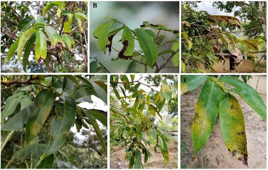
Figure 1 Leaf symptoms in fruit trees in the community of San Sebastián Nopalera, Oaxaca, Mexico. A-B. Symptoms of necrotic leaf in Cuajinicuil trees; C. Defoliation in cuajinicuil; D-E-F. Symptoms of lesions with yellowing and brown spots in soursop trees.
In cuajinicuil, the fungus subjected to Koch’s postulates induced symptoms 13 days after inoculation in plants, whereas in loose leaves, the symptoms appeared 10 days after inoculation. The leaves displayed round to irregular yellowish spots, characteristic of the original disease. When the fungi of both tests were reisolated, they were confirmed to be similar to the initially isolated fungi. The 8.5 cm growth of the culture was achieved in seven days. The culture’s characteristics are described in Table 1.
Table 1 Nomenclature, taxonomic assignment and characteristics of the cuajinicuil and soursop pathogenic fungi.
| Fruit tree | Strain/NCBI accession number | Genus | Culture characteristics | Colonies in PDA |
| Cuajinicuil | HCUA1/MW418007 | Diaporthe sp. (Sordariomycetes, Diaporthaceae) | Aerial mycelia growth in cream concentric rings with lobulated edge and the vegetative growth in gray. After few days, mycelium becomes cottony and dense, grayish sepia color on the reverse |

|
| Soursop | HGUA1/MW418003 | Lasiodiplodia sp. (Dothideomycetes, Botryosphaeriaceae) | White dense aerial mycelia. Few days later, the mycelia turns olive gray to dark gray. |

|
For soursop, the inoculated fungus induced similar symptoms to those initially observed in the field, 10 days after inoculation. The spots were maroon with yellow edges and irregularly shaped. When reisolating, the fungus displayed similarity with the initially isolated fungi. The culture’s growth characteristics (Table 1) coincide with those reported by Picos-Muñoz et al. (2015) described for the fungus Lasiodiplodia theobromae obtained from papaya.
The fungi isolated from cuajinicuil displayed ovoid, septated and hyaline conidia, whereas the conidia of the fungus found in soursop were subovoidal to ellipsoidal, with rounded, hyaline and aseptate apices (immature), with a septum and brown in colored (mature).
The DNA sequences obtained from the isolated fungi displayed percentages of identity of 99% after performing a BLAST in NCBI for the genera Diaporthe and Lasiodiplodia. Based on the phylogenetic analysis of the ITS region, the phylogenetic relation of the isolates with sequences deposited in the GenBank data base was determined, resulting in the identification of Diaporthe sp. from the fungus isolated from cuajinicuil, and Lasiodiplodia sp. for the fungus isolated from soursop (Figure 2). The nucleotidic sequences of both pathogens were deposited in the GenBank data base, where they were given an accession number (Table 1).

Figure 2 Maximum likelihood phylogenetic tree for the Diaporthe sp. fungus from cuajinicuil (HCUA1) and Lasiodiplodia sp. isolated from soursop (HGUA1). Alternaria alternata (KX377683.1) was used as an external group. The reliability in each node was evaluated using 1000 bootstrap replications. The isolations indicated in this study are marked blue.
The species of Diaporthe (Phomopsis) can be phytopathogenic, endophytic or saprophytic, in a wide range of hosts and new species were recently reported in diverse regions (Sun et al., 2021; Guo et al., 2020). The fungus causes the rotting of roots and fruits, canker, leaf spots, dieback and wilting (Gomes et al., 2013); in citrus fruits it causes melanosis, peduncle rot and gummosis (Guarnaccia and Crous, 2018; Mondal et al., 2007). To date there are no reports of this fungus affecting cuajinicuil. Diaporthe may have been related to the tree as an epiphyte, and in certain weather conditions, the fungus started to behave as a pathogen. In a thorough revision of recent investigations, no reports were found on the presence of Diaporthe on cuajinicuil crops causing leaf symptoms. An important aspect of this genus is that, being endophytic, they can occasionally behave as opportunist pathogens of the plants. An example of this is D. foeniculina, which is endophytic and an opportunist pathogenic in several weeds, ornamental plants and fruit trees (Mondal et al., 2007). This finding in cuajinicuil genus represents an important contribution for the management of the fungus in fruit trees in small rural communities of Oaxaca, Mexico.
Some of the symptoms caused by diverse Lasiodiplodia species have been reported to be canker, gummosis, dieback, leaf blight and crown damage in several crops (Shahbaz et al., 2009). Many pathogens have a latent stage in their life cycle and cause no symptoms in their hosts. Salvatore et al. (2020) mention that the duration of the latent stage is highly variable, and the pathogenic change may depend on changes in the susceptibility of the host (some kind of stress), that may unleash a more aggressive behavior in the pathogen. The members of Botryosphaeriaceae are known to be latent pathogens of many hosts (Sakalidis et al., 2011; Slippers et al., 2007). In addition, a characteristic of Lasiodiplodia spp. is that it easily adapts to tropical and subtropical areas (Mehl et al., 2017), such as the microclimates of Oaxaca; the increase in temperature can even generate the expansion of the range of hosts.
Lasiodiplodia theobromae has been reported to affect crops such as limes (Valle-De la Paz et al., 2019), cacao, avocado, banana, peach (Picos-Muñoz et al., 2015) and others. In the particular case of soursop, L. theobromae was identified as the causal agent of the rotting of fruits in Nayarit, Mexico by Cambero-Ayón et al. (2019), and in Mauritius island by Lutchmeah (1988), although these authors did not perform their studies on leaves. The morphological characteristics of Lasiodiplodia, isolated in this study coincide with those for the species L. theobromae, although morphologic (culture and conidial characteristics) and molecular studies are required, with the use of more genetic markers (26S rRNA, calmodulin and β-tubulin) to identify them at the level of species.
This study is the first approach to fungi that affect the leaves of soursop and cuajinicuil trees. The identification of the causal organisms of a disease initially helps design its integrated management. Both diseases are causing a severity of 50% in each crop. The scattering and emergence of phytopathogens in new regions in which there were no reports of its presence is a commitment to explore them, and after identifying them, establishing phytosanitary control plans to contribute to the integrated management of the crop. This work is a baseline to carry out more profound studies of fungi that limit the production of food in small communities in the country. It is therefore important to broaden the sampling to other fields of the same region, as well as the microscopic revision of more fungal isolations, whose genes that codify for the elongation factor 1-α (TEF 1-α) and β-Tubulin (β-Tub) will be analyzed.
According to Koch’s postulates and the molecular identification, Diaporthe sp. was associated as a leaf pathogen for cuajinicuil in Peña Negra, San Sebastián Nopalera, Oaxaca. On the other hand, leaf symptoms in soursop were identified as having been caused by Lasiodiplodia sp. in the same location of Oaxaca, where these are backyard crops.

