


 texto em
  Inglês (pdf)
 Artigo em XML
 Referências do artigo
 Enviar este artigo por email
 Citado por SciELO
  Similares em
SciELO
Similares em
SciELO 
 Permalink
The Phoenix palm or Canarian palm (Phoenix canariensis) is native to the Canary Islands (archipelago of Spain) and can reach 12 to 15 m in height (Broschat, 2013). It was widely introduced in several countries and is one of the most cultivated and appreciated ornamental species in the world (CABI, 2016). One of the main sanitary problems that affects this species is wilt, which has been documented in several countries around the world. In France (1973), the causal agent was identified as Fusarium oxysporum f. sp. canariensis; Likewise, it was reported in Italy, Japan (1977) (Arai and Yamamoto, 1977; Feather et al., 1979), the Canary Islands and California. In this last place it has been reported in palms planted in fields and in nurseries in Florida (Garofalo and McMillan, 2003). In the case of Italy, the presence of Phytophthora palmivora has been documented as the causal agent of bud rot of the Canarian palm (Pane et al., 2007).
Garofalo and McMillan (1999) indicated that bud rot is caused by Phytophthora sp., one of the most common pathogens of palms found in the humid tropics; and mention that among the susceptible species is P. canariensis. Other fungal species that have been reported to cause bud rot in palms, often as secondary infections late in disease development or just before palm death, include the genera Botryodiplodia, Chalara (Thielaviopsis) and Colletotrichum.
In America, this disease has spread to Panama, Costa Rica, Nicaragua, Ecuador, Brazil, Suriname, Peru and Venezuela (Franqueville, 2001). In the case of Mexico, according to information provided by Romero-Valencia (Personal comm. 2019), in the city of Santiago de Queretaro, the regressive death of the palm began to be observed since 2009, with precedents in Guanajuato; however, the causative agent has not been studied. The dieback of the Canarian palm has spread rapidly in the city of Santiago de Queretaro, killing dozens of palms in avenues, public parks, institutions, and private gardens, affecting specimens aged from four to more than 80 years according to records from the owners, regardless of the type of management. Due to the variation of the pathogens that have been found in different studies and places around the world, the objective of this work was to determine the causal agent(s) associated with the dieback of the Canariense palm (P. canariensis) in the metropolitan area of Santiago de Queretaro, Queretaro.
The study area was the city of Queretaro and surroundings, which includes the municipalities of Queretaro, El Marques and Corregidora. It was divided into four quadrants, taking the central zone as the midpoint and magnetic north as the reference. During the investigation, tours were carried out (September 2014 to August 2015) in the avenues, main boulevards, public parks, as well as privately owned sites when Canarian palms were observed. The tours began in quadrant I and ended in IV, thus considering healthy, dead palms and those with initial symptoms, classifying them as: a) palms with dead basal leaves, b) dead flag leaf (apical meristem) and c) the combination of both symptoms.
To carry out the sampling, diseased palms were felled in the four quadrants, so that at least two palms per quadrant were sectioned. The selected palms were felled with a Still MS660® chainsaw and with Felco® scissors, the leaves that presented the symptoms of color change in the rachis were cut (Figure 1 A and D). To obtain the apical meristem of the felled palms, they were sectioned to get the top (Figure 1 B and C). All samples were preserved in paper bags and in turn inside plastic bags in a cooler with frozen gel for microbiological analysis.

Figure 1 Canarian palm (Phoenix canariensis) associated with dieback collected in the metropolitan area of the city of Querétaro, Querétaro. A) Rachis with brown color change along the central part. B) Section of the apical meristem with soft rot. C) The longitudinal section of the apical meristem and dead flag leaf is observed. D) Cross section of the same rachis, showing a color with a pink tendency in the upper section.
The samples were analyzed in the Microbiology laboratory of Facultad de Ciencias Naturales de la Universidad Autónoma de Queretaro. Tissues from the leaf rachis and meristem (<0.8 cm) that presented the transition from healthy to diseased tissue were cut, which were disinfected with 2% sodium hypochlorite for 2 min and washed in sterile distilled water (three washes), subsequently, they were allowed to dry on sterile paper towels. Fragments were placed in humid chambers in Petri dishes with sterile filter paper and moistened with sterile distilled water. Of the total palms sampled, six humid chambers were prepared with five pieces of rachis per plant and three chambers with five pieces of apical meristem per plant. As control, two Petri dishes were prepared in a humid chamber with asymptomatic tissue, one of rachis tissue and the other of meristem. The humid chambers were incubated at room temperature for three days.
Once the presence of fungal growth was detected, slides were prepared using a Leica® Zoom 2000 dissection microscope and morphological determination was performed using the Barnett and Hunter (2003) and Booth (1971) keys. The fungal colonies were transferred by hyphal tip to Petri dishes with PDA (Potato Dextrose Agar) culture medium (Bioxon®), while the bacterial colonies obtained from the apical meristems with soft rot were cultured in Petri dishes with nutrient agar medium (Bioxon®).
For the fungi molecular identification, DNA extraction was performed using the method by Mirhendi et al. (2010). The following primers were used: ITS1-TCCGTAGGTGAACCTGCGG and ITS4-TCCTCCGCTTATTGATATGC; which amplifies a size of 500 bp of the ITS region of rDNA. PCR conditions were initial denaturation at 95 °C for 3 min, followed by 25 cycles at 58 °C for 30 seconds (alignment), 72 °C for 2 min (extension), 95 °C for 30 s (denaturation). and a final extension at 72 °C for 10 minutes (White et al., 1990). The amplified PCR products were sent for sequencing to the Laboratorio Nacional de Genómica para la Biodiversidad (LANGEBIO), of Centro de Investigacion y Estudios Avanzados (CINVESTAV), Irapuato, Guanajuato, Mexico for identification. The fungal sequences were compared with the database deposited in GenBank NCBI (National Center for Biotechnology Information). With the generated sequences in this work and those from the gene bank (NCBI), a matrix was built using the McClade 4.0 program (Maddison and Maddison, 2000). Dendrograms were constructed using the principles of parsimony, maximum likelihood, and Bayesian inference. For the construction of the parsimony tree, the PAUP 4.0b10 program (Swofford, 2002) was used, with a heuristic search of 1000 replicates, as well as bootstraps. The maximum likelihood tree was built with the RaxML program (Stamatakis, 2006), using the GTRGAMMA model, with 1000 replicates and a bootstrap of 1000 replicates. Finally, the Bayes tree was built using the MrBayes 3.1.2 program (Ronquist and Helsenbeck, 2003), with the GTR invgamma model, with four simultaneous runs and a sampling frequency of 100.
On the other hand, Koch’s postulates were carried out with one of the fungi strains obtained. For this, two-year-old P. canariensis seedlings were used, which were desinfected by immersion of the bare root in a 1% sodium hypochlorite solution for 2 min; subsequently, they were washed by immersion in sterile water and transplanted in sterile substrate. Irrigations were carried out with sterile water. 30 seedlings were inoculated by spraying with a solution of 1x10⁶ conidia in the roots, additionally a puncture was made in the rachis of the leaves to facilitate fungi infection, that is, the test specimens were double inoculated. Five seedlings were used as a control and were kept at room temperature in the laboratory. The seedlings were checked every third day until visible and characteristic symptoms of the disease were observed. Subsequently, samples were taken from the plants with symptoms to re-isolate the fungus.
For bacteria identification, basic identification tests were done using the manual of Schaad et al. (2001). Isolations were made in selective media such as King B medium and oxidation and fermentation tests (Huge and Leifson). Subsequently, Biolog® plates were used, with sugar acidification tests (salicin, melibiose, lactose, raffinose, sorbitol, maltose, and inositol). Additionally, complementary tests were performed: gas production from glucose, gelatin liquefaction, indole production, Simmons citrate agar, tolerance to 5% NaCl, growth at temperatures of 30 and 37 °C, and triple sugar iron agar. indicated by Schaad et al. (2001).
In addition, a meristem sample (dead flag leaf) and petiole were sent to the Centro Nacional de Referencia Fitosanitaria (CNRF) for analysis in the mycology, bacteriology, and virology laboratories. Finally, to determinant the correspondence of the three types of symptoms that the plants presented during the sampling, a Chi square test (X 2) was carried out to determine which symptom was the most frequent in the urban area.
During the rounds, 262 palms were sampled, of which 95 were asymptomatic (36.3%), 122 dead (46.6%) and 45 were observed to be sick (17.2%), only 42 presented combined symptoms of dead basal leaves and dead flag leaf, two of them presented only the dead basal leaves, and one presented the dead flag leaf. The Chi square test indicated that the combination of basal leaf death and flag leaf death are the symptoms of diseased plants with 95% confidence (0.03 ≤ 0.04). Of the 12 palms analyzed, 72 humid chambers were prepared for the search for fungi in the rachis and 36 for the search for bacteria in the apical meristem.
From the humid chambers, only 24 fungal growths were observed, and the isolation done by hyphal tip, the genus Fusarium was consistently obtained, corresponding to two isolates for each felled palm (12 isolates). Of the 12 isolates, only four were molecularly identified, which corresponded to Fusarium sp. in the F. incarnatum complex (F1) with a beige mycelial coloration, of conidia with pedicelled cells; while three of them corresponded to Fusarium sp. in the F. verticillioides complex violet mycelial color and microconidia formed in chains (F2, F3 and F4) (Figure 2). Likewise, an isolate was determined morphologically, identified also as F. solani with a pale purple mycelial color and abundant oval microconidia. Colony and morphological characteristics were correspondent as proposed by Booth (1971). Part of the plant material from which F. solani was obtained was isolated and confirmed by the Centro Nacional de Referencia Fitosanitaria (CNRF), who isolated and identified the same species of fungus.

Figure 2 Maximum likelihood tree, with the phylogenetic position of the four Fusarium strains (in bold). The values of the branches correspond to the support of parsimony bootstrap/maximum likelihood bootstrap/posterior probabilities.
This phytosanitary problem is distributed throughout the urban area of the city of Queretaro, since diseased and dead plants were found in the four quadrants. It is important to highlight that when the symptoms appear on the flag leaf, the damage is irreversible, that is, the death of the plant is imminent, as mentioned by Tomlinson (2012) who points out that within the crown itself, it is made up of tissues meristematic and differentiating, the latter derives from the former and consists of cells in a state of mitotic turnover and the cells that form the young tissues of the trunk; Thus, according to what was observed in this investigation, since the apical meristem is necrotic, the palm is considered dead.
On the other hand, the pathogenicity test was carried out only with F. solani due to the high frequency found in humid chambers. At 30 days, the inoculated seedlings showed the color change in the rachis of the leaves where the apical parts began to wither and later advanced towards the base of this; additionally, wilting was observed in the leaflets. All plants died 60 days after inoculation, while control plants remained healthy. The fungus was isolated again from the inoculated plants, confirming its presence of F. solani (Figure 3 A).
Although Elliott (2015) points to F. oxysporum f. sp. canariensis as the causal agent of wilt in P. canariesis, this study broadens the range of pathogens such as F. solani (pathogenicity test) and the fungi Fusarium spp. (without pathogenicity test) associated with the dieback of P. canariensis in the urban area of Queretaro. In this regard, Mansoori and Kord (2006) found that F. solani attacks the date palm (Phoenix dactylifera) in Iran, causing the “yellow death”; with the symptoms of severe generalized yellowing and dry leaves adhered to the plant; However, these symptoms do not agree with what was observed in this investigation, since the observed symptoms coincide with what was described by Broschat (2013) in the United States and Elliott (2015) in Florida, where they are reported to F. oxysporum f. sp. canariensis attacking this palm species.
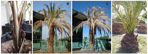
Figure 3 Symptoms of Canarian palm (P. canariensis). A) The color change in the rachis of the inoculated leaf is observed, which indicates an infection process. B) Specimen of P. canariensis affected in basal leaves and flag leaf. C) Same specimen one month later. D) Young palm with dead flag leaf.
On the other hand, during the dissection of the apical meristem of the analyzed palms, a fetid aroma was detected, as well as the arrival of flies from the families Muscidae and Calliphoridae, characteristic of soft rots of bacterial origin, as indicated by the studies by Pérez-Aragon et al. (2013), who determined Erwinia sp., which was identified in this research. This bacterium causes soft rot in the apical meristem, the palms in which it has access to the flag leaf, detach easily since its attachment to the apex of the stem becomes necrotic. In the case of planting the apical meristem to determine the presence of bacteria, 19 (19/36) colonies were obtained from the humid chambers, which corresponded to 11 of the 12 palms analyzed. Rivas and Herrera (2015) also identified Erwinia spp., in the oil palm (Elaeis guineensis) suggesting that the bacterium may contribute to the final phases of the rotting process of the plant apical meristem.
However, the sample of meristem sent to the CNRF for the detection of bacteria was positive by the PCR protocol for the detection of the phytoplasm of lethal yellowish of the coconut (ALC), which corresponds to what is reported by Harrison et al. (2002) where they detected high mortality of P. canariensis with similar symptoms to those of ALC (Texas Phoenix decline) in Texas. This phytosanitary problem is also studied by Gurr et al. (2016) in Puerto Rico and Florida of the American Union, as well as in Mexico, and corresponds to group IV-D of the 16Sr gene; although in the case of Mexico the detection analyzes were mainly in palms other than P. canariensis, as indicated by Aviña-Padilla et al. (2011) who analyzed positive samples for the phytoplasma in P. dactilyfera and Sabal mexicana. Mora-Aguilera et al. (2016) indicate that the typical symptoms of ALC consist of necrosis of the developing inflorescence, drop of small fruits, yellowing of the fronds starting from the lower ones, total defoliation, and death. Figure 3 (B and C) shows a specimen of the Canarian palm at least 30 years old, with the basal leaves and the flag leaf dead. Whereas in Figure 3 (D), a young palm (>8 years old) showing death of the flag leaf with asymptomatic basal leaves is observed.
The results of this study indicate that the dieback of P. canariensis in the metropolitan area of the city of Queretaro is associated with a microbial complex: phytoplasma of lethal yellowing of the coconut palm (confirmed by CNRF), Erwinia sp. as well as fungi, in a complex of Fusarium spp. and F. solani, the latter confirmed by Koch’s postulates. However, it is important to highlight that more studies are required to determine the etiology of this syndrome; as well as carrying out tests with all the isolates obtained in this study to evaluate the pathogenicity in the palms; likewise, the evaluation in a greater number of plants to corroborate the presence of the phytoplasma, since plants of all ages have died in the entire metropolitan area of the city of Queretaro.

