


Serviços Personalizados
Journal
Artigo
 texto em
  Inglês (pdf)
 Artigo em XML
 Referências do artigo
 Enviar este artigo por email
Indicadores
-
 Citado por SciELO
 -
 Acessos
Links relacionados
-
 Similares em
SciELO
Similares em
SciELO
Compartilhar

 Permalink
Revista mexicana de fitopatología
versão On-line ISSN 2007-8080versão impressa ISSN 0185-3309
Rev. mex. fitopatol vol.35 no.1 Texcoco Jan. 2017
https://doi.org/10.18781/r.mex.fit.1608-1
Phytopathological notes
First record of Fusarium solani and F. equiseti in plantations of Jatropha curcas in Mexico
1Instituto Nacional de Investigaciones Forestales, Agrícolas y Pecuarias (INIFAP), Campo Experimental Mocochá. Mérida, Yucatán, C.P. 97454, México.
2Instituto Tecnológico de Conkal. Avenida Tecnológico S/N Conkal, Yucatán. C.P. 97345, México.
3INIFAP, Campo Experimental Rosario Izapa. km. 18 de la carretera Tapachula-Cacahoatán, Tuxtla Chico, Chiapas, C.P. 30870, México.
4INIFAP, Campo Experimental Zacatepec, Zacatepec, Morelos, C.P. 62780, México.
Physic nut (Jatropha curcas) is a multipurpose plant that has acquired importance as biofuel; due to the oil extracted, its seed can be used to produce biodiesel. Vascular tissue necrosis, vascular wilting, root rot, dieback and shortening symptoms were observed in J. curcas plantations in Yucatan, Mexico. The objective this work was to identify the causal agent of symptoms using conventional and molecular techniques. Pathogenicity tests in the greenhouse, morphological characterization and sequence analysis showed that the causal agents of the disease were Fusarium solani and Fusarium equiseti. This is the first report of F. solani as causal agent of vascular tissue necrosis, vascular wilting and root rot, and also the first report of F. equiseti to cause dieback and shortening in J. curcas in Mexico.
Key words: Fusarium solani; Fusarium equiseti; phytopathogens; physic nut
El piñón (Jatropha curcas) es una planta multipropósitos que ha adquirido importancia debido a que el aceite que se extrae de sus semillas, se puede transformar en biodiesel. Desde el año 2012, en plantaciones de J. curcas ubicadas en Yucatán, México, se observaron síntomas de marchitez, necrosamiento de tejido vascular y acortamiento de entrenudos. El objetivo del trabajo fue identificar el agente causal de los síntomas descritos por medio de métodos convencionales y moleculares. Las pruebas de patogenicidad en invernadero, caracterización morfológica, así como el análisis de secuencias mostraron que los agentes causales de los síntomas de la enfermedad fueron Fusarium solani y Fusarium equiseti. Este es el primer reporte de F. solani como agente causal de necrosamiento de tejido vascular, marchitez y pudrición de raíz, y también es el primer reporte de F. equiseti como responsable de muerte descendente y acortamiento de entrenudos en plantas de J. curcas en México.
Palabras clave: Fusarium solani; Fusarium equiseti; fitopatógenos; piñón
Physic nut (Jatropha curcas) is a perennial multipurpose flowering plant that belongs to the family Euphorbiaceae. It is native to Central America but most likely to Mexico (Openshaw, 2000; Pecina-Quintero et al., 2014). Physic nut is distributed across tropical and subtropical regions of Asia, Africa and America, where it is grown for seed oil extraction and biodiesel production (Heller, 1996; Wang et al., 2008). Jatropha curcas is resistant to drought and, because of its toxicity, it is considered to be resistant and/or tolerant to plant pests and diseases (Heller, 1996; ZamarripaColmenero et al., 2009; Biswaset al., 2010).
The global expansion of J. curcas as a crop has contributed to disease outbreaks of unknown etiology in spite of the supposed resistance and/or tolerance attributed to this species (Machado and Pereira, 2012; Machado et al., 2014). According to literature, J. curcas is infested by 42 fungi, four bacteria and two viruses (Alonso and Lezcano, 2014). Among the most common diseases found in J. curcas plantations are those caused by fungi, including Colletotrichum gloeosporioides, C. capsici (anthracnosis); Passalora jatrophigena, Cercospora jatrophicola, Pseudocercospora jatrophae (leaf spot); Pseudoidium jatrophae (powdery mildew); Phakopsora arthuriana (rust); Lasiodiplodia theobromae (dieback); Fusarium solani (root rot); Macrophomina phaseolina (crown and root rot); Rhizoctonia bataticola (root rot); Botryosphaeria dothidea (canker) and other fungi (Kumar et al., 2011; Rao et al., 2011; Machado and Pereira, 2012; Ellison, 2015).
In Mexico, as a result of research conducted by different institutions, there have been reports of damage caused by plant pathogens to J. curcas. Valdez-Rodríguez et al. (2011) reported the presence of Pythium aphanidermatum in seedlings and seed cultivated in Veracruz, while Espinoza et al. (2012) found Alternaria alternata in inflorescences showing symptoms of necrosis in Sinaloa. Other J. curcas diseases reported in Mexico are caused by C. gloeosporioides, C. circinans, P. arthuriana, Pestalotiopsis sp., Fusarium sp., Lasiodiplodia sp., Pleurotus ostreatus, Ralstonia solanacearum (Valdez-Rodríguez et al., 2011; Espinoza et al., 2012; Nolasco-Guzmán et al., 2013; Quiroga-Madrigal et al., 2013; Salazar-Pinacho et al., 2016)
In Yucatán, Mexico, symptoms of wilting, necrosis, shortened internodes and dead plants have been observed since 2012 in some plantations. For this reason, this research was aimed at identifying the agent or agents causing symptoms of diseases in J. curcas plantations.
Samplings were conducted in two J. curcas experiment plantations of the Instituto Nacional de Investigaciones Forestales, Agrícolas y Pecuarias (INIFAP) in the Campo Experimental Mocochá (21° 06’ N and 89° 26’ O at 13 masl) and Sitio Experimental Uxmal, in Yucatán (20° 25’ N and 89° 46’ O, at 50 masl), respectively. The two plantations accounted for 3,000 plants; directed samplings of plants with symptoms of necrotic vascular tissue, wilting, root rot, dieback and shortened internodes were conducted at both sites. At least 2 % of plants were sampled in each plantation.
Samples with infected tissue were placed in brown paper bags and taken to the Laboratory of Plant Pathology of the Instituto Tecnológico de Conkal for testing purposes.
In the laboratory, to isolate fungi associated with root and internodes rot, 0.5 cm2 cuttings of diseased tissue were made, which then were disinfected with 2 % sodium hypochlorite for 1 min, washed twice with distilled water and placed on sterile absorbent paper to remove excess of moisture. Later, separately, each sample was sown in 90 mm Petri dishes (five cuts per Petri dish) containing Potato Dextrose Agar (PDA) culture medium. The samples were incubated at 28±1 °C until mycelium growth was observed. Finally, using a sterile needle, portions of mycelium were individually transferred to new Petri dishes with PDA.
The identification of the causal agent of J. curcas necrotic vascular tissue, wilting, root rot, dieback and internodes shortening was made through observation of temporary and permanent preparations of fungi using the methodology by Cristóbal-Alejo et al. (2013); the identification at the genus and species levels was made following the keys of Barnett and Hunter (2006) and Leslie and Summerell (2006), respectively. Pictures of the preparations observed under a compound microscope were taken with a Sony Cybershot digital camera.
The molecular identification of fungi was based on a sequence analysis of specific DNA regions (Seifert et al., 2007). The DNA extraction was carried out with a Kit ZR Fungal/Bacterial DNA MiniPrepTM, and the DNA integration was verified through 1.2 % agarose gel electrophoresis. The conventional polymerase chain reaction (PCR) technique was used to amplify the internal regions ITS1 and ITS2 between the ribosomal genes (rADN) 18S-5.8S and 28S with the primers ITS1 and ITS4 (Manter and Vivanco, 2007). The reaction mixture included the primers ITS1 and ITS4 (1 mM), MgCI2 (3 mM), dNTP (0.2 mM), Taq DNA Polymerase (2.5 units) and DNA (100 ng) for a final volume of 50 µL. The amplification was performed in a Thermocycler (TECHNE®-312, Minnesota, USA) under the following conditions: initial denaturation of 2 min at 95 ºC, followed by 30 cycles (denaturation at 94 °C for 1 min, alignment at 54 °C for 30 s and an extension of 1 min at 72 °C) with a final extension of 5 min at 72 °C (White et al., 1990).
The final products of the amplification were visualized in 2% agarose gel (Ultra PureTM). The amplified fragments were delivered to the company Macrogen (USA) to be sequenced. The sequences obtained were analyzed and compared with the sequences stored at the gene bank of NCBI (National Center for Biotechnology Information) using BLASTn. The sequences were submitted to the GenBank database to obtain their access number.
The Koch’s postulates were used to confirm the pathogenicity of fungi isolates obtained consistently from asymptomatic tissue of plants of J. curcas. For the isolate fungus of plants with necrotic vascular tissue, wilting and root rot, seeds of J. curcas were disinfected with 1 % sodium hypochlorite for 2 min, washed twice with sterile distilled water and sown in 1L polystyrene cups containing sterile soil. After seed germination, twenty 40-day-old plants were selected; 2mm-deep cuts in the plant neck or bottom were made and inoculated by spraying a 1 x 10⁶ mL-1 spore concentration. Twenty plants sprayed with sterile distilled water were used as control. The cuts of all the plants were covered with Parafilm® paper and left in the greenhouse until symptoms appeared. For the fungus isolate of plants showing dieback and shortened internodes, the same methodology was used, but the fungus inoculation was performed 15 cm above the plant’s neck or base.
From plants of J. curcas with necrotic roots, wilting and necrotic vascular tissue, Fusarium solani was isolated and identified (Figure 1A, B y C). These symptoms were also observed by Sharma et al. (2001), Wu and Yu (2011), Machado and Pereira (2012), and Machado et al. (2012). Fusarium solani is the causal agent of root rot symptoms in J. curcas plantations in China (Wuet al., 2011), whose teleomorph was identified as Nectria haematococca. According to literature reviews, this is the first report in J. curcas plantations in Mexico.
Fusarium solani presented cottony mycelium, cream colored septate that changed from blue to green; unicellular and egg-kidney-shaped micronidia 2.7-8.9 μm long x 2-4 μm wide produced in large conidiophores, single and in twos; canoe- or crescent-shaped 9.1-38.9 μm x 2.2-4.4μm in diameter with 1-4 septates, thick cylindrical walls, back and ventral surface parallel along its entire length, blunt and rounded apical cell with a slot at the base, globose chlamydospores, single and in twos, 6.2-11.1μm in diameter (Figure 1D and E). These morphological characteristics agree with those described by Barnett and Hunter (2006) and Leslie and Summerell (2006) for F. solani.
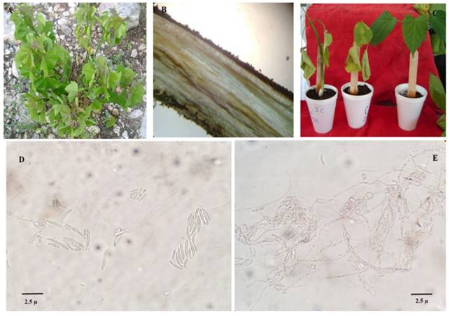
Figure 1. Symptoms caused by Fusarium solani in Jatropha curcas, and microscopic characteristics of Fusarium solani. (A) Plants with wilting symptoms. (B) Root system necrosis. (C) Dissemination of wilting symptoms in seedlings. (D) Microconidia and macroconidia. (E) Chlamydospores formation.
Results of search for homology using BLASTn with nucleotide sequences reported by NBCI were 99 % similar to the sequence of the F. solani fungus (Access Number U733636). The nucleotides sequence was submitted to the GenBank database with Access Number KY013237.
In plants of J. curcas inoculated with F. solani, wilting symptoms were observed seven days after inoculation (dpi), followed by a loss of turgidity in plants and chlorosis in leaves, which gradually turned brown (Figure 1C). Root extraction of J. curcas at 13 dpi showed necrosis in the vascular system; from the symptoms observed, F. solani was isolated. The control plants did not show any symptom. The symptoms observed correspond to those reported by Wu et al. (2011) in J. curcas plantations in China; the authors found that 2 months after inoculating seedlings of J. curcas, they showed symptoms of root rot, leaf chlorosis, leaves falling and necrotic and rotten roots. Similar symptoms were also reported by Herrera-Parra et al. (2011) in wilted plants of Thevetia peruviana, as well as by Hasan et al. (2014) in plants of Hibiscuss abdariffa with symptoms of root rot and wilting.
In plants of J. curcas with symptoms of dieback and stems with shortened internodes, Fusarium equiseti was isolated and identified (Figure 2A, B, D and E). There are other reports of F. equiseti but as causal agent of damping-off, root rot and wilting in Pinus halepensis, H. sabdariffa and Panax quiquefolius (Punja et al., 2007; Hasan et al., 2014; Lazreg et al., 2014), as well as inducer of floral necrosis and young fruit falling in Carica papaya (Vásquez et al., 2012). However, this is the first report of dieback and internodes shortening in stems of J. curcas.
This fungus in a PDA culture medium was characterized by abundant mycelia growth, which was initially white to pale yellow and then turned brown; 53-80 x 4.0-4.5 μm oval micronidia with a pedicellate foot cell and chlamydospores in chains and aggregates (Figure 2F); and the development of sporodochia which were initially orange and then turned dark. Sporodochia showed plenty of macronidia developed in monophialides or in dorsoventral curved branched conidiophores, usually with five to seven 25-120μm long marked septates, a thick wall slightly arched at the ventral part and abruptly arched at the dorsal part, with a foot-like basal cell and a stranded apical part (Figure 2G). The morphological characteristics previously mentioned are in accordance with those described by Barnett and Hunter (2006) and Leslie and Summerell (2006) for F. equiseti.

Figure 2. Symptoms caused by Fusarium equiseti in Jatropha curcas, and macroscopic and microscopic characteristics of Fusarium equiseti. (A) Shortening of internodes in branches. (B) Branch dieback. (C) Control plant without symptoms. (D) Plant inoculated with dieback. (E) Cutting of a necrotic stem. (F) Development in PDA culture medium. (G) Macroconidia.
Sequencing results show that the nucleotides sequence was 100 % similar to that of the F. equiseti fungal species (Access Number KJ412501) reported by NCBI. The nucleotides sequence was also submitted to the GenBank database with Access Number KY012795.
In plants of J. curcas inoculated with F. equiseti, symptoms of stem necrosis and dieback were observed at 20 dpi (Figure 2D and E); from these, F. equiseti was re-isolated. The control plants did not show any symptom (Figure 2C).
According to Machado and Pereira (2012), in areas with prolonged dry seasons, J. curcas shows a higher incidence of root and neck rot. For this reason, it is believed that water stress is the main environmental condition that favors the disease development; such condition was observed during samplings in J. curcas plantations in Yucatán.
In this research, F. solani is reported for the first time as the causal agent of wilting and vascular tissue necrosis, and F. equiseti as inducer of internodes shortening and dieback in J. curcas in Yucatán, Mexico. This disease may become a serious problem for J. curcas cultivation in Mexico since there are no control and monitoring strategies in place. Therefore, further studies to know the environment conditions that favor the disease are needed, as well as strategies for its integrated management.
Conclusions
Based on morphological characterization, rDNA ITS sequences analyses and pathogenicity tests, this is the first time F. solani is reported as the causal agent of vascular tissue necrosis, root rot and wilting in plants of J. curcas in Yucatán, Mexico. Also, for the first time, F. equiseti is reported as the causal agent of internodes shortening and dieback in J. curcas.
Acknowldgements
The authors express their appreciation to Secretaría de Agricultura, Ganadería, Desarrollo Rural, Pesca y Alimentación (SAGARPA) for its support to the Project “Potential pests and diseases of Physic Nut and Castor Bean that affect the biofuel production in the Mexican tropic”.
REFERENCES
Alonso O y Lezcano JC. 2014. Pathogen microorganisms in Jatropha curcas Linnaeus. Potential strategies for their management. Pastos y Forrajes 37:131-137. http://www.redalyc.org/articulo.oa?id=269131791001. [ Links ]
Barnett LH and Hunter BB. 2006. Illustrated Genera of Imperfect Fungi. Fourth Edition. The American Phytopathological Society Press. St. Paul, Minnesota, USA. 234 p. [ Links ]
Biswas PK, Pohit S and Kumar R. 2010. Biodiesel from Jatropha: can India meet the 20% blending target. Energy Policy 38:1477-484. http://doi:10.1016/j.enpol.2009.11.029. [ Links ]
Cristóbal-Alejo J, Navarrete-Mapen Z, Herrera-Parra E, Mis-Mut M, Tun-Suárez JM and Ruiz-Sánchez E. 2013. Hifomicetos asociados a plantas tropicales del estado de Yucatán, México: Identificación genérica y evaluación de fungicidas para su control. Revista de Protección Vegetal 28:138-144. http://scielo.sld.cu/pdf/rpv/v28n2/rpv07213.pdf. [ Links ]
Ellison CA. 2015. First Report of Colletotrichum truncatum causing stem cankers on Jatropha curcas in Burkina Faso. Plant Disease 99:14-20. http://dx.doi.org/10.1094/PDIS-02-14-0181-RE. [ Links ]
Espinoza MA, Santos AE, Fernández E, Espinoza MG, Chávez JA, Bermúdez EM, Martínez AL, Martínez J and Leyva NE. 2012. First report of Alternaria alternata (Fr.) Keissler causing inflorescence blight in Jatropha curcas in Sinaloa, México. Canadian Journal of Plant Pathology 34:455-458. http://10.1080/07060661.2012.688770. [ Links ]
Hassan N, Shimizu M and Hyakumachi M. 2014. Occurrence of root rot and vascular wilt diseases in roselle (Hibiscus sabdariffa L.) in upper Egypt. Mycobiology 42:66-72. http://dx.doi.org/10.5941/MYCO.2014.42.1.66. [ Links ]
Heller J. 1996. Physic nut. Jatropha curcas L. Institute of Plant Genetics and Crop Plant Research. Gatersleben, International Plant Genetic Resources Institute. Rome, Italy. 66 p. http://www.bioversityinternational.org/uploads/tx_news/Physic_nut__Jatropha_curcas_L._161.pdf. [ Links ]
Herrera-Parra E, Bacab-Pérez I, Cristóbal-Alejo J, Tún-Suárez JMA and Ruiz-Sanchez E. 2011. Patogenicidad de Fusarium solani (Mart.) Sacc. y Alternaria alternata (Fries) Keissler en Thevetia peruviana (Pers.) K. Schum. y su control in vitro. Fitosanidad 15:231-236. http://www.redalyc.org/articulo.oa?id=209123682005. [ Links ]
Kumar S, Sharma S, Pathak DV and Beniwal J. 2011. Integrated management of Jatropha root rot caused by Rhizoctonia bataticola. Journal of Tropical Forest Science 23:35-41. http://www.jatropha.pro/PDF%20bestanden/Rootrot.%20Kumar.pdf. [ Links ]
Lazreg F, Belabid L, Sanchez J, Gallego E, Garrido-Cardenas JA and Elhaitoum A. 2014. First report of Fusarium equiseti causing damping-off disease on aleppo pine in Algeria. Plant Disease 98:1268. http://dx.doi.org/10.1094/PDIS-02-13-0194-PDN. [ Links ]
Leslie JF and Summerell BA. 2006. The Fusarium Laboratory Manual. Blackwell Publishing. Ames, Iowa, USA. 388 p. [ Links ]
Machado AR and Pereira OL. 2012. Major diseases of the biofuel plant, physic nut (Jatropha curcas). Pp:59-75. In: Fang Z (ed.). Biodiesel: Feedstocks, Production and Applications. Intech, Croatia.75p. http://dx.doi.org/10.5772/52336. [ Links ]
Machado AR, Pinho BD and Pereira LO. 2014. Phylogeny, identification and pathogenicity of the Botryosphaeriaceae associated with collar and root rot of the biofuel plant Jatropha curcas in Brazil, with a description of new species of Lasiodiplodia. Fungal Diversity 67:231-247. http://link.springer.com/article/10.1007%2Fs13225-013-0274-1. [ Links ]
Machado AR, Pinho DB, Dutra DC and Pereira OL. 2012. Collar and root rot caused by Neoscytalidium dimidiatum in the biofuel plant Jatropha curcas. Plant Disease 96:1697. http://dx.doi.org/10.1094/PDIS-05-12-0504-PDN. [ Links ]
Manter KD, Vivanco MJ. 2007. Used of the ITS primers, ITS1F and ITS4, to characterize fungal abundance and diversity in mixed Template samples by qPCR and length heterogeneity analysis. Journal of Microbiological Methods 71: 7.14. http://www.elsevier.com/located/jmicmeth. [ Links ]
Nolasco-Guzmán, V, Escobar VA, Tovar-Pedraza JM, Ríos-López EG, Calyecac-Cortero HG and Rangel AM. 2013. Primer reporte de Phakopsora arthuriana en Jatropha curcas en México. Revista Mexicana de Fitopatología 31:70-73. http://www.redalyc.org/articulo.oa?id=61230974008. [ Links ]
Openshaw K. 2000. A review of Jatropha curcas: An oil plant of unfulfilled promise. Biomass and Bioenergy 19:1-15. http://dx.doi.org/10.1016/S0961-9534(00)00019-2. [ Links ]
Pecina-Quintero V, Anaya-López JL, Zamarripa-Colmenero A, Núñez-Colí CA, Montes-García N, Solís-Bonilla JL and Jiménez-Becerril MF. 2014. Genetic structure of Jatropha curcas L. in Mexico and probable centre of origin. Biomass and Bioenergy 60:147-155. http://dx.doi.org/10.1016/j.biombioe.2013.11.005. [ Links ]
Punja ZK, Wan A, Goswami RS, Verma N, Rahman M, Barasubiye T, Seifert KA and Lévesque CA. 2007. Diversity of Fusarium species associated with discolored ginseng roots in British Columbia. Canadian Journal of Plant Pathology 29:340-353. http://dx.doi.org/10.1080/07060660709507480. [ Links ]
Quiroga-Madrigal RR, Rosales Esquinca MA, Rincón Espinosa MP. Garrido Ramírez E, Holguín Meléndez F, González Pinacho JA, Salazar Pinacho WM y Sol Hernández G. 2013. Enfermedades de J. curcas. Pp:83-102. In: Zamarripa CA y Solís Bonilla JL (eds.). Jatropha curcas L. Alternativa Bioenergética en México. Ave Dos Taller Creativo. Tapachula, Chiapas, México.157p. [ Links ]
Rao SC, Kumari PM,Wani SP and Marimuthu S. 2011. Occurrence of black rot in Jatropha curcas L. plantations in India caused by Botryosphaeria dothidea. Current Science 100:1547-1549.http://www.currentscience.ac.in/Volumes/100/10/1547.pdf. [ Links ]
Salazar-Pinacho WM, Quiroga-Madrigal RR, Rosales-Esquinca MA, Garrido-Ramírez ER y Holguín-Meléndez F. 2016. Etiología de la marchitez y pudrición basal de Jatropha curcas en Arriaga, Chiapas, México. Revista Mexicana de Fitopatología 34:110-115. http://dx.doi.org/10.18781/R.MEX.FIT.1507-5. [ Links ]
Seifert KA, Samson RA, DeWaard, JR, Houbraken J, Levesque CA, Moncalvo JM, Seize GL and Hebert PDN. 2007. Prospects for fungus identification using CO1 DNA barcodes, with Penicillium as a test case. PNAS, The National Academy of Sciences of the USA. 104: 3901-3906. http://dx.doi.org/10.1073/pnas.0611691104. [ Links ]
Sharma S, Kaushik JC and Kaushik N. 2001. Fusarium moniliforme causing root rot of jatropha. Indian Phytopathology 54:275-277. http://epubs.icar.org.in/ejournal/index.php/IPPJ/article/viewFile/18956/9311. [ Links ]
Valdéz-Rodríguez OA, García ER, Sánchez SO and Pérez VA. 2011. Isolation and pathogenicity of a possible Pythium aphanidermatum in Jatropha curcas L. non toxic. Tropical and Subtropical Agroecosystems 14:649-660. http://www.redalyc.org/articulo.oa?id =93918231028. [ Links ]
Vásquez A, Hernández CE, Mora AA, Nava DC. 2012. Sánchez GF. Etiología y epidemiología de la necrosis de flores y frutos juveniles del papayo (Carica papaya L.) en Guerrero, México. Agrociencia 46:757-767. http://www.redalyc.org/articulo.oa?id=30225072001. [ Links ]
Wang ZY, Lin JM and Xu ZF. 2008. Oil contents and fatty acid composition in Jatropha curcas seeds collected from different regions. Nan Fang Yi Ke Da Xue Xue Bao 28:1045-1046. http://www.j-smu.com/pdf2/200806/2008061045.pdf. [ Links ]
White TJ, Bruns T, Lee S and Taylor J.1990. Amplification and direct sequencing of fungal ribosomal RNA genes for phylogenetics. Pp:315-321. In: Innis MA, Gelfand DH, Sninsky JJ and White TJ (eds). PCR Protocols: A Guide to Methods and Applications. Academic Press. San Diego, CA, USA.345p. [ Links ]
Wu YK and Yu GT. 2011. Study on the sexual stage of the root rot disease pathogen in Jatropha curcas L. Chinese Agricultural Science Bulletin 27:297-301.http://www.casb.org.cn/casb/ch/reader/view_abstract.aspx?file_no=2011-1795. [ Links ]
Wu YK, Ou GT and Yu JY. 2011. First report of Nectria haematococca causing root rot disease of physic nut (Jatropha curcas) in China. Australasian Plant Disease. Notes 6:39-42. http://dx.doi.org/10.1007/s13314-011-0014-x. [ Links ]
Zamarripa-Colmenero A, Ruiz Cruz PA, Solís Bonilla JL, Martínez Herrera J, Olivera De los Santos A and Martínez Valencia B. 2009. Biocombustibles: perspectivas de producción de biodiesel a partir de Jatropha curcas L., en el trópico de México. Campo Experimental Rosario Izapa, INIFAP. Folleto Técnico No. 12. Tuxtla Chico, Chiapas, México. 46 p. http://biblioteca.inifap.gob.mx/portal/index.php/component/content/article/78colecciones-inifap/96-folleto-tecnico-busquedas. [ Links ]
Received: September 04, 2016; Accepted: December 21, 2016
 Este es un artículo publicado en acceso abierto bajo una licencia Creative Commons
