


Servicios Personalizados
Revista
Articulo
 texto en
  Inglés (pdf)
 Artículo en XML
 Referencias del artículo
 Enviar artículo por email
Indicadores
-
 Citado por SciELO
 -
 Accesos
Links relacionados
-
 Similares en
SciELO
Similares en
SciELO
Compartir

 Permalink
Revista mexicana de fitopatología
versión On-line ISSN 2007-8080versión impresa ISSN 0185-3309
Rev. mex. fitopatol vol.35 no.1 Texcoco ene. 2017
https://doi.org/10.18781/r.mex.fit.1607-4
Review articles
Powdery mildews in agricultural crops of Sinaloa: Current status on their identification and future research lines
1Universidad de Occidente, Unidad Los Mochis, Departamento de Ciencias Biológicas, Blvd. Macario Gaxiola y Carretera Internacional s/n, CP 81223. Los Mochis, Sinaloa, México.
2Instituto Politécnico Nacional (IPN), Departamento de Biotecnología Agrícola, CIIDIR-Sinaloa. Blvd. Juan de Dios Bátiz Paredes No 250, CP 81101. Guasave, Sinaloa, México.
3Universidad de Occidente, Unidad Guasave, Departamento de Ciencias Biológicas, Av. Universidad s/n, CP 81120. Guasave, Sinaloa, México.
4Laboratorio de Microscopía Electrónica de Barrido, Facultad de Ciencias, Universidad Nacional Autónoma de México. CP 04510, Coyoacán, D.F., México.
The present review aims to discuss the evolution of the procedures for the identification of the powdery mildew pathogens. The morphometric characteristics deciphered by light microscopy and scanning electron microscopy, and the availability of molecular tools have contributed to the identification of the anamorph in different species of Erysiphales in various regions around the world. In Sinaloa, the identification of these plant pathogens started in 2005. Applying morphometric studies to the teleomorph and anamorph of the powdery mildew fungus in squash and cucumber allowed the identification of Podosphaera xanthii as the causal agent; the anamorph of the same species was also identified in melon, watermelon, bottlegourd, and husk tomato. In subsequent studies, in addition to the morphometric characteristics, the ITS of rDNA was used for the identification of the anamorph of Erysiphe diffusa, Erysiphe quercicola, and Podosphaera pannosa on bean, mango, and roses, respectively. The known species of Erysiphales at the present time in Sinaloa might be only a segment of many of them infecting cultivated and wild species of plants; thus, there is an immense opportunity to focus research lines on the etiology, epidemiology, and control of this type of diseases.
Keywords: Anamorph; teleomorph; light microscopy; scanning electron microscopy; morphometry
La presente revisión tiene como objetivo abordar la evolución de los procedimientos para identificar los agentes causales de las cenicillas. Las características morfométricas que se descifran mediante la utilización de microscopía de luz y microscopía electrónica de barrido, así como el rango de hospedantes y herramientas moleculares han permitido la identificación del anamorfo en diferentes especies de Erysiphales, en diversas regiones del mundo. En Sinaloa, México, la identificación de este tipo de patógenos inició en 2005. Los estudios morfométricos del teleomorfo y anamorfo en calabaza y pepino permitieron la identificación de Podosphaera xanthii en estos hospedantes; el anamorfo del mismo hongo se identificó en melón, sandía, bule y tomatillo recurriendo a las mismas técnicas. En estudios subsiguientes, además de la morfometría se recurrió a los espaciadores transcritos internos (ITS; por su abreviatura en inglés) para la identificación de los anamorfos de Erysiphe diffusa, Erysiphe quercicola y Podosphaera pannosa. Las especies de Erysiphales conocidas a la fecha en Sinaloa son sólo un segmento de muchas de ellas que actualmente atacan a plantas cultivadas y silvestres; por lo que existe espacio para el surgimiento de líneas de investigación direccionadas hacia la etiología, epidemiología y el manejo de este tipo de enfermedades.
Palabras clave: Anamorfo; teleomorfo; microscopía de luz; microscopía electrónica de barrido; morfometría
In Mexico, Sinaloa is the main vegetableproducing state. In Sinaloa, during 2014, the surface dedicated to the planting of zucchini (Cucurbita pepo L.), tomato (Solanum lycopersicum L.), Mexican husk tomato (Physalis philadelphica Lam.) and cucumber (Cucumis sativus L.) was 31,716 ha, whereas the surface for beans (Phaseolus vulgaris L.) and mango (Mangifera indica L.) was 122,412 ha and 31,180 ha, respectively (SAGARPA, 2014). Although Sinaloa is not one of the main producers of roses, the surface planted has increased due to the demand for this ornamental plant. In Sinaloa, fungal diseases constitute one of the main limiting factors in these crops (León-Gallegos, 1988; Cruz-Ortega et al., 1998; Ramírez-Villapudúa et al., 2006; Ramírez-Villapudúa and Sáinz-Rodríguez, 2006) and among the diseases that affect foliage, powdery mildew stands out, since it appears in every agricultural cycle, and its incidence and severity vary with the prevalent weather conditions. Powdery mildew is caused by a group of diverse fungi, complex in shape, reproductive structures, range of hosts, and geographic distribution (Bélanger and Labbé, 2002). They are found in the family Erysiphaceae of the order Erysiphales; they are obligate parasites (biotrophes) and they parasitize around 9,838 species of plants that belong only to angiosperms (Amano, 1986). 93 % of host plants are dicotiledons, while 7 % are monocotiledons (Takamatsu, 2013).
Erysiphales produce spherical asci called chasmothecia (previously called cleistothecia), as well as conidiophores and hyaline hyphae, uninuclear septate hyphae, and conidia that form a white ash-like powder when developing in large amounts, making them easily recognizable. These structures are mainly produced on the leaves, buds, flowers, and fruits, and produce haustoria on the epidermal cells of their hosts (Boesewinkel, 1980). They damage plants slowly and are vulnerable to fungicides, due to the epiphytic habit in its interaction with the host (Yarwood, 1973). Although mildew is common and causes considerable damage in cool areas; they are even more common and severe in areas with warm, dry climates, because in these conditions the wind easily detaches and spreads the conidia (Romero-Cova, 1988); also, it has been established that mildew presents itself in the succulent tissue of the host in cool, shady areas (Yarwood, 1973). The lives of conidia are brief and they are favored by high relative humidity, but not by rain and inmersion in water (Sivapalan, 1993a; Sivapalan, 1993b). In general, the mycelium is epiphitic, except in genuses Leveillula, Phyllactinia, Pleochaeta where the mycelium is hemi-endophytic, and occasionally in species of Cystotheca; in this case, hyphae penetrate the leaves through the stomata and form mycelia inside, although the mycelia of the species of Leveillula are more abundant inside the leaf of the host (Braun et al., 2002).
The development of the taxonomy of Erysiphales is vast and controversial and has been widely discussed in many publications (Braun, 1999; Braun et al., 2002; Braun y Cook, 2012). In recent years, based on molecular phylogenetics, it has been shown that Erysiphales belong to the Leotiomycetes (unoperculated Discomycetes) and not to the Pirenomycetes, as believed for many years (Glawe, 2008). With the classical works on the Erysiphales (Yarwood, 1957), it was imposible to refer to the anamorph of the causal agents of powdery mildew; however, reseach carried out by Boesewinkel (1980) and Braun and Cook (2012) contributed to the knowledge of the anamorph and its relation with the teleomorph; these authors introduced new characteristics and a wider and more natural concept of species, while showing that many powdery mildew species have a wider range of hosts than is known.
As established by Boesewinkel (1980), powdery mildews in nature are mainly found in their asexual phase. Occasionally, researchers underestimate a wide variety of morphological characteristics in the anamorphs, which are useful for identifying species. The structures considered include: the location of the mycelium in the host; the diameter and color of the hyphae; the characteristics of the haustoria, appressoria and their location in the mycelium; simple or branched conidiophores; individual conidia production or in chain; characteristics of the basal cell of the conidiophores; the shape of the conidia which varies from oval to cylindrical or lanceolated; as well as the presence of fibrosin bodies in the conidia (Boesewinkel, 1980). In most species, conidia are monomorphic, although the species Leveillula, Pleochaeta (Braun et al., 2002) and Phyllactinia (Liberato, 2007) produce dimorphic conidia.
In Erysiphales, many species are related to certain plant families and genuses, which is also useful in the process of identifying anamorphs. The morphological characteristics were combined and codes were created to identify members of the family Erysiphaceae (Boesewinkel, 1980). Later, the scanning electron microscope showed that the wall of the turgid conidia presents a wide variation (from smooth to diversely ornamented) and when they are dehydrated their sinuous wrinkling patterns can be longitudinal or transverse, reticulated, linear, or rectangular, among others (Braun et al., 2002). Likewise, different patterns were found in the ornamentations of the end of the conidia, which differ from those of the rest of the conidia (Cook et al., 1997; Braun et al., 2002; Braun and Cook, 2012). Some authors claim that the cell wall characteristics are constant, since they form from its inner layers (Plumb and Turner, 1972), which contrasts with the apendices of the chasmothecium, which are modifiable by age and environmental factors (Cook et al., 1997).
The Erysiphales taxonomy manual (Braun and Cook, 2012) locates tribes and genera and considers the morphology of the teleomorph, conidial germination and the characteristics of their walls under the electron microscope; it contains codes based on the characteristics of the anamorph , as well as codes for the anamorph only. In an orderly fashion, it shows codes for species in each genus, as well as tabular codes for species based on the families and genuses of the hosts. The location of the tribes and genuses is based on the recent phylogenetic classification of Erysiphales and describe 873 species that include 853 figures. This work undoubtedly contributes to the identification of the causal agents of mildew.
Identifying Powdery mildews can be difficult, depending on the reproductive state og the fungus (Braun and Cook, 2012), making molecular tools very useful, particularly if applied to specimens in which the teleomorph is not observed, as well as in herbarium material (Cunnington et al., 2003). In taxonomical studies at a molecular level, the region of the internal transcript spacers (ITS) of the ribosomal DNA (rDNA) can be very useful to relate the anamorph of the specimens with their respective teleomorphs. Studies on this indicate that sequencing the regions of the ITS in 25 anamorphic specimens, and comparing them with the ITS of their potential teleomorphs gave, in most cases, similarities of over 99 %; although the study was not conclusive for some specimens, this technique provides information that, when complemented with the morphology and range of hosts, contributes to the identification of causal agents of mildew, as indicated in previous studies (Cunnington, et al., 2003; Monkhung et al., 2011). The current taxonomical systems resort to the morphological characteristics, the range of hosts, and to the molecular phylogenetic information (Glawe, 2008). The use of ITS and 18S sequences of the rDNA to infer phylogenetic relations of Erysiphales began in the 1990’s (Saenz et al., 1994; Saenz and Taylor, 1999).
On the other hand, as in the fungal taxonomy, the nomenclature codes have also changed. In the symposium “One fungus = which name” in Amsterdam in 2012, changes in the nomenclature of pleomorphic fungi were addressed; the conclusion reached was that this type of fungi will be treated as plants and other types of organisms (Braun, 2012), and therefore, the principle “one fungus = one name” is valid as specified in the new Melbourne Code (ICN), Art. 59 (McNeill et al., 2012). This principle is applicable to the nomenclature of the species that cause mildew and the assignment of only the teleomorph in the species of Erysiphales (Braun, 2013) is proposed, implying that only one name will be assigned to each species.
Below is an analysis of the information available on the current situation of mildew in economically important crops in Sinaloa, and we propose lines of investigation on this type of diseases.
Ethiology of mildew in platations in Sinaloa
Cucurbit Powdery Mildew
Although previous studies had confirmed Erysiphe cichoracearum as a causal agent of the cucurbit powdery mildew (Alvarez, 1976; Cebreros et al., 1991), the first paper that included morphometry and the teleomorph for the identification of cucurbit mildew in Sinaloa was published in 2005. Using the signs of the pathogen collected from various types of pumpkin, melon, watermelon, and gourd (Lagenaria siceraria (Molina) Standley), a morphometric analysis was carried out on both the anamorph and teleomorph, resorting to light microscopes and scanning electron microscopes (Figure 1. A-F). The images of Euoidium conidia under the scanning electron microscope, the type of host, and the characteristics of the anamorph helped obtain a preliminary identification of Podosphaera (sect. Sphaerotheca) xanthii (Castagne) U. Braun and N. Shishkoff, (sin. Sphaerotheca fuliginea (Schelechtend.:Fr.) Pollacci) as the species related to cucurbit mildew (Félix-Gastélum et al., 2005). Studies on the teleomorph of different types of pumpkin helped confirm the identity of the fungus, as performed in California, U.S.A (Kontaxis, 1978), although this phase of the organism was not found in melon, gourd, or watermelon, even though commercial plantations of these crops were damaged by mildew and were sometimes adjacent to cucumber or pumpkin plantations where the teleomorph was found (Félix-Gastélum et al., 2005). Later studies on the cucumber mildew in the Valley of Culiacán, Sinaloa helped identify the anamorph and teleomorph of P. xanthii in cucumbers, by means of morphological and molecular studies. Also, the presence of physiological breeds 1, 2F, 4, and 5 were also confirmed (Bojórquez-Ramos et al., 2012). According to Ballantyne (1975)Erysiphe cichoracearum (sin. Golovinomyces cichoracearum (DC.) V.P. Heluta), Leveillula taurica (Lév.) G. Arnaud, Erysiphe communis (Wallr.) Schltdl., Erysiphe polygoni DC. and Erysiphe polyphaga Hammarl. have been found to be related to the disease in other parts of the world; however, these species were not found in the different cucurbits studied in four agricultural cycles (2000 to 2004) in northern Sinaloa (Félix-Gastélum et al., 2005), which coincides with studies on cucumber mildew in the Valley of Culiacán (Bojórquez-Ramos et al., 2012). However, in the Czech Republic P. xanthii and G. cichoracearum were found in one species of cucurbit (Lebeda et al., 2004).

Figure 1. Micrography with a scanning electron microscope of Podosphaera xanthii in pumpkin: A) Smooth conidium wall, B) Terminal part of the same structure with faint concentric rings and lobular projections, C) Partially dehydrated conidium with longitudinal and transversal wrinkles. D) Casmotecios with mycelial appendices with septa. E) Immature hyaline ascus in a partially open casmotecio. F) Ascus with ascospores inside.
The absence of the teleomorph in melon, gourd, and watermelon in Sinaloa is difficult to explain; however, the fungus could be heterotallic, as is the case in P. xanthii (McGrath, 1994) and other mildew-causing species (Yarwood, 1935; Schnathorst, 1959; Smith, 1970, Coyier, 1972). Chasmothecia are not formed in all the species and varieties of cucurbitáceas (Khan and Khan, 1970), and in Sinaloa these were found in different types of pumpkins and cucumbers at the end of the cycle (Félix-Gastélum et al., 2005). Previous studies indicate that the formation of chasmothecia begins when the formation of conidia decreases or stops due to the presence of the host, or otherwise by environmental and handling factors, such as limited nourishment conditions, low humidity levels and low temperatures (Yarwood, 1935). Environmental conditions that promote the development of mildew also influence the development of the teleomorph, since a greater foliar area covered by the fungus increases the probabilities of reproduction, and therefore the formation of chasmothecia (Schnathorst, 1965).
Mildew in tomatillo
(Physalis philadelphica Lam.). The causal agent of mildew in tomatillo was identified as P. Xanthii with a study on the morphometric characteristics of the anamorph and images of the conidia under the compound microscope (Figure 2 A and B) and the scanning electron microscope (Fig. 2 C); no structures were found that revealed the presence of the teleomorph of the fungus in infected plants (Félix-Gastélum et al., 2007). Once again it was proven that the characteristics of the anamorph contributed towards the preliminary identification of mildew in tomatillo, as mentioned by other authors (Boesewinkel, 1980; Braun et al., 2002; Braun y Cook, 2012). It is worth pointing out that in the bibliography there are only two reports of this disease: one in California, U.S.A. (Koike and Smith, 1988) and another in Taiwan (Cheng et al., 2006). Preliminary results indicate that the causal agent of pumpkin mildew does not attack tomatillo, nor does mildew in tomatillo attack pumpkin (Félix-Gastélum, results not published), which corresponds with previous studies, which mention that most species that cause powdery mildew are specific to certain hosts; however, there are various species of this group of fungus that have a wide range of hosts, such as Erysiphe quercicola S. Takam. and U. Braun, which attacks temperate climate plants and some species of tropical trees (Takamatsu et al., 2007).

Figure 2. A) Mildew Euoidium conidiophora in tomatillo mildew with immature conidia and crenellated edges, typical of Podosphaera (sect. Sphaerotheca) xanthii. B) Conidia with forked germinative tube originating in the middle section of the conidium. C) Micrography with a scanning electron microscope of a mature conidium with a smooth wall.
Mildew in beans (Phaseolus vulgaris L.)
Regional literature has referred to E. polygoni as a causal agent of the bean plant (León-Gallegos, 1988). The first study to mention the morphometry of the anamorph and de molecular identification of the causal agent of the disease in Sinaloa was published recently (Félix-Gastélum et al., 2011). Faced with the absence of the teleomorph, we resorted to morphometric studies of the anamorph, which was complemented with images of conidia under a scanning electron microscope (Figure 3. A and B). In addition, the ITS amplification of the rDNA confirmed the causal agent as Pseudoidium (Cook et al., 1997), anamorph of Erysiphe diffusa (Cooke and Peck) U. Braun and S. Takam. (Takamatsu et al., 2007).
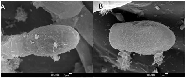
Figure 3. Micrography with a scanning electron microscope of Erysiphe diffusa in bean plants: A) Immature conidium joined to the Pseudoidium conidium, and B) Mature conidium, with a cross-link on its wall.
Fortunately, mildew is not an important disease in Azufrado Higuera, the predominant bean variety in the region, but it is severe in creeping bean varieties, which account for less than 10% of the surface planted in Sinaloa. As in Brazil (Almeida et al., 2008), the E. diffusa teleomorph has been observed in Sinaloa, therefore some alternate hosts are inferred to play an important part in the life cycle and survival of the pathogen. Some wild legumes could act as a bridge for the survival of the pathogen in the absence of bean plants. This line of investigation, once explored, will surely contribute to understanding the biology of the pathogen and the epidemiology of the disease.
Mildew in mango
(Mangifera indica L.). Mildew in mango is one of the most common and widespread diseases in the world (Nasir et al., 2014), causing losses of up to 90 % when it appears during flowering (Misra, 2001). In Sinaloa, although sometimes underestimated by producers, this disease can cause losses of up to 70 % when it occurs at the beginning of the flowering period, during the months of January and February. The fungus attacks young tissues, mainly, including leaves, petioles, flower scales, flower buds, and fruits in initial development stages (Singh, 1960), symptoms which have also been observed in Sinaloa for decades. Regional literature referred to Oidium mangiferae Berthet as a causal agent of the disease for many years (Ramírez-Villapudúa et al., 2006).
Based on morphometric studies, images of conidia under a scanning electron microscope (Figure 4 A y B), as well as molecular studies and phylogenetic analyses of the anamorph, the causal agent of the disease in Sinaloa was established to be Pseudoidium anacardii (F. Noack) U. Braun and R.T.A. Cook with E. quercicola as a teleomorph (Félix-Gastélum et al., 2013). As in other mangoproducing areas of the world (Nasir et al., 2014), in northern Sinaloa the teleomorph was not found in specimens collected in mango vars. Kent and Keith in commercial or backyard orchard trees (Félix-Gastélum et al., 2013). In other parts of the world, the fungus hybernates as mycelia or haustoria in buds or where it can remain in the dense tree canopies (Nasir et al., 2014); its form of survival during the summer in Sinaloa is unknown. Dilucidating the form of survival of the P. anacardii anamorph remains as a future line of research. On the other hand, studies carried out in Guerrero and Michoacan indicated that the maximum severity of mildew in mango coincided with periods with temperatures of 20 °C and a relative humidity of 2260 % in the first two flowering flushes; however, the third flush coincided with temperatures of 20-25 °C and a relative humidity of 60-80 %. Phenological stages 1 to 6 (swollen bud and elongation of panicles) behaved as tolerant, whereas stages 7 and 8 (floral opening) were considered susceptible and 9, 10 and 11 (full bloom and fruit growth) as highly susceptible (Guillen-Sánchez et al., 2004). The efficient handling of powdery mildew in mango in Sinaloa requires the implementation of this type of studies, which integrate the relationship between relative humidity, temperature and the phenology of the host with the severity of the disease. Also, the factor inherent to the varieties should be contemplated, since although the Kent variety is predominant, other cultivars such as Ataulfo, Haden, Keith and Tommy Atkins have been included in the production of mango in recent years.

Mildew in rosebushes
(Rosa spp.). On a global scale, mildew is the most common and important disease in rosebushes (Leus et al., 2006) in greenhouses and in open fields (Hosseini et al., 2014). However, no studies had been carried out in Sinaloa to elucidate the identity of the causal agent. Faced with the absence of the teleomorph, the anamoorph was identified using morphometry, light microscopes, and scanning electron microscopes, as well as the amplification of the ITS region of the rDNA. The conclusion was that the causal agent of the disease is Oidium (=Euoidium) leucoconium with Bary Podosphaera pannosa (Wallr.) as a teleomorph (Félix-Gastélum et al., 2014). Although the teleomorph has not been observed in Sinaloa, it has been confirmed in Korea (Shin, 1999; Lee et al., 2011) and Yugoslavia (Ranković and Čomić, 1997). In other lattitudes, in rosebushes planted in the open, the fungus hybernates as a mycelium in leaf bud primordia; in the spring, when the new tissue emerges from the buds, the fungus produces plenty of inoculant, which is dispersed and infects healthy tissue. Although chasmothecia have been found on damaged tissue, its presence is erratic, therefore its efficiency as a means of survival is unlikely (Horst, 1983). In Sinaloa, the survival of the fungus in the summer could take place in bud primordia or leaves which remain attached after trimming, in such a way that in the winter and spring, the disease presents itself again in the rosebushes.
Mildew in tomato
(Solanum lycopersicum L.). This disease was first reported in Sinaloa in the 1979-1980 cycle (Sánchez-Castro, 1983). The causal agent was identified as Oidiopsis taurica (Lév.) E.S. Salmon, which was only supported using a morphometry of the anamorph, since the teleomorph was not found in the mentioned cycle or in the following ones. According to Hirata (quoted by Correl et al., 1987) the pathogen infects 710 plant species included in 290 genuses of 59 families, predominantly annual, although it also infects perennial plants such as olives and others. The disease has also been found in eggplant in the Valley of Culiacán where it is considered less important, since it presents itself sporadically, and because the damages it causes are not significant (Cebreros-Sánchez and Sánchez-Castro, 1998). It is worth pointing out that the symptoms of mildew, similar to those observed in tomato and eggplant, have also been observed in the ornamental plant known as the Jerusalem cherry (Solanum pseudocapsicum L.) and the ruderal plant known as the silverleaf nightshade (Solanum elaeagnifolium Cav.) (Félix-Gastélum, not published). Although mildew has appeared at epidemic levels in tomato, there are no studies that support the identification of the pathogen in plants cultivated plants, as well as in the ornamental and ruderal plants mentioned above; also, its role in the ecology of the pathogen and the epidemiology of the disease are unknown.
Future lines of investigation on powdery mildew in Sinaloa
The knowledge of mildew in Sinaloa and the rest of Mexico is limited. Even after having identified the anamorph of some species, considering their morphometry, including images of conidia under the scanning electron microscope, as well as molecular studies in some of them, there is much room for exploration in regards to identifying the causal agents of this type of diseases; for example, in melon, watermelon, gourd, and tomatillo, the fungus related to mildew was identified prelimnarly as the anamorph of P. xanthii. However, additional studies that include molecular an phylogenetic analyses are justified as support elements for the identification of the pathogen.
In virtue of the fact that the teleomorph of the causal agent of mildew in bean, mango, and rosebush has not been described, either, the identity of a broader group of specimens related to their respective hosts must be developed in Sinaloa, since preliminary works included an average of ten specimens in northern Sinaloa. Also, it is important to research its alternate potential hosts or other forms of survival in the absence of economically important hosts, since this will contribute to the knowledge on the life cycle of these pathogens, crucial for the design of disease management strategies.
Although the teleomorph in many species of Erysiphales is unknown, mainly in tropical and subtropical areas (Monkhung et al., 2011), the characteristics of the anamorph, as described by Boesewinkel (1980), as well as the images of conidial walls under the scanning electron microscope (Cook et al., 1997; Braun et al., 2002), the range of hosts (Cook et al., 1997), as well as molecular and phylogenetic analyses (Cunnington et al., 2003) are elements that contribute thoroughly to the identification of species that cause mildew, which is important for the management of this type of diseases and in the genetic breeding programs aimed at obtaining hybrids and resistant varieties.
The host range of Erysiphales has been studied broadly and it is known that a limited number of species hold a wide range of hosts, such as in the cases of L. taurica (Braun and Cook, 2012; Palti, 1988) and E. polygoni (Salmon, 1900). Based on these precedents, it is important to determine the pathogenicity of tomatillo P. xanthii in cucurbits and vice-versa, since both hosts present symptoms of the disease in the autumn-winter agricultural cycle in northern Sinaloa. Usually, a plant species is only host to one species of mildew, but Erysiphe trina Harkn., Microsphaera alni (DC.) G. Winter, and Phyllactinia corylea (Pers.) P. Karst. were identified in species of Quercus (Yarwood, 1973).
In Sinaloa in the two only studies in several species of cucurbits, only P. xanthii is found (Félix-Gastélum et al., 2005; Bojórquez-Ramos et al., 2012), although for many years G. cichoracearum was mentioned as a causal agent of the disease (León-Gallegos, 1988). Nowadays, the reason for, and ways in which, P. xanthii displaced G. cichoracearum in the region are unknown, or perhaps previous efforts were not made to correctly identify the pathogen. The presence of P. xanthii as the only species related to cucurbits in Sinaloa (Félix-Gastélum et al., 2005; Bojórquez-Ramos et al., 2012) coincides with earlier reports that record its presence in Spain, Israel, and Turkey as the only species present in cucurbits, whereas in some countries G. cichoracearum and P. xanthii were found in coinfections or individually (Křístková et al., 2009).
Although P. xanthii chasmothecia have been found in diverse types of pumpkin and cucumber in Sinaloa, there is no knowledge on its potential as a source of primary inoculant for the outbreak of epidemics in cucurbits in the following agricultural cycle. There are few species of causal agents of mildew in which the infection process of ascospores has been determined (Jarvis et al., 2002), as in the case of Erysiphe necator Schwein., the ascospores of which germinated, produced appressoria 12 h after inoculation at 20 °C, and immediately afterwards, somatic structures typical of the pathogen appeared on the inoculated leaves (Pearson and Gadoury, 1987). The survival of the chasmothecia during the summer after its incorporation with the stubble that takes place in postharvest is questionable in Sinaloa, since unlike sclerotia, such structures of sexual origin are not capable of surviving on the ground in summer, although its survival in wild cucurbits must also be taken into account. In this sense, in Sinaloa mildew symptoms have been observed in buffalo gourd (Cucurbita foetidissima Kunth.), as in the Czech Republic (Lebeda et al., 2004). Studies on its role and that of other wild cucurbits as potential sources of inoculants are worthy of attention, since P. xanthii has been found to infect species of the families Asteracea, Lamiaceae, Scrophulariaceae and Solanaceae (Pérez-García et al., 2009). In recent years, the fungus has been found in association with papaya and bitter melon (Momordica charantia L.) (Huang and Wuang, 2007; Tsay et al., 2011; Joa et al., 2013; Liu and Kirschner, 2015), as well as with members of Solanaceae which include Petunia (Kiss et al., 2008; Brielmaier-Liebetanz et al., 2015) and eggplant (Liu et al., 2015). By virtue of the fact that several P. xanthii hosts, recorded in other parts of the world, are found in Sinaloa, the association of the pathogen with such hosts must be determined in this region.
Basic molecular biology studies involving pathogenesis processes in powdery mildew deserve attention in Sinaloa; for example, the pre-penetration and post -penetration of the fungus into the host requires high-level de novo protein biosynthesis (Both 2005). In this case specific proteomic aspects in the formation of haustoria and other phases of pathogenesis should be included (Noir et al., 2009).
On the other hand, studies on mildew in zucchini caused by P. xanthii indicate that the combination of high concentrations of CO2 (800 ppm) and high temperatures (28 °C in the day and 22 °C at night) stimulate the development and severity of the pathogen (Pugliese et al., 2012). Studies of this type in Sinaloa could explain if the increase in temperature derived from climate change could be the cause of the presence of P. xanthii instead of G. cichoracearum, as it occurred in some regions of the Czech Republic (Lebeda et al., 2009).
The spatial and temporary distribution of species and breeds that cause mildew and their respective virulence in different cucurbit hybrids and other crops in Sinaloa should also be determined. This is essential in studies on ecology, epidemiology, and particularly in the development of hybrids resistant to this disease (Coffey et al., 2006).
Considering that the studies on the biological effectiveness of mildew control-friendly substances have taken place mainly in greenhouse conditions (Pérez-Ángel et al., 2010; Yáñez et al., 2012), it is important to carry out investigations aimed at the search of endemic antagonistic organisms and determine their biological effectiveness in greenhouse and open fields and that these studies be used to determine the effect of climatic parameters that affect the control of the disease.
Conclusions
Powdery mildews (Ascomycota, Erysiphales) are diseases that appear frequently in cultivated and wild plants worldwide. Although these diseases affect the yield and quality of crops such as tomato, tomatillo, chili, cucurbits, and others, knowledge on their ethiology, epidemiology, and control in and the rest of Mexico is limited. The appearance of molecular tools and the use of light microscopy and scanning electron microscopy have contributed significantly to identifying the anamorph of the causal agents of mildew in regions where the fungal teleomorph is not found. There are several lines of investigation that need to be explored regarding this type of diseases in Sinaloa. For example, the search for potential teleomorphs of mildew in tomato, tomatillo, chili, mango, rosebush, and some cucurbits such as watermelon, melon, and gourd will contribute to the knowledge of the life cycle of these pathogens in these crops. The determination of the host of range of the causal agents of this type of disease also requires special attention, since this will contribute to the knowledge of the ecology and epidemiology of mildews, which would support more efficient measures for their control, in which the use of environment-friendly measures, such as organic and inorganic products, as well as biocontrol agents, endemic of Sinaloa.
Literatura citada
Almeida AMR, Binneck E, Piuga FF, Marin SSR, Ribeiro do Valle PRZ and Silveira CA. 2008. Characterization of powdery mildews strains from soybean, bean, sunflower and weeds in Brazil using rDNA-ITS sequences. Tropical Plant Pathology 33:20-26. http://dx.doi.org/10.1590/S1982-56762008000100004 [ Links ]
Amano K. 1986. Host range and geographical distribution of the powdery mildew fungi. Japan Scientific Societies Press, Tokyo. 741p. Disponible en línea: https://www.cabdirect.org/cabdirect/abstract/19861318791 [ Links ]
Ballantyne BJ. 1975. Powdery Mildew, of Cucurbitaceae: Identity, Distribution, host range and source Resistance. Proceedings of the Linnean Society of New South Wales 99:100-120. [ Links ]
Bélanger RR and Labbé C. 2002. Control of powdery mildews without chemicals: Prophylactic and biological alternatives for horticultural crops. Pp. 256-267. In: Bélanger RR, Bushnell WR, Dik AJ, and Carver TLW (eds.) The powdery mildews: a comprehensive treatise. Academic Press. American Phytopathological Society. St. Paul Mn, USA. 292 p. [ Links ]
Boesewinkel HJ. 1980. The morphology of the imperfect stages of powdery mildews (Erysiphaceae). Bot Rev (Lancaster) 46: 167-224. http://dx.doi.org/10.1007/BF02860869 [ Links ]
Bojórquez-Ramos C, León-Félix J, Allende-Molar R, Muy-Rangel MD, Carrillo-Facio JA, Valdez-Torres JB, López-Soto FSM and García-Estrada RS. 2012. Characterization of powdery mildew in cucumber plants under greenhouse conditions in the Culiacan Valley, Sinaloa, Mexico. African Journal of Agricultural Research 7:3237-3248. http://dx.doi.org/10.5897/AJAR11.2093 [ Links ]
Both M, Eckert SE, Csukai M, Müller E, Dimopoulos G and Spanu PD. 2005. Transcript profiles of Blumeria graminis development during infection reveal a cluster of genes that are potential virulence determinants. Molecular Plant-Microbe Interactions 18:125-133. http://dx.doi.org/10.1094/MPMI-18-0125 [ Links ]
Braun U. 1982. Taxonomic notes on some powdery mildews. Mycotaxon 15:138-154. Disponible en línea: http://www.mycotaxon.com/vol/abstracts/109/109-21.html [ Links ]
Braun U. 1999. Some critical notes on the classification and the generic concept of the Erysiphaceae. Schlechtendalia 3:48-54. [ Links ]
Braun U, Cook RTA, Inman AJ and Shin HD. 2002. The taxonomy of the powdery mildew fungi. Pp. 13-55. In: Bélanger RR, Bushnell WR, Dik AJ and Carver TLW (eds.) The powdery mildews: a comprehensive treatise. American Phytopathological Society Press. St. Paul Mn., USA. 292 p. [ Links ]
Braun U. 2012. The impact of the discontinuation of dual nomenclature of pleomorphic fungi: the trivial facts, problems and strategies. IMA fungus 3:81-86. http://dx.doi.org/10.5598/imafungus.2012.03.01.08 [ Links ]
Braun U and Cook RTA. 2012. Taxonomic manual of the Erysiphales (powdery mildews). CBS-KNAW Fungal Biodiversity Centre, Utrecht, the Netherlands. 707 p. [ Links ]
Braun U. 2013. (2210-2232) Proposals to conserve the teleomorph-typified name Blumeria against the anamorphtypified name Oidium and twenty-two teleomorph typified powdery mildew species names against competing anamorph-typified names (Ascomycota: Erysiphaceae). Taxon 62:1328-1331. http://dx.doi.org/10.12705/626.20 [ Links ]
Brielmaier-Liebetanz U, Field AE, Warfield CY and Braun U. 2015. Powdery mildew Erysiphaceae) on Calibrachoa hybrids in Germany, Nicaragua and the USA. Plant Pathology and Quarentine 5: 1-5. Disponible en línea: http://www.plantpathologyquarantine.org/volume-5/issue-1.html [ Links ]
Catlin N. 2012. Powdery mildew on Petunia. E-Gro Alert 1(12), 1-2. Disponible en línea: http://www.e-gro.org/pdf/Petunia_Powdery_Mildew.pdf [ Links ]
Cebreros-Sánchez F, Sánchez-Castro MA y Acosta-M. I. 1991. Supervivencia de Erysiphe cichoracearum De Candolle causante de la cenicilla de las cucurbitáceas en el Valle de Culiacán. Memorias del XVIII Congreso Nacional de la Sociedad Mexicana de Fitopatología. Puebla, Puebla, México. 120 p. [ Links ]
Cebreros-Sánchez F y Sánchez-Castro MA. 1998. Enfermedades de la berenjena. P. 161-175. In: Cruz-Ortega JE, García-Estrada RS y Carrillo-Facio JA (coordinadores). Enfermedades de las hortalizas. Universidad Autónoma de Sinaloa. Culiacán, Rosales, Sinaloa, México. 234 p. [ Links ]
Chávez-Solís AL, Pedroza-Sandoval A, Nava-Díaz C, Cano-Ríos P y Castro-Franco R. 2014. Control de la cenicilla del melón (Podosphaera xanthii) mediante el uso de extracto de Larrea tridentata (D.C.) Coville (L.). Revista Chapingo Serie Zonas Áridas 13:103-113. http://dx.doi.org/10.5154/r.rchsza.2012.08.038 [ Links ]
Cheng CW, Chen RS, Chang WH and Tsay JG. 2006. The occurrence of powdery mildew on Physalis angulata caused by Podosphaera xanthii. Plant Protection Bulletin 48:4151. Disponible en línea: https://www.cabdirect.org/?target=%2fcabdirect%2fabstract%2f20063166427 [ Links ]
Coffey MD, McCreight JD and Miller T. 2006. New races of the cucurbit powdery mildew Podosphaera xanthii present in California. Phytopathology 96:S25 (Abstr.). Disponible en línea: http://apsjournals.apsnet.org/doi/pdf/10.1094/PHYTO.2006.96.6.S1 [ Links ]
Cook RTA, Inman AJ and Billings C. 1997. Identification and classification of powdery mildew anamorphs using light and scanning electron microscopy and host range data. Mycological Research 101:975-1002. http://dx.doi.org/10.1017/S095375629700364X [ Links ]
Coyier DL. 1972. Heterothalism in the apple powdery mildew, Podosphaera leucotricha. Phytopathology 62:1102. http://dx.doi.org/10.1094/Phyto-64-246 [ Links ]
Cruz-Ortega JE, García-Estrada RS y Carrillo-Facio JA. 1998. Enfermedades de las hortalizas. Universidad Autónoma de Sinaloa. Culiacán, Rosales, Sinaloa, México. 234 p. [ Links ]
Cunnington JH, Takamatsu S, Lawrie AC and Pascoe IG. 2003. Molecular identification of anamorphic powdery mildews (Erysiphales). Australasian Plant Pathology 32:421-428. http://dx.doi.org/10.1071/AP03045 [ Links ]
Félix-Gastélum R, Apodaca-Sánchez MA, Martínez-Valenzuela MC y Espinosa-Matías S. 2005. Podosphaera (sect. Sphaerotheca) xanthii (Castagne) U. Brawn & N. Shishkoff en Cucurbitáceas en el Norte de Sinaloa, México. Revista Mexicana de Fitopatología 23:162-168. Disponible en línea: http://www.redalyc.org/articulo.oa?id=61223209 [ Links ]
Félix-Gastélum R, Ávila-Díaz JA, Valenzuela-Cota BO, Trigueros-Salmerón JA y Longoria-Espinoza RM. 2007. Identificación y control químico de los agentes causales de la mancha foliar y la cenicilla del tomatillo (Physalis ixocarpa Brot.) en el Norte de Sinaloa México. Revista Mexicana de Fitopatología 25:1-10. Disponible en línea: http://www.scielo.org.mx/pdf/rmfi/v25n1/v25n1a1.pdf [ Links ]
Félix-Gastélum R, Maldonado-Mendoza IE, Herrera-Rodríguez G, Martínez-Valenzuela C, Espinosa-Matías S, Cordero-Ramírez JD and Martínez-Álvarez JC. 2011. Powdery mildew on common bean (Phaseolus vulgaris L.) in Northern Sinaloa, Mexico. Sydowia 63:169-182. [ Links ]
Félix-Gastélum R, Herrera-Rodríguez G, Martínez-Valenzuela C, Longoria-Espinoza RM, Maldonado-Mendoza IE, Quiroz-Figueroa FR, Martínez-Álvarez JC, García-Pérez LM and Espinoza-Matías S. 2013. First report of powdery mildew (Pseudoidium anacardii) of mango trees in Sinaloa, Mexico. Plant Disease 97:994. http://dx.doi.org/10.1094/PDIS-11-12-1014-PDN [ Links ]
Félix-Gastélum R, Herrera-Rodríguez G, Martínez-Valenzuela C, Maldonado-Mendoza IE, Quiroz-Figueroa FR, Brito-Vega H and Espinosa-Matías S. 2014. First report of powdery mildew (Podosphaera pannosa) of roses in Sinaloa, Mexico. Plant Disease 98:1442. http://dx.doi.org/10.1094/PDIS-06-14-0605-PDN [ Links ]
Glawe DA. 2008. The powdery mildews: A review of the world ́s most familiar (yet poorly known) plant pathogens. Annual Review of Phytopathology 46:27-61. http://dx.doi.org/10.1146/annurev.phyto.46.081407.104740 [ Links ]
Guillen-Sánchez D, Téliz-Ortiz D, Mora-Aguilera A, Nieto-Angel D, Cárdenas-Soriano E, Siebe-Grabach C y Villanueva-Jiménez JA. 2004. La severidad de la cenicilla (Oidium mangiferae Berthet) del mango (Mangifera indica L.) y su relación con las emisiones de ceniza de una central termoeléctrica. Revista Mexicana de Fitopatología 2:90-99. Disponible en línea: http://www.redalyc.org/articulo.oa?id=61222112 [ Links ]
Horst RK. 1983. Compendium of rose diseases. American Phytopathological Society Press. St. Paul, Minessota, USA. 50 p. [ Links ]
Hosseini MH, Dewitte A, Van Bockstaele E, Van Huylenbroeck J and Leus L. 2014. Roses exhibit pathotypespecific resistance responses to powdery mildew. Journal of Phytopathology 62:107-115. http://dx.doi.org/10.1111/jph.12159 [ Links ]
Huang JH and Wang YH. 2007. The races of Podosphaera xanthii causing melon powdery mildew in Taiwan. Journal of Taiwan Agricultural Research 56:307-315. Disponible en línea: http://ir.tari.gov.tw:8080/bitstream/345210000/323/1/56-4-6.pdf [ Links ]
Jarvis WR, Gubler WD and Grove GG. 2002. Epidemiology of powdery mildew in agricultural pathosystems. Pp.169-199. In: Bélanger RR, Bushnell WR, Dik AJ and Carver TLW (eds.) The powdery mildews: a comprehensive treatise. American Phytopathological Society Press. St. Paul Mn., USA. 292 p. [ Links ]
Joa JH, Chung BN, Han KS, Cho SE and Shin HD. 2013. First report of powdery mildew caused by Podosphaera xanthii on papaya in Korea. Plant Disease 97:1514. http://dx.doi.org/10.1094/PDIS-06-13-0581-PDN [ Links ]
Khan MW and Khan AM. 1970. Studies on the cucurbit powdery mildew. I Perithecial production in cucurbit powdery mildew in northern India. Indian Phytopathology 23:497-502. Disponible en línea: https://www.cabdirect.org/cabdirect/abstract/19711606353 [ Links ]
Kiss L, Jankovics T, Kovács GM and Daughtrey ML. 2008. Oidium longipes, a new powdery mildew fungus on petunia in the USA: A potential threat to ornamental and vegetable solanaceous crops. Plant Disease 92:818-825. http://dx.doi.org/10.1094/PDIS-92-5-0818 [ Links ]
Koike ST and Smith RF. 1988. First report of powdery mildew caused by Sphaerotheca fusca on tomatillo in California. Plant Disease 82:711 http://dx.doi.org/10.1094/PDIS.1998.82.6.711C [ Links ]
Kontaxis DG. 1978. Cleistothecia of cucurbit powdery mildew in California. A new record. Plant Disease Reporter 63:278. [ Links ]
Křístková E, Lebeda A and Sedláková B. 2009. Species spectra, distribution and host range of cucurbit powdery mildews in the Czech Republic, and in some other European and Middle Eastern countries. Phytoparasitica 37:337-350. http://dx.doi.org/10.1007/s12600-009-0045-4 [ Links ]
Lebeda A, Sedláková B and Křistková E. 2004. Distribution harmfulness and pathogenic variablitiy of cucurbit powdery mildew in the Czech Republic. Acta fytotechnica et zootechnica 7:174-176. Disponible en línea: https://www.researchgate.net/publication/267785272_DISTRIBUTION_HARMFULNESS_AND_PATHOGENIC_VARIABILITY_OF_CUCURBIT_POWDERY_MILDEW_IN_THE_CZECH_REPUBLIC [ Links ]
Lebeda A, Sedláková B, Křistková E and Vysoudil M. 2009. Long-lasting changes in the species spectrum of cucurbit powdery mildew in the Czech Republic - influence of air temperature changes or random effect?. Plant Protection Science 45:S41-S47 Disponible en línea: http://www.agriculturejournals.cz/publicFiles/13967.pdf [ Links ]
Lee SH, Han KS, Park JH and Shin HD. 2011. Occurrence of Podosphaera pannosa teleomorph on Rosa rugosa from Korea. The Plant Pathology Journal 27:398. http://dx.doi.org/10.5423/PPJ.2011.27.4.398 [ Links ]
León-Gallegos HM. 1988. Enfermedades de cultivos en el Estado de Sinaloa. Tercera Edición. CAEVACU-CIAPAN-INIFAP. Culiacán, Sinaloa, México. 262 p. [ Links ]
Leus L, Dewitte A, Van Huylenbroeck J, Vanhoutte N, Van Bockstaele E, and Höfte M. 2006. Podosphaera pannosa (syn. Sphaerotheca pannosa) on Rosa and Prunus spp.: characterization of pathotypes by differential plant reactions and ITS sequences. Journal of Phytopathology 154:23-28. http://dx.doi.org/10.1111/j.1439-0434.2005.01053.x [ Links ]
Liberato JR. 2007. Taxonomic notes on two powdery mildews: Phyllactinia chorisiae and Ovulariopsis wissadulae (Erysiphaceae: Phyllactinieae). Mycotaxon 101:29-34. Disponible en línea: http://www.mycotaxon.com/vol/abstracts/101/101-29.html [ Links ]
Liu Sh.-Y, Men X-Y and Li Y. 2015. First report of powdery mildew caused by Podosphaera xanthii on Solanum melongena (Eggplant) in China. Plant Disease 9:1856. http://dx.doi.org/10.1094/PDIS-02-15-0176-PDN [ Links ]
Liu WA and Kirschner R. 2015. First report of powdery mildew caused by Podosphaera xanthii on wild bitter gourd in Taiwan. Plant Disease 9:726. http://dx.doi.org/10.1094/PDIS-09-14-0910-PDN [ Links ]
McGrath MT. 1994. Heterothalism in Sphaerotheca fuliginia. Mycologia 86:517-523. http://dx.doi.org/10.2307/3760745 [ Links ]
McNeill J, Barrie FR, Buck WR, Demoulin V, Greuter W, Hawksworth DL, Herendeen PS, Knapp S, Marhold K, Prado J, Prud’homme Van Reine WF, Smith GF, Wiersema JH and Turland NJ (eds.). 2012. International Code of Nomenclature for algae, fungi and plants (Melbourne Code) adopted by the Eighteenth International Botanical Congress Melbourne, Australia. (Regnum Vegetabile, 154). XXX, 240 p. [ Links ]
Misra AK. 2001. Powdery mildew a serious disease of mango. Journal of Applied Horticulture 3:63-68. Disponible en línea: https://www.researchgate.net/profile/Ak_Misra3/publication/281590887_Powdery_mildew_-A_serious_disease_of_mango/links/55ef0b5d08ae0af8ee1b0920.pdf [ Links ]
Monkhung S, To-anun C and Takamatsu S. 2011. Molecular approach to clarify taxonomy of powdery mildew on chilli plants caused by Oidiopsis sicula in Thailand. Journal of Agricultural Technology 7:1081-1808 Disponible en línea: http://www.ijat-aatsea.com/pdf/v7_n6_11_November/30_IJAT%202011_7_6__Dr.%20Chaiwat_FX.pdf [ Links ]
Nasir M, Mughal SM, Mukhtar T and Awan MZ. 2014. Powdery mildew of mango: A review of ecology, biology, epidemiology and management. Crop Protection 64:19-26. http://dx.doi.org/10.1016/j.cropro.2014.06.003 [ Links ]
Noir S, Colby T, Harzen A, Schmidt J and Panstruga R. 2009. A proteomic analysis of powdery mildew (Blumeria graminis f.sp. hordei) conidiospores. Molecular Plant Pathology 10:223-236. http://dx.doi.org/10.1111/j.1364-3703.2008.00524.x [ Links ]
Palti J. 1988. The Leveillula mildews. Botanical Review 54:423-535. Disponible en línea: http://link.springer.com/article/10.1007/BF02858418 [ Links ]
Pearson RC and Gadoury DM. 1987. Cleistothecia, the source of primary inoculum for grape powdery mildew in New York. Phytopathology 77:1509-1514. http://dx.doi.org/10.1094/Phyto-77-1509 [ Links ]
Pérez-Ángel R, García-Estrada RS, Carrillo-Fasio JA, Angulo-Escalante MA, Valdez-Torres JB, Muy-Rangel MD, García-López AM y Villarreal-Romero M. 2010. Control de Cenicilla (Sphaerotheca fuliginea Schlechtend.:Fr, Pollaci) con aceites vegetales y sales minerales en pepino de invernadero en Sinaloa, México. Revista Mexicana de Fitopatología. 28:17-24. Disponible en línea: http://www.scielo.org.mx/pdf/rmfi/v28n1/v28n1a2.pdf [ Links ]
Pérez-García A, Romero D, Fernández-Ortuño D, López-Ruíz F, De Vicente E and Tóres JA. 2009. The powdery mildew fungus Podosphaera fusca (synonym Podosphaera xanthii), a constant threat to cucurbits. Molecular Plant Pathology 10:153-160. http://dx.doi.org/10.1111/J.1364-3703.2008.00527.X [ Links ]
Plump RT and Turner RH. 1972. Scanning electron microscopy of Erysiphe graminis. Transaction of the British Mycological Society 59:149-150. http://dx.doi.org/doi:10.1016/S0007-1536(72)80052-4 [ Links ]
Pugliese M, Liu J, Titone P, Garibaldi A and Gullino ML. 2012. Effects of elevated CO2 and temperature on interactions of zucchini and powdery mildew. Phytopathologia Mediterranea 51:480−487. Disponible en línea: http://www.fupress.net/index.php/pm/article/download/9801/11450 [ Links ]
Ramírez-Villapudúa J, Sáinz-Rodríguez RA y Quiñonez-Félix JA. 2006. Cultivo, enfermedades y plagas del mango bajo el sistema convencional y orgánico. Gobierno del Estado de Sinaloa. Culiacán, Sinaloa, México. 256 p. [ Links ]
Ramírez-Villapudúa J y Sáinz-Rodríguez RA. 2006. Manejo integrado de las enfermedades del tomate. 1a edición. Once ríos editors. Culiacan, Sinaloa. 360 p. [ Links ]
Ranković B and Čomić L. 1997. Contribution to the knowledge of fungi of the genus Sphaerotheca in Yugoslavia. Mycotaxon 63:301-305. [ Links ]
Romero-Cova S. 1988. Hongos fitopatógenos. Universidad Autónoma Chapingo. Chapingo, Estado de México. México. 347 p. [ Links ]
Saenz GS, Taylor JW and Gargas A. 1994. 18S rRNA gene sequences and supraordinal classification of the Erysiphales. Mycologia 86:212-216. http://dx.doi.org/10.2307/3760639 [ Links ]
Saenz GS and Taylor JW. 1999. Phylogeny of the Erysiphales (powdery mildews) inferred from internal transcribed spacer ribosomal DNA sequences. Canadian Journal of Botany 77:150-168. http://dx.doi.org/10.1139/b98-235 [ Links ]
Salmon ES. 1900. A monograph of Erysiphaceae. Memoirs of the Torrey Botanical Club 91:1-292. Disponible en línea: https://www.jstor.org/stable/pdf/43383647.pdf [ Links ]
Sánchez-Castro MA. 1983. La cenicilla del tomate, causada por Oidiopsos taurica (Lév.) Salmon, una enfermedad en el Estado de Sinaloa, México. Revista. Mexicana de Fitopatología. 2:3-6. [ Links ]
Secretaría de Agricultura, Ganadería, Desarrollo Rural, Pesca y Alimentación (SAGARPA). 2014. Anuarios estadísticos de la producción agrícola. Servicio de Información Agroalimentaria y Pesquera (SIAP). México, D. F. Disponible en línea: Disponible en línea: http://www.siap.sagarpa.gob.mx (consultado el 09 de junio de 2016). [ Links ]
Schnathorst WC. 1959. Heterothallism in the lettuce strain of Erysiphe cichoracearum. Mycologia 51:708-711. http://dx.doi.org/10.2307/3755898 [ Links ]
Schnathorst WC. 1965. Environmental relationships in the powdery mildews. Annual Review of Phytopathology 3:343-366. http://dx.doi.org/10.1146/annurev.py.03.090165.002015 [ Links ]
Shin HD. 1999. Teleomorph of Sphaerotheca pannosa on Durian Rose in Korea. Mycotaxon 72:1-5. [ Links ]
Singh LB. 1960. The mango: botany, cultivation and utilization. London, L. Hill; New York Interscience Publishers 438 p. [ Links ]
Sivapalan A. 1993a. Effects of water on germination of powdery mildew conidia. Mycological Research 97:71-76. http://dx.doi.org/10.1016/S0953-7562(09)81115-5 [ Links ]
Sivapalan A. 1993b. Effects of impacting rain drops on the growth and development of powdery mildew fungi. Plant Pathology 42:256-263. http://dx.doi.org/10.1111/j.1365-3059.1993.tb01498.x [ Links ]
Smith CG. 1970. Production of powdery mildew cleistocarps in a controlled environment. Transactions of the British Mycological Society 55:355-365. http://dx.doi.org/10.1016/S0007-1536(70)80057-2 [ Links ]
Takamatsu S, Braun U, Limkaisang S, Kom-Un S, Sato Y and Cunnington JH. 2007. Phylogeny and taxonomy of the oak powdery mildew Erysiphe alphitoides sensu lato. Mycological Research 111:809-826. http://dx.doi.org/10.1016/j.mycres.2007.05.013 [ Links ]
Takamatsu S. 2013. Origin and evolution of the powdery mildews (Ascomycota, Erysiphales). Mycoscience 54:75-86. http://dx.doi.org/10.1016/j.myc.2012.08.004 [ Links ]
Tsay JG, Chen RS, Wang HL, Wang WL and Weng BC. 2011. First report of powdery mildew caused by Erysiphe diffusa, Oidium neolycopersici, and Podosphaera xanthii on papaya in Taiwan. Plant Disease 95:1188. http://dx.doi.org/10.1094/PDIS-05-11-0362 [ Links ]
Yáñez JMG, León RJF, Godoy ATP, Gastélum LR, López MM, Cruz OJE y Cervantes DL. 2012. Alternativas para el control de la cenicilla (Oidium sp.) en pepino (Cucumis sativus L.). Revista Mexicana de Ciencias Agrícolas 3:259-270. Disponible en línea: http://cienciasagricolas.inifap.gob.mx/editorial/index.php/agricolas/article/view/733/704 [ Links ]
Yarwood CE. 1935. Heterothalism of sunflower powdery mildew. Science 82:417-418. http://dx.doi.org/10.1126/science.82.2131.417 [ Links ]
Yarwood CE. 1957. Powdery mildews. Botanical Review 233:235-301. http://dx.doi.org/10.1007/BF02872581 [ Links ]
Yarwood CE. 1973. Pyrenomycetes: Erysiphales. Pp. 71-86. In: Ainsworth GC, Sparrow FK and Sussman AS (eds.). The fungi. An advanced treatise. Vol. IV A taxonomic review with keys: ascomycetes and fungi imperfecti. Academic Press. New York, USA. 621 p. [ Links ]
Received: July 16, 2016; Accepted: October 20, 2016
 Este es un artículo publicado en acceso abierto bajo una licencia Creative Commons
