


 nueva página del texto (beta)
 Inglés (pdf)
 Artículo en XML
 Referencias del artículo
 Enviar artículo por email
 Citado por SciELO
  Similares en
SciELO
Similares en
SciELO 
 Permalink
Introduction
Procyrnea spp. (Chabaud, 1958) has a vast geographical distribution and has been found parasitizing birds of prey (Zhang et al., 2004, 2011). Procyrnea spp., has been associated with lesions and ulcers of the gastric mucosa of infected birds (Santoro et al., 2010). Nevertheless, infections are generally subclinical and hence not much information is available about the potential pathological lesions that can be caused by this parasite in birds. Furthermore, most reports describe its prevalence values in certain bird species without any reference to pathological findings. In the Americas there are a few reports of Procyrnea: P. anterovulvata in Piciformes birds from Brazil (Pinto et al., 1996); P. brevicaudata in a Cryptellus cinnamomeus (Passeriformes), P. mawsonae in a Rupornis magnirostris (Falconiformes), P. mclennanae in a Heliomaster constantii (Apodiforme), and Procyrnea sp. in a Campephilus guatemalensis (Piciforme) from Costa Rica (Zhang et al., 2004); P. pileata in a black-backed woodpecker (Picoides arcticus) from California (Siegel et al., 2012). Most reports in Falconiformes are from Europe or Asia. P. leptoptera has been found in Buteo buteo, Falco tinnunculus and Pernis apivorus (Kutzer et al., 1980). In a study made by Sanmartín et al., (2004), 285 birds were sampled from 14 species (both Falconiformes and Strigiformes), high prevalence of Procyrnea was assessed in Buteo buteo. However, significantly lower prevalence was found in Accipiter nisus, Falco tinnunculus and Accipiter gentiles. In Southern Italy, Santoro et al. (2010) found P. leptoptera in Buteo buteo and Falco tinnunculus (57% and 24% respectively) and Procyrnea spp. in Accipiter nisus and Pernis apivorus (43.7% and 12.5% respectively).
Histopathologic descriptions have been limited to the gizzard, where the nematodes may be found embedded in the underlying koilin membrane, distorting and dilating the mucosal glands (Siegel et al., 2012). In Mexico, there is a lack of information about the impact of this nematode in raptor birds.
Roadside hawk's habitat is diverse and may include lowlands, forested mountains, humid forests, and forest near open non-farmland, edges of tropical rainforest and farmlands (Ferguson-Lees & Christie, 2001). The habitat distribution of this raptor species ranges from the Southwestern state of Jalisco to the northern Yucatan peninsula in the Southeast. The bird size is from 33 to 40.5 cm, with a wingspan of 68 to 79 cm; varies between 230 and 440g. Males are approximately 20% smaller than females (Howell & Webb, 1995; Ferguson-Lees & Christie, 2001). The plumage is brown-gray in both head and body. Gray-brown upperparts and rufous belly with white to buff coarse bars. Tail is banded with a white tip (Ferguson-Less & Christie, 2001; Wheeler & Clark, 1996). The roadside hawk feeds on insects, reptiles and small mammals. Here, we report on a nematode from the genus Procyrnea found in the proventriculus of roadside hawk adult female (Rupornis magnirostris) from Veracruz, México.
Material and methods
An adult female roadside hawk was admitted to a Migration Study Center for raptor birds, which is part of the "Veracruz River Rapture Center" program in Veracruz State. The bird showed severe trauma on its left wing and upon physical examination it was revealed that there were muscular hemorrhages and perforation in the pectoral muscles, multiple open fractures of the humerus, coracoids, and left clavicle due to impact from bullet or shot pellets. Additionally, midriasis, pale mucosa, hypothermia and tachypnea were observed. Due to the severe damage to these bones and muscles, and the unfavorable prognosis, it was decided to euthanize the bird using sedation followed by an injection of 4.4 mg/kg Xilazine (Prosin® - Pfizer Animal Health Laboratories Mexico City, Mexico), according to the Guidelines for Euthanasia (AVMA, 2013) and to the Norma Oficial Mexicana (SAGARPA, 2001).
Over the lining of the proventriculus, five dark red nodules, soft on touch were found with a diameter of 0.3 to 0.6 cm (Fig. 1). Dissection of these nodules revealed a moderate amount of sanguineous fluid and round, white and curved-shaped helminthes. Three specimens of helminthes were collected, preserved in 70% ethanol, cleared in lactophenol (Doster & Goater, 1997), and identified according to Chabaud (1975). Proventricular samples were collected and fixed in 10% buffered formalin for 24 hours, followed by routine histology techniques. Three-micrometer-thick tissue sections were sliced and stained with hematoxylin an eosin (H&E).

Results
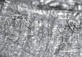
Using light microscopy, the nematodes were identified as spiruroids of the genus Procyrnea (Spiruroidea: Habronematidae) according to the following characteristics: the body presented fine transverse striations, buccal region consisting of two pseudolabia and dorsal and ventral labia each consisted of two submedial lobels and small teeth on the border of pseudolabia, thick-walled buccal cavity, esophagus divided into a short anterior muscular part and a long posterior glandular part, a coiled male tail with caudal alae, unequal spicules, presence of a gubernaculum, and a vulva in females near their midbody (Chabaud, 1975). Two specimens were females measuring 5.82 × 0.40 mm and 7.5 × 0.58 mm (average, 6.66 × 0.49 mm) and one male measured 4.8 × 0.52 mm (Fig. 2). The length of the muscular and glandular esophagus was 320-480 and 1.45-1.7 mm, respectively. The nerve ring was located in the middle of the muscular esophagus and the excretory pore immediately posterior to nerve ring. The tail was curved and ended in a tip point. The male had 2 unequal sized spicules in its tail: the left spicule was bigger (1.45 mm) than the right one (220), the ratio of right spicule-left spicule was 1:6.5, neither was splintered, and both distal ends were blunt (Fig. 3). One uterus had embryonated eggs with oval shape and smooth thick shells that measured 45 μm × 27.5 μm in average (Fig. 4). Analysis of proventricular histological sections indicated that the submucosa contaided parasitic structures which pressed and atrophied the adenomeres adjacent to the parasite (Fig. 5).
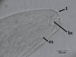
Figura 2 Procyrnea sp. Anterior end of a male showing teeth on the border of pseudolabia (t); bucal cavity (bc), and esophagus (es).
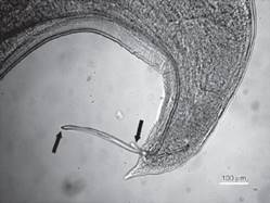
Figura 3 Lateral view of the posterior end of male Procyrnea sp, where unequal size blunt pointed spicules can be seen (arrows).

Discussion
This is the first documented case of Procyrnea sp. from a Rupornis magnirostris in Mexico. Most references of this parasites have been published from Europe and a few from the US, Costa Rica, and Brazil (Siegel et al., 2012; Pinto et al., 1996; Zhang et al., 2004). While in the current report the general characteristics of the nematodes and the eggs found in the female uterus are in agreement with those of nematodes from the order Spirurida and the genus Procyrnea (Zhang et al., 2004; Quentin et al., 1983; Vicente et al., 1995), there is no previous publication in which a Procyrnea species is associated with this kind of gross and microscopic proventricular lesions described herein. Most reports have identified this parasite to infect the ventriculus (Pinto et al., 1966; Vicente et al., 1995; Zhang et al., 2004, 2011), but only few cases have mentioned the proventriculus, besides the gizzard, as site of infection (Sanmartín et al., 2004; Salcedo & Villa, 2001) and very few cases have described proventricular lesion associated to Procyrnea (Salcedo & Villa, 2001). In the case described herein, no gross lesions were detected in the gizzard. Furthermore, the characteristics identified in the male parasite found in the present case differ from others previously documented: both spicules had a blunt end, whereas in other cases one or both spicules had sharp or barbed ends. The present study described the presence of adult male and female Procyrnea sp. detected in the proventricular submucosa and serosa of Rupornis magnirostris. The effects of Procyrnea in wild birds have not been clearly elucidated. Salcedo & Villa (2011) observed epithelial destruction in the proventriculus and gizzard in Palawan hillmyna infected with P. graculae. Foster et al., (2002) found macroscopic changes in texture, color and integrity of the cornified epithelium in the gizzard of red-bellied woodpeckers associated with P. pileata infection.
More than 40 species have been reported in this genus from multiple Falconiformes species. P. leptoptera has been reported from the gizzard and proventriculus of the European honey buzzard (Pernis apivorus), Red kite (Milvus milvus), Eurasian hobby (Falco subbuteo) in Spain (Sanmartín et al., 2004); in Euroasian sparrowhawk (Accipeter nisus) (Cordero del Campillo et al., 1994); in Buteo buteo, Falco tinnunculus and Pernis apivorus in Germany (Kutzer et al., 1980; Krone, 2000). P. beveridgei was reported in Accipiter nisus and Buteo buteo; and P. strialata in Accipiter virgiatus nisoides in China (Zhang et al., 2011). P. mawsonae was found in a Rupornis magnirostris in Costa Rica (Zhang et al., 2004). In this study Procyrnea sp. was found in a Roadside hawk too; however, P. mawsonae differs from the nematode reported in this study (Procyrnea sp.) in having a bigger body size (male 9.05 mm × 191 μm, female 13.94 mm × 275 μm) whereas this species (Procyrnea sp.) has a shorter body size (male 4.8 × 0.52 mm, female 6.66 × 0.49 mm); P. mawsonae has smaller spicules, left spicule 665-874 μm long, right spicule 247-323 μm long, and Procyrnea sp. presented the left spicule 1.45mm long and right spicule 220 μm long. The ratio of right spicule-left spicule is smaller in P. mawsonae (1:2.6) than in Procyrnea sp., (1:6.5). The distal end of the left spicule is divided into 2 branches and the distal end of the right spicule is hook like in P. mawsonae, whereas both spicules have blunt ends in Procyrnea sp. Finally Procyrnea sp. differs from P. mawsonae by having shorter eggs (35.1 long × 19.0 wide and 45 long × 27.5 μm respectively).
Nematodes have a complex and varied population dynamics, which is determined by factors associated with the biology, ecology, and the evolution of the parasite host (Esch & Fernández, 1993). Among helminths that parasite falconiformes, there is a clear association with the bird diet. Procyrnea uses arthropods as intermediate host (Anderson, 2000; Siegel et al., 2012).
Similarly, Sanmartin et al. (2004) reported an association between the prevalence of P. leptoptera and the host invertebrates' diet. In a study made in Argentina by Beltzer (1990), the stomach contents of 22 roadside hawks were analyzed throughout one year, and it was determined that these birds consumed mostly insects (77% orthopterans). In the current case the gizzard was empty, probably due to the bird injury and incapacity to hunt.
Several nematodes species from the Spirurida Order can parasite raptor birds. In the wild, these infections often reach equilibrium, where a few or no lesions are caused to the host (Lacina & Bird, 2000; Krone & Cooper, 2002).
Although it was not possible to identify the species of the specimens, the authors believe this finding is relevant and should be reported due to this is the first documented case of Procyrnea sp., in a Rupornis magnirostris in Mexico. Furthermore the characteristics of these parasites, as mentioned, do not coincide with previous Procyrnea species reports in raptors; nor the site of localization and kind of injuries in the proventriculus. In this case the lesions associated with the presence of Procyrnea sp. were found confined to the proventriculus and were seen dark red nodules over the lining of the proventriculus where the parasite was located in their adult phase; so possibly these specimens correspond to a distinct species of Procyrnea.
The contribution of this paper is to report the finding and the main distinguishing features of these specimens; and injuries associated with their presence as an example of the diversity of nematode parasites of the genus Procyrnea that can be found in birds of prey in Mexico.
Further information is, however, needed to gain deeper understanding about the parasite biology as well as its epidemiology in wild birds.

