SciELO - Scientific Electronic Library Online

 
vol.61 número3Epigenetica en carcinomas pulmonares de células no pequeñasNecesidades de cobertura y atención del cáncer pulmonar en México índice de autoresíndice de materiabúsqueda de artículos
Home Pagelista alfabética de revistas  

Servicios Personalizados

Revista

Articulo

Indicadores

Links relacionados

  • No hay artículos similaresSimilares en SciELO

Compartir


Salud Pública de México

versión impresa ISSN 0036-3634

Salud pública Méx vol.61 no.3 Cuernavaca may./jun. 2019  Epub 03-Mar-2020

https://doi.org/10.21149/10115 

Artículos de revisión

Squamous cell lung cancer: genomic evolution and personalized therapy

Cáncer pulmonar de células escamosas: evolución genómica y terapia personalizada

Andrés F Cardona, MSc, PhD1  2  3 

Luisa Ricaurte, MD2 

Zyanya Lucía Zatarain-Barrón, MSc, PhD4 

Oscar Arrieta, MD, MSc4 

1 Grupo Oncología Clínica y Traslacional, Clínica del Country. Bogotá, Colombia.

2 Fundación para la Investigación Clínica y Molecular Aplicada del Cáncer. Bogotá, Colombia.

3 Grupo Oncología Molecular y Sistemas Biológicos en Cáncer, Universidad El Bosque. Bogotá, Colombia

4 Unidad de Oncología Torácica, Instituto Nacional de Cancerología. Mexico City, Mexico.


Abstract:

Objective:

To review the state-of-the-art in relation to the current information on squamous cell lung cancer (SCLC). We describe the genetic anomalies reported, their effect, and finally the most promising therapeutic agents.

Materials and methods:

We reviewed published articles in peer-reviewed journals as well as current treatment guidelines from local and international resources.

Results:

SCLC represents a smaller proportion of the global burden of disease for lung cancer compared to its more frequent presentation, the adenocarcinoma. However, more than 400 000 cases are reported annually, a substantial population for whom therapeutic options are scarce and with limited efficacy. Several groups have been given the task of elucidating the mechanisms that lead to the development of SCLC, including molecular anomalies that can be used as targets for drug design.

Conclusion:

There are potential therapeutic targets for SCLC, which must be studied in clinical trials for validation.

Keywords: carcinoma, squamous cell; personalized therapy; targeted therapy; genomic signature; lung cancer; genomics

Resumen:

Objetivo:

Revisar el estado del arte en relación con la información actual sobre el cáncer de pulmón de células escamosas (CPCE) y describir las anomalías genéticas reportadas, su efecto y los agentes terapéuticos más prometedores.

Material y métodos:

Se realizó una revisión de artículos publicados en revistas indizadas, así como las guías de tratamiento publicadas por instancias locales e internacionales.

Resultados:

El CPCE representa una proporción menor de la carga mundial de la enfermedad por cáncer pulmonar en comparación con su presentación más frecuente, el adenocarcinoma. Sin embargo, más de 400 000 casos son reportados anualmente, una población sustancial para quienes las opciones terapéuticas son escasas y con una eficacia limitada. Diversos grupos se han dado a la tarea de elucidar los mecanismos que conllevan al desarrollo del CPCE, incluyendo anomalías moleculares que puedan servir como blancos para el diseño de fármacos.

Conclusiones:

Existen blancos terapéuticos potenciales para el CPCE que deben ser estudiados en ensayos clínicos para ser validados.

Palabras clave: carcinoma de células escamosas; terapia personalizada; terapia dirigida; firma genómica; cáncer de pulmón; genómica

Introduction

Despite concrete global efforts on primary and secondary prevention, lung cancer is the most lethal malignancy worldwide, causing over 1.6 million deaths yearly. It is also the most common cancer in men worldwide (1.2 million), and is considered a global public health problem.1 Interestingly, the incidence differs according to geographic regions, with the highest incidence rates present in Europe and Asia.1 The landscape of this malignancy is even more complex given the enormous healthcare burden, high costs and expenditure associated with the disease,2 especially because most (58%) lung cancer cases occur in underdeveloped countries.3 Another distinguishing factor of lung cancer is that it displays the lowest five years survival rate (5-18%), compared to other highly incident malignancies (breast, colorectal, prostate, skin and stomach cancer). In fact, lung cancer mortality more than doubles that of colorectal cancer, the second most common cause of cancer related deaths.1,2

Lung cancer is closely related to tobacco use; however, this varies according to the histologic subtypes. ADC of the lung arises mostly in patients with no previous significant tobacco exposure; in contrast SCC arises almost exclusively in former or current smokers. 4 To further complicate this relationship, recent changes of cigarette composition are thought to cause differences in both risks associated with smoking and lung cancer, and in the histologic subtypes associated with exposure.5,6All of this together added to the decreases in smoking prevalence since the 1950s7 have led to significant changes in the lung cancer histology distribution, with ADC leading as the most common. Nevertheless, SCC is responsible for 400 000 incident cases per year worldwide; as a consequence, research in this field has focused on the molecular background of the disease in order to identify potential drug targets.8,9

Due to the predominant role tobacco plays on SCC carcinogenesis, this malignancy is characterized by genomic complexity and high overall mutational load. The Cancer Genome Atlas (TCGA) has identified various genomically defined subsets of SCC. This has led to the development and investigation of targeted agents that can impact the therapeutics of this disease.8 Additionally, despite multiple studies, to date there are no accepted prognostic gene signatures for risk stratification in terms of recurrence or survival, consequently limiting the selection of patients that would require adjuvant therapy.4

In this review, we highlight the current knowledge of molecular targets, ongoing clinical trials of targeted agents, and actionable mutations with matching drugs in lung SCC.

Gross molecular biology of SCC precursors and preinvasive lesions

The molecular background in SCC has been thoroughly studied. Most of the studies investigate the process of genetic changes that appear in pulmonary squamous precursor and pre-invasive lesions (carcinoma in situ, CIS). Immunohistochemical studies have demonstrated presence of cell cycle associated proteins such as proliferating cell nuclear antigen (PCNA), MIB1 and michrosome maintenance protein 2 (MCM2) on squamous dysplasia (SD) and CIS, indicating an increased cell cycle activity.9 Also, smoking status seems to have an effect on cell cycle activity, an increasing tobacco consumption is correlated with increased cell cycle activity while smoking cessation causes it to diminish.10

Not much is known about the activity of EGFR and its signaling pathways (RAS/RAF/ MAPK, JAK/STAT and PI3K/AKT/mTOR) during the transition from dysplasia to CIS and squamous neoplasia. EGFR protein is present in normal bronchial epithelium, as well as in bronchial basal cell hyperplasia and metaplasia. Tobacco smoke phosphorylates and amplifies PI3K causing it to activate, and upregulates AKT in severe bronchial squamous dysplasia.11 In comparison to oncogene activity on SD/CIS, there is much mature data for loss of tumor suppressor genes (TSG) activity in the precursor lesions of SCC. TP53 is a tumor suppressor gene that has a role in controlling cell cycle activity, by promoting p21; CDK4/Cyclin D1 complex is repressed, which in turn promotes Rb phosphorylation and thus cell cycle activity is suppressed.12 Despite the heterogeneity of studies, the deregulation of P53 in the progression from normal epithelium to hyperplasia, SD and CIS has been a consistent finding. The P53 alteration causes bcl2 expression deregulation and a decrease in bax, increasing cell cycle activity and resistance to apoptotic mechanisms. Interestingly, the actual loss of P53 gene (by mutation or chromosomal deletion in 17p [LOH, loss of heterozygocity]) is much less common in SD/CIS than in SCC, and is detected in only 10% of high grade lesions.13,14

Abnormalities in p53 related genes and proteins are less studied in SD and CIS. p63 overexpression and P63 gene amplification is frequently present in high grade SD/CIS or initially invasive disease.15 On the other hand, there is insufficient data to make any conclusion regarding p21, mdm2 or p14 expression in SD/CIS.16 In contrast, p16 (suppressor of multiple growth promoter complexes) is commonly lost in SD/CIS through decreased protein expression and/or diminished gene function caused by deletion of 9p21 or epigenetic silencing by P16 gene promoter hypermethylation.17 Although there is great variability between studies, there is a trend of increasing p16 deregulation as the grade of atypia rises. Cyclin D1 overexpression is present initially in early inflammatory stages of SD, and upsurges as the number of high-grade lesions increases.18 In the case of Rb protein, the loss of Rb is a frequent alteration in small cell lung cancer (SCLC), but it happens to be rare in SD/CIS or in SCC.19 Other putative tumor suppressor genes of uncertain role are also lost in SD/CIS, specially loss of expression of retinoic acid receptor (RAR-B) and fragile histidine triad (FHIT).20

As dysplasia progresses, the molecular milieu becomes favorable of cell survival. Telomerase activation prevents cellular ageing, with the human telomerase reverse transcriptase (hTERT) increasing by 40 times in invasive SCC.21 Altogether, the increase of TERT, altered bax/bcl2 ratio and decreased p53 expression converge in the common purpose of cell survival, and development of SD/CIS.22,23

Angiogenesis stimulation is also relevant to the development and establishment of lesions. For example, the subepithelial stroma in SD/CIS becomes more vascular in relation to the grade of the lesion.24 Neuropilin, KDR and ftl1, all vascular endothelial growth factor receptor (VEGFR) ligands, are increased in SD/CIS, and together with an increased expression of vascular endothelial growth factor (VEGF) contribute to the increase in vascularization.24,25COX-2 is also a proangiogenic factor that appears to be increased in higher grades of SD/CIS.26,27

Other molecular alterations in SCC genesis include those related to cell invasion and migration, which include the increased expression of matrix metalloproteinases (MMP3, 9 and 11), a decrease in MMP1 and its inhibitors TIMP1 and MMP7, thus promoting tissue invasion in CIS.28 Additional alterations include transcription factors such as Upstream Stimulatory Factors USF 1 and 2 and NF-KappaB,29 and heat shock proteins 10 and 60, which are lost throughout the transformation process.30

Other global genetic studies of SC/CIS reported aneuploidy and specific chromosomal aneusomy of chromosomes 3, 5, 6, 7 and 8.31,32 Chromosomal gains and losses in chromosomes 1q, 3p, 8p, 8q, 12q, and 17q have been shown previously in CIS using comparative genomic hybridization.32,33,34 LOH studies have shown a wide spectrum of changes in several loci, especially in 3p. This highlights that a number of possible TSG are altered in the initial process of bronchial carcinogenesis. These changes are aparent even in the bronchial epithelium in smokers but increase in extent, number and frequency as SD/CIS evolves.31,35 These very early losses are present in: 3p14.2 (FHIT), 3p21 (RASSF1A, FUS1, SEMA3B), 3p22-24 (BAP1), 3p25, 9p21 (p16). Later changes include alterations in 17p13 (p53) and deletions in 8q21-23 related to focal losses in 3p and 9p.36,37

Genomic landscape of lung SCC

Whilst comprehensive genome-scale characterization has been performed in ADC,38 genetic alterations in SCC are less understood. Lung SCC is strongly associated to tobacco use with most cohorts reporting a rate of tobacco exposure in excess of 90%.39 SCC displays a somatic mutation rate and spectrum comparable to that of patients with small cell lung cancer or other smoking-related cancers. The high mutation rate in SCC is likely to result in expression of a large complement of tumor antigens, and is similar across populations from North and South America, Europe and Asia.40,41,42Interestingly, the genomic alterations in lung SCC are strikingly similar to those found in Human Papilloma Virus (HPV) negative head and neck cancers.43

Several genetic changes have been related to SCC, including amplification of p63, PI3KCA, PDGFRA, SOX2, or FGFR1 and mutation in p53, EGFRvIII, PI3KCA, NRF2, PTEN, and DDR2.38 In 2012, The TCGA identified that lung SCC is characterized by complex genomic alterations, with a mean of 165 genomic rearrangements, 360 exonic mutations and 323 segments of copy number alterations in every single tumor. The TCGA also identified statistically recurrent mutations in 11 genes (TP53, CDKN2A, PTEN, PIK3CA, KEAP1, MLL2, HLA-A, NRF2, NOTCH1, RB1 and HLA-A) and somatic copy number alterations of chromosomal segments containing SOX2, PDGFRA and/or KIT, EGFR, FGFR1 and/or WHSC1L1, CCND1, CDKN2A, NFE2L2, MYC, CDK6, MDM2, BCL2L1, EYS, FOXP1, PTEN, and NF1.8 Recent results have confirmed the TCGA findings using multiplex PCR sequencing. Most important findings in this population were driver mutations in PIK3CA, PTEN and DDR2, as well as FGFR1 amplification. Overall, 60% of patients were found to have druggable targets (figure 1).44,45

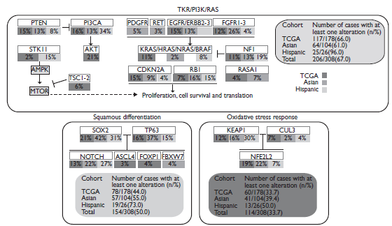
PDGFR: Platelet-derived growth factor receptor; EGFR: Epidermal Growth Factor Receptor; FGFR: Fibroblast growth factor receptors; PI3CA: Phosphoinositide 3-kinases; STK11: Serine/Threonine Kinase 11; AMPK: 5’ AMP-activated protein kinase; AKT: Serine/threonine-protein kinases 1-3, MTOR: Mammalian target of rapamycin.

Figure 1  Diagram integrating three different core pathways of lung squamous cell carcinoma (considering information of substitution mutations, truncating mutations, amplifications and deletions) from the information provided by the TCGA and by Kim and colleagues.44 

Novel cytotoxic agents

Novel cytotoxic agents for SCC include Nab-paclitaxel, an agent which is combined to a human albumin molecule in order to achieve higher intratumoral concentration compared to previous taxanes due to the use of the albumin receptor 60-kDa glycoprotein (gp60). Previous clinical trials have shown that nab-paclitaxel produce higher response rates (33% vs. 25; response rate ratio, 1.313; 95%CI 1.082 to 1.593; p=0.005). Unfortunately, there was no significant benefit in terms of PFS or OS. Notably, in the patients with SCC the nab-paclitaxel regimen achieved much higher ORR than the control group (41% vs. 24; response rate ratio, 1.680; 95%CI, 1.271 to 2.221; p<0.001).46 SCC has an aberrant caveolin-1 expression; this may explain the better efficacy of nab-paclitaxel in the squamous histology.47 There is an ongoing clinical trial (NCT00729612) that is evaluating the role of SPARC, ceveolin-1, and microRNAs as predictive biomarkers in patients with advanced SCC treated with carboplatin and nab-paclitaxel. Multiple clinical trials have proven the clinical relevance of nab-paclitaxel in the treatment of NSCLC, some with special emphasis on SCC in particular populations such as the elderly.48,49

Another promising compound for SCC is the second-generation platinum compound Nedaplatin, which has shown a better toxicity profile with lower rates of renal toxicity and emesis compared with cisplatin. Nedaplatin has shown to have better response rates in the SCC subtype compared to non-squamous histologies (ORR, 55.6% in squamous histology vs. 34.4% in non-squamous histology). These results suggest there is a more potent activity of nedaplatin against SCC. In a randomized phase 3 study for advanced SCC patients, nedaplatin plus Docetaxel achieved a significantly prolonged OS compared to cisplatin plus Docetaxel (median OS 13.6 vs. 11.4 months, respectively; HR 0.81; P=0.037), though no significant differences were observed between nedaplatin and cisplatin arms in terms of PFS (median PFS, 4.9 vs. 4.5 months; HR 0.83; P=0.050) and response rates (56 vs. 53%).50

Novel targeted agents for SCC

Anti-EGFR therapy

To date, it is known that EGFR activating mutations are exceptionally rare in SC, though, loyal to its epithelial cancer nature, it keenly expresses EGF and a subset demonstrates EGFR amplification.8 Cetuximab, the monoclonal antibody (mAbs) against the EGFR protein, in combination with cisplatin/vinorelbine had a minimal survival benefit compared with chemotherapy alone in patients with stage IIIB/IV NSCLC in the FLEX study (OS 11.3 vs. 10.1 months; P=0.044).51 In a preplanned subset analysis, a minimal survival benefit was seen in SCC (OS, 9.0 vs. 8.2 months). High tumor EGFR expression (immunohistochemistry score [H-score] ≥200) and amplification, were more common in SCC than in non-SCC histology, seem to serve as predictive biomarkers for the correct patient selection of patients that would obtain a clinical benefit from treatment with chemotherapy plus cetuximab [FISH+ (11.8 versus 6.4 months; HR 0.56; P=0.01); high EGFR expression H-score ≥200 9.3 versus 6.9 months; HR 0.64; P=0.03), and both FISH+ and high-EGFR expressing tumors (12.3 versus 4.9 months; HR 0.32; P=0.0004).52

Necitumumab is another anti-EGFR mAbs. It was evaluated specifically in lung SCC in a large phase III trial (SQUIRE).53 1 093 patients with advanced SCC were randomized to gemcitabine-cisplatin with or without necitumumab. OS was significantly increased in necitumumab-treated patients (11.5 versus 9.9 months, HR 0.84, 95%CI 0.74-0.96) and PFS was also significantly increased (HR 0.85, 95%CI 0.74-0.98). A further analysis showed that addition of necitumumab to chemotherapy resulted in significant improvements in OS (11.7 versus 10.0 months; HR 0.79; 95%CI 0.69, 0.92; p = 0.002) for the vast majority of patients whose tumors expressed EGFR (95%), but not in patients without EGFR expression (5% of patients; HR 1.52). FISH status was determined for 51% of patients in the study, there was also a trend for greater necitumumab benefit in patients with EGFR FISH+ tumors (12.6 versus 9.2 months; HR 0.70; 95%CI 0.52, 0.96), but no benefit in patients with EGFR FISH- tumors (11.1 versus 10.7 months; HR 1.02; 95%CI 0.80, 1.29).53,54,55

Use of mAbs against EGFR is limited in the SCC setting, due to limited efficacy and lack of robust biomarkers for patient selection.56 However, ESMO guidelines include necitumumab combined with cisplatin-gemcitabine as an option for first-line treatment of SCC.57

Other studies, including the LUX-Lung 8 phase III trial, have focused on SCC patients with wild-type EGFR. In this study, previously treated SCC patients were randomized to receive either erlotinib or Afatinib, an irreversible pan-EGFR blocker. Results from this study showed that patients treated with Afatinib had a significantly longer PFS and disease control rate (DCR). Further, stratification of patients using Veristrat (VeriStrat Good, VS-G) demonstrated a significant benefit in terms of OS, which was maintained in a multivariate analysis independently of ECOG or best-response to first line chemotherapy.58,59Other secondary analyses have also identified that among LUX-Lung 8 study patients, those with mutations in the ERBB family assessed by NGS and presence of mutations in HER2 had a favorable PFS and OS when treated with Afatinib compared to erlotinib.60

Antiangiogenics

Ramucirumab (antiangiogenic VEGFR2 mAbs antagonist) is used in combination with Docetaxel for NSCLC patients with a good ECOG PS (0-2) based on the results from the phase III REVEL study,61 which showed an improvement in OS present in both the squamous and non-squamous subtypes. As opposed to bevacizumab, the use of ramucirumab was not associated with increased rates of hemoptysis or other pulmonary hemorrhage events in the SCC subgroup.

FGFR

Several mechanisms result in imbalanced FGFR signaling, which can lead to NSCLC development by either supporting tumor angiogenesis or promoting cell proliferation. Main mechanisms include overexpression caused by amplification or aberrant transcriptional regulation, interchanging between alternatively spliced isoforms, FGFR mutations, FGFR fusion proteins (more than 10 fusion proteins have been identified for FGFR1), increased ligand availability, and impaired down regulation of FGFR activity.62 In NSCLC cell lines with 8p12 amplification, small-molecule FGFR kinase inhibitors arrested cell growth and stimulated apoptosis, which confirms the oncogenic activity of FGFR1.63,64AZD4547 is a potent and selective inhibitor of FGFR 1-3, it is active against FGFR-deregulated and amplified tumors in preclinical models.65 The phase 1b trial, includes 15 patients with previously treated stage IV FGFR1-amplified lung SCC that were treated with AZD4547 80 mg oral bid. The most common treatment related adverse events were gastrointestinal and dermatologic. A single patient with high FGFR amplification (FISH ratio > 2.8) achieved a partial response and 4 had stable disease.65 More recently, results of a sub-study from the lung-MAP clinical trial were reported. This phase II study evaluated AZD4547 in patients with FGFR-altered chemotherapy refractory SCC. The majority of the patients had FGFR1 amplification. AZD4547 showed an acceptable toxicity profile but minimal activity. Only one of the 28 patients who received AZD4547, harbored an FGFR3 S249C mutation and showed an unconfirmed partial response (PR).66 This study was closed for futility, but at least two more phase II trials continue to evaluate the role of this compound in FGFR altered lung SCC. Other compounds currently under study for FGFR include BGJ398 [NCT01004224], LY2874455 and BAY1163877, they have a similar toxicity profile and have managed marginal stability of the disease in few patients.67 GSK3052230 is a novel FGF ligand trap that binds to all of the mitogenic FGF ligands, it blocks FGF cell proliferation in vitro and FGF- (and VEGF-) induced angiogenesis in vivo. The phase 1 trial included 39 patients with advanced solid tumors (three with NSCLC) treated with escalating doses of weekly doses of GSK3052230 for four weeks.68 Preliminary results showed that GSK3052230 elicited a good tolerance with most frequent AEs being diarrhea, fatigue, and nausea. In addition, 41.7% achieved stable disease.68

Multiple multikinase non-selective FGFR inhibitors including nintedanib, cediranib, ponatinib (AP24534), lucitanib (E3810), pazopanib, regorafenib, brivanib, orantinib, ENMD-2076, FGF401 and ODM-203 are in development, some with promising early results, others with negative phase 3 clinical trials.

PI3K pathway inhibitors

The PI3K pathway is a major signaling pathway that plays a role in cell survival and proliferation.8 Somatic mutation or amplification of PIK3CA, that affects the p110 catalytic subunit, is more prevalent in lung SCC than ADC.69 The incidence of PIK3CA mutation is approximately 8-16% and amplification is 33-43% in SCC. In phase I trials, response rate to PI3K/AKT/mTOR pathway inhibitors was significantly higher for patients with PIK3CA mutations than for those without documented mutations (35 vs. 5%, P< 0.001). Recent results among SCC showed that taselisib generates a PR in patients with PIK3CA E545K gene alteration (5% RR, 95%CI 1-24%) while 13 additional patients experienced disease stability. Median PFS was 2.5 months (95%CI 1.7-4.5) and grade 3 AEs included five patients each with hyperglycemia or diarrhea, and three with lymphopenia.70 The tumor suppressor PTEN has phosphatase activity and inhibits the PI3K/AKT/mTOR signaling pathway. In the case of SCC, the incidence of PTEN mutation/deletion was reported to be 15-28%.71,72This alteration is associated with a poorer response to erlotinib in EGFR-mutant NSCLC as well as with intrinsic resistance to immunotherapy.72,73

PI3K/AKT/mTOR pathway inhibitors may be effective in tumors with alterations in PTEN. PIK3CB has been reported in PTEN-deficient tumors.69 Further, in preclinical models of solid tumors, the inhibition of PIK3CB reduced Akt phosphorylation and prevented tumor progression; suggesting that the selective inhibition of PIK3CB may be effective in the treatment of PTEN-deficient tumors.74 Parallel inhibition of PI3K through AKT has also generated intriguing results in silico and in vivo models. Previously, Lara et al. reported the efficacy and safety of erlotinib plus MK-2206 (highly selective AKT inhibitor) in advanced previously treated NSCLC. In a combined population of patients with wild type EGFR, a disease control rate (DCR) between 40 and 47% was seen. Median PFS was of 4.6 months, and mean AEs were rash, diarrhea, fatigue, and Mucositis.74 More research is required to elucidate the role of PI3K and AKT inhibitors in lung SCC.

PDGFRA amplification/mutation

Platelet-derived growth factor receptor (PDGFR) is a tyrosine kinase, two subtypes are currently known: PDGFRA and PDGFRB. It plays a critical role in cell proliferation and angiogenesis. The amplification of the chromosomal segment of 4q12 harboring PDGFR- has been reported in ∼9% of lung SCCs.8,75 Several multi-targeted TKIs targeting PDGFRA, such as sorafenib, sunitinib, and imatinib, have been tested previously in NSCLC. Disappointingly, the addition of sorafenib to platinum-based chemotherapy failed to improve survival, and actually increased mortality in a subset of lung SCC, in a randomized phase III study.76

PARP-1

Poly (ADP-ribose) polymerase-1 (PARP-1) is an important DNA repair enzyme, implicated in base excision repair and single-strand break (SSB) repair. When PARP-1 is inhibited, accumulated unrepaired SSBs are converted to double-strand breaks (DSB), hence inducing cell death.77 PARP-1 is significantly up-regulated at the mRNA level in multiple cancer types, including lung SCC. Paul and colleagues showed that BRCA1-deficient NSCLC cells BRCA1-deficient were more sensitive to PARP-1 inhibition. Furthermore, he demonstrated that BRCA-deficient, platinum-resistant cells still remained sensitive to PARP inhibition. The authors propose that BRCA-deficient cells bypass the BAX and BAK apoptotic proteins, and that cell death occurs independently of mitochondrial induced apoptosis.78 Several PARP inhibitors are currently undergoing clinical trials in lung cancer. Recently, the results of a randomized, placebo controlled phase II study in NSCLC were reported. Patients with advanced or metastatic NSCLC are randomized to receive carboplatin/paclitaxel plus veliparib or placebo, 77 of the 158 patients enrolled had lung SCC. For the intent-to-treat population median PFS and OS was higher with veliparib, however the differences were not statistically significant.79 The improvement in PFS and OS was only present in patients with squamous histology (6.1 versus 4.1 months [HR 0.77] and 10.3 vs. 8.4 months [HR 0.71], respectively).

DDR2

The receptor tyrosine kinase Discoidin Domain Receptor Tyrosine kinase 2 (DDR2) is mutated in approximately 4% of patients with lung SCC. DDR2 is a receptor for extracellular collagens that activates a complex signaling network involving SHP-2 as well as SRC and MAP kinases.80 It regulates epithelial-mesenchymal transitions (EMT). Multitargeted kinase inhibitors like dasatinib, imatinib, nilotinib, and ponatinib suppres the proliferation DDR2 on cancer cell line models.81 Dasatinib, the most potent of these inhibitors, has been studied in multiple lung cancer clinical trials.82 While some responses to dasatinib have been reported in patients with the DDR2 S768R mutation, its associated toxicity has limited its investigation.

BRAF

BRAF mutation is oncogenic since it causes constitutional serine/threonine kinase activation. According to a recent meta-analysis, there was no significant difference in BRAF mutation rate in former or current smokers and never smokers (OR: 0.95, 95%CI 0.45-2.02). Nevertheless, several studies reported an association between BRAF mutation and tumor histology. BRAF mutations were detected 4% of adenocarcinomas and 0.58% of lung SCC.83 Paik and colleagues found no difference in OS for BRAF-mutated patients when compared with other EGFR mutated, ALK-mutated, or KRAS-mutated subpopulations.84 Marchetti and colleagues, described that patients with the BRAF V600E mutation had shorter disease free survival (DFS) and OS compared with wild-type and nonV600E mutations.85 In vitro preclinical studies have identified that vemurafenib and trametinib are effective single agents in BRAF V600E mutant cells, and trametinib in non-V600E mutants also.85 The combination of vemurafenib and trametinib increased tumor cell death, showing that the combination is more effective at least in this particular model. Two other MEK inhibitors (PD0325901 and CI-1040) have also shown activity in in vitro and in vivo preclinical models of NSCLC with BRAF V600E. Recently, Planchard et al. published the results of a phase II trial that included 36 patients treated with first-line dabrafenib plus trametinib, with an ORR in 23 (64%, 95%CI 46-79), with 6% of the cases achieving a CR and 58% a PR. Unfortunately, all cases presented one or more AEs and 69% had one or more grade 3 or 4 event, including hypertension and liver enzyme increase.86

Conclusion

The recent expansion of our knowledge on the genomic landscape of lung SCC has led to the identification of potential driver mutations, including FGFR1 amplification, PIK3CA mutation, PTEN mutation/deletion, PDGFRA amplification/mutation, DDR2 mutation, and BRAF mutation. The impact of these mutations on the response to the matching targeted therapy should be validated in the near future.

References

1. Jemal A, Siegel R, Xu J, Ward E. Cancer statistics, 2010. CA Cancer J Clin. 2010;60(5):277-300. https://doi.org/10.3322/caac.20073 [ Links ]

2. GBD 2015 Disease and Injury Incidence and Prevalence Collaborators. Global, regional, and national incidence, prevalence, and years lived with disability for 310 diseases and injuries, 1990-2015: a systematic analysis for the Global Burden of Disease Study 2015. Lancet. 2016;388(10053):1545-602. https://doi.org/10.1016/S0140-6736(16)31678-6 [ Links ]

3. Ferlay J, Soerjomataram I, Dikshit R, Eser S, Mathers C, Rebelo M, et al. Cancer incidence and mortality worldwide: sources, methods and major patterns in GLOBOCAN 2012. Int J Cancer. 2015;136(5):E359-86. https://doi.org/10.1002/ijc.29210 [ Links ]

4. Gandara DR, Hammerman PS, Sos ML, Lara PN Jr, Hirsch FR. Squamous cell lung cancer: from tumor genomics to cancer therapeutics. Clin Cancer Res. 2015;21(10):2236-43. https://doi.org/10.1158/1078-0432.CCR-14-3039 [ Links ]

5. Khuder SA, Mutgi AB. Effect of smoking cessation on major histologic types of lung cancer. Chest. 2001;120(5):1577-83. https://doi.org/10.1378/chest.120.5.1577 [ Links ]

6. Burns DM, Anderson CM, Gray N. Do changes in cigarette design influence the rise in adenocarcinoma of the lung? Cancer Causes Control. 2011;22(1):13-22. https://doi.org/10.1007/s10552-010-9660-0 [ Links ]

7. Burns DM, Anderson CM, Gray N. Has the lung cancer risk from smoking increased over the last fifty years? Cancer Causes Control. 2011;22(3):389-97. https://doi.org/10.1007/s10552-010-9708-1 [ Links ]

8. Cancer Genome Atlas Research Network. Comprehensive genomic characterization of squamous cell lung cancers. Nature. 2012;489(7417):519-25. https://doi.org/10.1038/nature11404 [ Links ]

9. Sousa V, Espirito-Santo J, Silva M, Cabral T, Alarcao AM, Gomes A, et al. EGFR/erB-1, HER2/erB-2, CK7, LP34, Ki67 and P53 expression in preneoplastic lesions of bronchial epithelium: an immunohistochemical and genetic study. Virchows Arch. 2011;458(5):571-81. https://doi.org/10.1007/s00428-011-1062-5 [ Links ]

10. Lee JJ, Liu D, Lee JS, Kurie JM, Khuri FR, Ibarguen H, et al. Long-term impact of smoking on lung epithelial proliferation in current and former smokers. J Natl Cancer Inst. 2001;93(14):1081-8. https://doi.org/10.1093/jnci/93.14.1081 [ Links ]

11. Massion PP, Taflan PM, Shyr Y, Rahman SM, Yildiz P, Shakthour B, et al. Early involvement of the phosphatidylinositol 3-kinase/Akt pathway in lung cancer progression. Am J Respir Crit Care Med. 2004;170(10):1088-94. https://doi.org/10.1164/rccm.200404-487OC [ Links ]

12. Shabnam MS, Srinivasan R, Wali A, Majumdar S, Joshi K, Behera D. Expression of p53 protein and the apoptotic regulatory molecules Bcl-2, Bcl-XL, and Bax in locally advanced squamous cell carcinoma of the lung. Lung Cancer. 2004;45(2):181-8. https://doi.org/10.1016/j.lungcan.2004.01.021 [ Links ]

13. Kohno H, Hiroshima K, Toyozaki T, Fujisawa T, Ohwada H. p53 mutation and allelic loss of chromosome 3p, 9p of preneoplastic lesions in patients with nonsmall cell lung carcinoma. Cancer. 1999;85(2):341-7. https://doi.org/10.1002/(SICI)1097-0142(1999 0115)85:2<341::AID-CNCR11>3.0.CO;2-S [ Links ]

14. Boyle JO, Lonardo F, Chang JH, Klimstra D, Rusch V, Dmitrovsky E. Multiple high-grade bronchial dysplasia and squamous cell carcinoma: concordant and discordant mutations. Clin Cancer Res. 2001;7(2):259-66. [ Links ]

15. Massion PP, Taflan PM, Jamshedur Rahman SM, Yildiz P, Shyr Y, Edgerton ME, et al. Significance of p63 amplification and overexpression in lung cancer development and prognosis. Cancer Res. 2003;63(21):7113-21. [ Links ]

16. Brambilla E, Gazzeri S, Lantuejoul S, Coll JL, Moro D, Negoescu A, et al. p53 mutant immunophenotype and deregulation of p53 transcription pathway (Bcl2, Bax, and Waf1) in precursor bronchial lesions of lung cancer. ClinCancer Res . 1998;4(7):1609-18. [ Links ]

17. Lamy A, Sesboue R, Bourguignon J, Dautreaux B, Metayer J, Frebourg T, et al. Aberrant methylation of the CDKN2a/p16INK4a gene promoter region in preinvasive bronchial lesions: a prospective study in high-risk patients without invasive cancer. Int J Cancer. 2002;100(2):189-93. https://doi.org/10.1002/ijc.10474 [ Links ]

18. Brambilla E, Gazzeri S, Moro D, Lantuejoul S, Veyrenc S, Brambilla C. Alterations of Rb pathway (Rb-p16INK4-cyclin D1) in preinvasive bronchial lesions. Clin Cancer Res . 1999 ;5(2):243-50. [ Links ]

19. Lantuejoul S, Salameire D, Salon C, Brambilla E. Pulmonary preneoplasia--sequential molecular carcinogenetic events. Histopathology. 2009;54(1):43-54. https://doi.org/10.1111/j.1365-2559.2008.03182.x [ Links ]

20. Sozzi G, Pastorino U, Moiraghi L, Tagliabue E, Pezzella F, Ghirelli C, et al. Loss of FHIT function in lung cancer and preinvasive bronchial lesions. Cancer Res . 1998;58(22):5032-7. [ Links ]

21. Capkova L, Kalinova M, Krskova L, Kodetova D, Petrik F, Trefny M, et al. Loss of heterozygosity and human telomerase reverse transcriptase (hTERT) expression in bronchial mucosa of heavy smokers. Cancer. 2007;109(11):2299-307. https://doi.org/10.1002/cncr.22683 [ Links ]

22. Shibuya K, Fujisawa T, Hoshino H, Baba M, Saitoh Y, Iizasa T, et al. Increased telomerase activity and elevated hTERT mRNA expression during multistage carcinogenesis of squamous cell carcinoma of the lung. Cancer. 2001;92(4):849-55. https://doi.org/10.1002/1097-0142(20010815)92:4<849::AID-CNCR1392>3.0.CO;2-4 [ Links ]

23. Zalewska-Ziob M, Dobija-Kubica K, Biernacki K, Adamek B, Kasperczyk J, Brulinski K, et al. Clinical and prognostic value of hTERT mRNA expression in patients with non-small-cell lung cancer. Acta Biochim Pol. 2017;64(4):641-6. https://doi.org/10.18388/abp.2017_1618 [ Links ]

24. Fontanini G, Calcinai A, Boldrini L, Lucchi M, Mussi A, Angeletti CA, et al. Modulation of neoangiogenesis in bronchial preneoplastic lesions. Oncology reports. 1999 ;6(4):813-7. https://doi.org/10.3892/or.6.4.813 [ Links ]

25. Merrick DT, Haney J, Petrunich S, Sugita M, Miller YE, Keith RL, et al. Overexpression of vascular endothelial growth factor and its receptors in bronchial dysplasia demonstrated by quantitative RT-PCR analysis. Lung Cancer. 2005;48(1):31-45. https://doi.org/10.1016/j.lungcan.2004.07.049 [ Links ]

26. Mascaux C, Martin B, Verdebout JM, Ninane V, Sculier JP. COX-2 expression during early lung squamous cell carcinoma oncogenesis. Eur Respir J. 2005;26(2):198-203. https://doi.org/10.1183/09031936.05.00001405 [ Links ]

27. Cavarga I, Kocan P, Boor A, Belak J, Zak V, Kluchova Z, et al. Immunohistochemical markers of proliferation and vascularisation in preneoplastic bronchial lesions and invasive non-small cell lung cancer. Neoplasma. 2009;56(5):414-21. https://doi.org/10.4149/neo_2009_05_414 [ Links ]

28. Galateau-Salle FB, Luna RE, Horiba K, Sheppard MN, Hayashi T, Fleming MV, et al. Matrix metalloproteinases and tissue inhibitors of metalloproteinases in bronchial squamous preinvasive lesions. Human Pathol. 2000;31(3):296-305. https://doi.org/10.1016/S0046-8177(00)80242-7 [ Links ]

29. Tang X, Liu D, Shishodia S, Ozburn N, Behrens C, Lee JJ, et al. Nuclear factor-kappaB (NF-kappaB) is frequently expressed in lung cancer and preneoplastic lesions. Cancer. 2006;107(11):2637-46. https://doi.org/10.1002/cncr.22315 [ Links ]

30. Cappello F, Di Stefano A, David S, Rappa F, Anzalone R, La Rocca G, et al. Hsp60 and Hsp10 down-regulation predicts bronchial epithelial carcinogenesis in smokers with chronic obstructive pulmonary disease. Cancer. 2006;107(10):2417-24. https://doi.org/10.1002/cncr.22265 [ Links ]

31. Romeo MS, Sokolova IA, Morrison LE, Zeng C, Baron AE, Hirsch FR, et al. Chromosomal abnormalities in non-small cell lung carcinomas and in bronchial epithelia of high-risk smokers detected by multi-target interphase fluorescence in situ hybridization. J Mol Diagn. 2003;5(2):103-12. https://doi.org/10.1016/S1525-1578(10)60459-X [ Links ]

32. Hilbe W, Auberger J, Dirnhofer S, Schmid T, Erdel M, Duba HC. High rate of molecular alteration in histologically tumour-free bronchial epithelium of NSCLC patients detected by multicolour fluorescence in situ hybridisation. Oncol Rep. 2006;15(5):1233-40. https://doi.org/10.3892/or.15.5.1233 [ Links ]

33. Woenckhaus M, Grepmeier U, Wild PJ, Merk J, Pfeifer M, Woenckhaus U, et al. Multitarget FISH and LOH analyses at chromosome 3p in non-small cell lung cancer and adjacent bronchial epithelium. Am J Clin Pathol. 2005;123(5):752-61. https://doi.org/10.1309/C4BK-7GQV-8E5X-U2TL [ Links ]

34. Dong XY, Lu YJ, Tong T, Wang YJ, Guo SP, Bai JF, et al. Molecular cytogenetic alterations in the early stage at human bronchial epithelial cell carcinogenesis. J Cell Biochem Suppl. 1997;28-29:74-80. https://doi.org/10.1002/(SICI)1097-4644(1997)28/29+<74::AID-JCB8>3.0.CO;2-T [ Links ]

35. Jeanmart M, Lantuejoul S, Fievet F, Moro D, Sturm N, Brambilla C, et al. Value of immunohistochemical markers in preinvasive bronchial lesions in risk assessment of lung cancer. Clin Cancer Res 2003;9(6):2195-203. [ Links ]

36. Endo C, Sato M, Fujimura S, Sakurada A, Aikawa H, Takahashi S, et al. Allelic loss on 17p13 (TP53) and allelic loss on 3p21 in early squamous cell carcinoma of the lung. Surg Today. 2000;30(8):695-9. https://doi.org/10.1007/s005950070079 [ Links ]

37. Wistuba II, Behrens C, Milchgrub S, Bryant D, Hung J, Minna JD, et al. Sequential molecular abnormalities are involved in the multistage development of squamous cell lung carcinoma. Oncogene. 1999 ;18(3):643-50. https://doi.org/10.1038/sj.onc.1202349 [ Links ]

38. Lee W, Jiang Z, Liu J, Haverty PM, Guan Y, Stinson J, et al. The mutation spectrum revealed by paired genome sequences from a lung cancer patient. Nature. 2010;465(7297):473-7. https://doi.org/10.1038/nature09004 [ Links ]

39. Govindan R, Ding L, Griffith M, Subramanian J, Dees ND, Kanchi KL, et al. Genomic landscape of non-small cell lung cancer in smokers and never-smokers. Cell. 2012;150(6):1121-34. https://doi.org/10.1016/j.cell.2012.08.024 [ Links ]

40. Imielinski M, Berger AH, Hammerman PS, Hernandez B, Pugh TJ, Hodis E, et al. Mapping the hallmarks of lung adenocarcinoma with massively parallel sequencing. Cell. 2012;150(6):1107-20. https://doi.org/10.1016/j.cell.2012.08.029 [ Links ]

41. Clinical Lung Cancer Genome Project, Network Genomic Medicine. A genomics-based classification of human lung tumors. Sci Transl Med. 2013;5(209):209ra153. https://doi.org/10.1126/scitranslmed.3006802 [ Links ]

42. Liu L, Liu J, Shao D, Deng Q, Tang H, Liu Z, et al. Comprehensive genomic profiling of lung cancer using a validated panel to explore therapeutic targets in East Asian patients. Cancer Sci. 2017;108(12):2487-94. https://doi.org/10.1111/cas.13410 [ Links ]

43. Stransky N, Egloff AM, Tward AD, Kostic AD, Cibulskis K, Sivachenko A, et al. The mutational landscape of head and neck squamous cell carcinoma. Science. 2011;333(6046):1157-60. https://doi.org/10.1126/science.1208130 [ Links ]

44. Kim Y, Hammerman PS, Kim J, Yoon JA, Lee Y, Sun JM et al. Integrative and comparative genomic analysis of lung squamouscell carcionomas in East Asian Patients. J Clin Oncol. 2014;32(2):121-8. https://doi.org/10.1200/JCO.2013.50.8556 [ Links ]

45. Paik PK, Hasanovic A, Wang L, Rekhtman N, Ladanyi M, Kris MG. Multiplex testing for driver mutations in squamous cell carcinomas of the lung. J Clin Oncol . 2012;15(suppl):7505. https://doi.org/10.1200/jco.2012.30.15_suppl.7505 [ Links ]

46. Socinski MA, Bondarenko I, Karaseva NA, Makhson AM, Vynnychenko I, Okamoto I, et al. Weekly nab-paclitaxel in combination with carboplatin versus solvent-based paclitaxel plus carboplatin as first-line therapy in patients with advanced non-small-cell lung cancer: final results of a phase III trial. J Clin Oncol . 2012;30(17):2055-62. https://doi.org/10.1200/JCO.2011.39.5848 [ Links ]

47. Yoo SH, Park YS, Kim HR, Sung SW, Kim JH, Shim YS, et al. Expression of caveolin-1 is associated with poor prognosis of patients with squamous cell carcinoma of the lung. Lung Cancer. 2003;42(2):195-202. https://doi.org/10.1016/S0169-5002(03)00287-3 [ Links ]

48. Chen Y, Li J, Chen S, Zhang Y, Hu Y, Zhang G, et al. Nab-paclitaxel in combination with cisplatin versus docetaxel plus cisplatin as first-line therapy in non-small cell lung Cancer. Sci Rep. 2017;7(1):10760. https://doi.org/10.1038/s41598-017-11404-9 [ Links ]

49. Miyauchi E, Inoue A, Usui K, Sugawara S, Maemondo M, Saito H, et al. Phase II study of modified carboplatin plus weekly nab-paclitaxel in elderly patients with non-small cell lung cancer: North Japan lung cancer study group trial 1301. Oncologist. 2017;22(6):640-e59. https://doi.org/10.1634/theoncologist.2017-0059 [ Links ]

50. Shukuya T, Yamanaka T, Seto T, Daga H, Goto K, Saka H, et al. Nedaplatin plus docetaxel versus cisplatin plus docetaxel for advanced or relapsed squamous cell carcinoma of the lung (WJOG5208L): a randomised, open-label, phase 3 trial. Lancet Oncol. 2015;16(16):1630-8. https://doi.org/10.1016/S1470-2045(15)00305-8 [ Links ]

51. Pirker R, Pereira JR, Szczesna A, von Pawel J, Krzakowski M, Ramlau R, et al. Cetuximab plus chemotherapy in patients with advanced non-small-cell lung cancer (FLEX): an open-label randomised phase III trial. Lancet. 2009;373(9674):1525-31. https://doi.org/10.1016/S0140-6736(09)60569-9 [ Links ]

52. Pirker R, Pereira JR, von Pawel J, Krzakowski M, Ramlau R, Park K, et al. EGFR expression as a predictor of survival for first-line chemotherapy plus cetuximab in patients with advanced non-small-cell lung cancer: analysis of data from the phase 3 FLEX study. Lancet Oncol. 2012;13(1):33-42. https://doi.org/10.1016/S1470-2045(11)70318-7 [ Links ]

53. Thatcher N, Hirsch FR, Luft AV, Szczesna A, Ciuleanu TE, Dediu M, et al. Necitumumab plus gemcitabine and cisplatin versus gemcitabine and cisplatin alone as first-line therapy in patients with stage IV squamous non-small-cell lung cancer (SQUIRE): an open-label, randomised, controlled phase 3 trial. Lancet Oncol. 2015;16(7):763-74. https://doi.org/10.1016/S1470-2045(15)00021-2 [ Links ]

54. Genova C, Socinski MA, Hozak RR, Mi G, Kurek R, Shahidi J, et al. EGFR gene copy number by FISH may predict outcome of necitumumab in squamous lung carcinomas: analysis from the SQUIRE Study. J Thorac Oncol. 2018;13(2):228-36. https://doi.org/10.1016/j.jtho.2017.11.109 [ Links ]

55. Paz-Ares L, Socinski MA, Shahidi J, Hozak RR, Soldatenkova V, Kurek R, et al. Correlation of EGFR-expression with safety and efficacy outcomes in SQUIRE: a randomized, multicenter, open-label, phase III study of gemcitabine-cisplatin plus necitumumab versus gemcitabine-cisplatin alone in the first-line treatment of patients with stage IV squamous non-small-cell lung cancer. Ann Oncol. 2016;27(8):1573-9. https://doi.org/10.1093/annonc/mdw214 [ Links ]

56. Bonomi PD, Gandara D, Hirsch FR, Kerr KM, Obasaju C, Paz-Ares L, et al. Predictive biomarkers for response to EGFR-directed monoclonal antibodies for advanced squamous cell lung cancer. Ann Oncol. 2018;29(8):1701-9. https://doi.org/10.1093/annonc/mdy196 [ Links ]

57. Novello S, Barlesi F, Califano R, Cufer T, Ekman S, Levra MG, et al. Metastatic non-small-cell lung cancer: ESMO Clinical Practice Guidelines for diagnosis, treatment and follow-up. Ann Oncol. 2016;27(suppl 5):v1-27. https://doi.org/10.1093/annonc/mdw326 [ Links ]

58. Soria JC, Felip E, Cobo M, Lu S, Syrigos K, Lee KH, et al. Afatinib versus erlotinib as second-line treatment of patients with advanced squamous cell carcinoma of the lung (LUX-Lung 8): an open-label randomised controlled phase 3 trial. Lancet Oncol. 2015;16(8):897-907. https://doi.org/10.1016/S1470-2045(15)00006-6 [ Links ]

59. Gadgeel S, Goss G, Soria JC, Felip E, Georgoulias V, Lu S, et al. Evaluation of the VeriStrat((R)) serum protein test in patients with advanced squamous cell carcinoma of the lung treated with second-line afatinib or erlotinib in the phase III LUX-Lung 8 study. Lung Cancer. 2017;109:101-8. https://doi.org/10.1016/j.lungcan.2017.05.010 [ Links ]

60. Goss GD, Felip E, Cobo M, Lu S, Syrigos K, Lee KH, et al. Association of ERBB mutations with clinical outcomes of afatinib- or erlotinib-treated patients with lung squamous cell carcinoma: secondary analysis of the LUX-Lung 8 randomized clinical trial. JAMA Oncol. 2018;4(9):1189-97. https://doi.org/10.1001/jamaoncol.2018.0775 [ Links ]

61. Garon EB, Ciuleanu TE, Arrieta O, Prabhash K, Syrigos KN, Goksel T, et al. Ramucirumab plus docetaxel versus placebo plus docetaxel for second-line treatment of stage IV non-small-cell lung cancer after disease progression on platinum-based therapy (REVEL): a multicentre, double-blind, randomised phase 3 trial. Lancet . 2014;384(9944):665-73. https://doi.org/10.1016/S0140-6736(14)60845-X [ Links ]

62. Turner N, Grose R. Fibroblast growth factor signalling: from development to cancer. Nat Rev Cancer. 2010;10(2):116-29. https://doi.org/10.1038/nrc2780 [ Links ]

63. Weiss J, Sos ML, Seidel D, Peifer M, Zander T, Heuckmann JM, et al. Frequent and focal FGFR1 amplification associates with therapeutically tractable FGFR1 dependency in squamous cell lung cancer. Sci Transl Med . 2010;2(62):62ra93. https://doi.org/10.1126/scitranslmed.3001451 [ Links ]

64. Dutt A, Ramos AH, Hammerman PS, Mermel C, Cho J, Sharifnia T, et al. Inhibitor-sensitive FGFR1 amplification in human non-small cell lung cancer. PloS one. 2011;6(6):e20351. https://doi.org/10.1371/journal.pone.0020351 [ Links ]

65. Paik PK, Shen R, Berger MF, Ferry D, Soria JC, Mathewson A, et al. A Phase Ib open-label multicenter study of AZD4547 in patients with advanced squamous cell lung cancers. Clin Cancer Res 2017;23(18):5366-73. https://doi.org/10.1158/1078-0432.CCR-17-0645 [ Links ]

66. Aggarwal C, Redman MW, Lara P, Borghaei H, Hoffman P, Bradley J, et al. Phase II study of the FGFR inhibitor AZD4547 in previously treated patients with FGF pathway-activated stage IV squamous cell lung cancer (SqNSCLC): LUNG-MAP sub-study SWOG S1400D. J Clin Oncol . 2017;15(suppl):9055. https://doi.org/10.1200/JCO.2017.35.15_suppl.9055 [ Links ]

67. Hyman D, Tran B, Corral-Jaime J, Garralda E, Machiels JP, Schellens HMJ, et al. Phase Ib study of BGJ398 in combination with BYL719 in patients (pts) with select advanced solid tumors. J Clin Oncol . 2016;34(suppl):2500. https://doi.org/10.1200/JCO.2016.34.15_suppl.2500 [ Links ]

68. Tolcher AW, Papadopoulos KP, Patnaik A, Wilson K, Thayer S, Zanghi J, et al. A phase I, first in human study of FP-1039 (GSK3052230), a novel FGF ligand trap, in patients with advanced solid tumors. Ann Oncol. 2016;27(3):526-32. https://doi.org/10.1093/annonc/mdv591 [ Links ]

69. Yamamoto H, Shigematsu H, Nomura M, Lockwood WW, Sato M, Okumura N, et al. PIK3CA mutations and copy number gains in human lung cancers. Cancer Res . 2008;68(17):6913-21. https://doi.org/10.1158/0008-5472.CAN-07-5084 [ Links ]

70. Wade JL, Langer C, Redman M, Aggarwal M, Bradley JD, Crawford J, et al. A phase II study of GDC-0032 (taselisib) for previously treated PI3K positive patients with stage IV squamous cell lung cancer (SqNSCLC): LUNG-MAP sub-study SWOG S1400B. J Clin Oncol . 35:15(suppl):9054. https://doi.org/10.1200/JCO.2017.35.15_suppl.9054 [ Links ]

71. Jin G, Kim MJ, Jeon HS, Choi JE, Kim DS, Lee EB, et al. PTEN mutations and relationship to EGFR, ERBB2, KRAS, and TP53 mutations in non-small cell lung cancers. Lung Cancer. 2010;69(3):279-83. https://doi.org/10.1016/j.lungcan.2009.11.012 [ Links ]

72. Sos ML, Koker M, Weir BA, Heynck S, Rabinovsky R, Zander T, et al. PTEN loss contributes to erlotinib resistance in EGFR-mutant lung cancer by activation of Akt and EGFR. Cancer Res . 2009;69(8):3256-61. https://doi.org/10.1158/0008-5472.CAN-08-4055 [ Links ]

73. Peng W, Chen JQ, Liu C, Malu S, Creasy C, Tetzlaff MT, et al. Loss of PTEN Promotes Resistance to T Cell-Mediated Immunotherapy. Cancer Discov. 2016;6(2):202-16. https://doi.org/10.1158/2159-8290.CD-15-0283 [ Links ]

74. Lara PN Jr, Longmate J, Mack PC, Kelly K, Socinski MA, Salgia R, et al. Phase II study of the AKT inhibitor MK-2206 plus erlotinib in patients with advanced non-small cell lung cancer who previously progressed on erlotinib. Clin Cancer Res . 2015;21(19):4321-6. https://doi.org/10.1158/1078-0432.CCR-14-3281 [ Links ]

75. Ramos AH, Dutt A, Mermel C, Perner S, Cho J, Lafargue CJ, et al. Amplification of chromosomal segment 4q12 in non-small cell lung cancer. Cancer Biol Ther. 2009;8(21):2042-50. https://doi.org/10.4161/cbt.8.21.9764 [ Links ]

76. Scagliotti G, Novello S, von Pawel J, Reck M, Pereira JR, Thomas M, et al. Phase III study of carboplatin and paclitaxel alone or with sorafenib in advanced non-small-cell lung cancer.J Clin Oncol . 2010;28(11):1835-42. https://doi.org/10.1200/JCO.2009.26.1321 [ Links ]

77. Byers LA, Wang J, Nilsson MB, Fujimoto J, Saintigny P, Yordy J, et al. Proteomic profiling identifies dysregulated pathways in small cell lung cancer and novel therapeutic targets including PARP1. Cancer Discov . 2012;2(9):798-811. https://doi.org/10.1158/2159-8290.CD-12-0112 [ Links ]

78. Paul I, Savage KI, Blayney JK, Lamers E, Gately K, Kerr K, et al. PARP inhibition induces BAX/BAK-independent synthetic lethality of BRCA1-deficient non-small cell lung cancer. J Pathol. 2011;224(4):564-74. https://doi.org/10.1002/path.2925 [ Links ]

79. Ramalingam SS, Blais N, Mazieres J, Reck M, Jones CM, Juhasz E, et al. Randomized, placebo-controlled, phase II study of veliparib in combination with carboplatin and paclitaxel for advanced/metastatic non-small cell lung cancer. Clin Cancer Res . 2017;23(8):1937-44. https://doi.org/10.1158/1078-0432.CCR-15-3069 [ Links ]

80. Terai H, Tan L, Beauchamp EM, Hatcher JM, Liu Q, Meyerson M, et al. Characterization of DDR2 inhibitors for the treatment of DDR2 mutated nonsmall cell lung cancer. ACS Chem Biol. 2015;10(12):2687-96. https://doi.org/10.1021/acschembio.5b00655 [ Links ]

81. Hammerman PS, Sos ML, Ramos AH, Xu C, Dutt A, Zhou W, et al. Mutations in the DDR2 kinase gene identify a novel therapeutic target in squamous cell lung cancer. Cancer Discov . 2011;1(1):78-89. https://doi.org/10.1158/2159-8274.CD-11-0005 [ Links ]

82. Xu C, Buczkowski KA, Zhang Y, Asahina H, Beauchamp EM, Terai H, et al. NSCLC driven by DDR2 mutation is sensitive to dasatinib and JQ1 combination therapy. Mol Cancer Ther. 2015;14(10):2382-9. https://doi.org/10.1158/1535-7163.MCT-15-0077 [ Links ]

83. Chen D, Zhang LQ, Huang JF, Liu K, Chuai ZR, Yang Z, et al. BRAF mutations in patients with non-small cell lung cancer: a systematic review and meta-analysis. PloS one . 2014;9(6):e101354. https://doi.org/10.1371/journal.pone.0101354 [ Links ]

84. Paik PK, Arcila ME, Fara M, Sima CS, Miller VA, Kris MG, et al. Clinical characteristics of patients with lung adenocarcinomas harboring BRAF mutations.J Clin Oncol . 2011;29(15):2046-51. https://doi.org/10.1200/JCO.2010.33.1280 [ Links ]

85. Marchetti A, Felicioni L, Malatesta S, Grazia-Sciarrotta M, Guetti L, Chella A, et al. Clinical features and outcome of patients with non-small-cell lung cancer harboring BRAF mutations.J Clin Oncol . 2011;29(26):3574-9. https://doi.org/10.1200/JCO.2011.35.9638 [ Links ]

86. Planchard D, Smit EF, Groen HJM, Mazieres J, Besse B, Helland A, et al. Dabrafenib plus trametinib in patients with previously untreated BRAF(V600E)-mutant metastatic non-small-cell lung cancer: an open-label, phase 2 trial. Lancet Oncol. 2017;18(10):1307-16. https://doi.org/10.1016/S1470-2045(17)30679-4 [ Links ]

Received: October 10, 2018; Accepted: April 30, 2019

Corresponding author: PhD. Andrés F Cardona. Clínica del Country. Calle 116 No. 9 - 72, cons 318/7th floor. Bogotá, Colombia. E-mail: a_cardonaz@yahoo.com

Declaration of conflict of interests. The authors declare that they have no conflict of interests.

Creative Commons License This is an open-access article distributed under the terms of the Creative Commons Attribution License