SciELO - Scientific Electronic Library Online

 
vol.59 número5Modificaciones en el nivel de anión superóxido en leche materna, según la ingesta de flavonoides y carotenoidesVariantes genéticas de CYP1A1, CYP1B1, GSTM1 y GSTT1, y riesgo de cáncer mamario en mujeres mexicanas índice de autoresíndice de materiabúsqueda de artículos
Home Pagelista alfabética de revistas  

Servicios Personalizados

Revista

Articulo

Indicadores

Links relacionados

  • No hay artículos similaresSimilares en SciELO

Compartir


Salud Pública de México

versión impresa ISSN 0036-3634

Salud pública Méx vol.59 no.5 Cuernavaca sep./oct. 2017

https://doi.org/10.21149/8318 

Original Articles

Micronuclei and nuclear anomalies in Mexico’s indigenous population

Micronúcleos y anormalidades nucleares en población indígena de México

Blanca Patricia Lazalde-Ramos, PhD1  * 

Ana Lourdes Zamora-Pérez, PhD2 

Martha Sosa-Macías, PhD3 

Carlos Galaviz-Hernández, PhD3 

Guillermo Moisés Zúñiga-González, PhD4 

1 Unidad Académica de Ciencias Químicas, Universidad Autónoma de Zacatecas. Zacatecas, México.

2 Centro Universitario de Ciencias de la Salud Instituto de Investigación en Odontología, Universidad de Guadalajara. Guadalajara, Jalisco, México.

3 Centro Interdisciplinario de Investigación para el Desarrollo Integral Regional, Unidad Durango, Instituto Politécnico Nacional. Durango, México.

4 Laboratorio de Mutagenesis, Centro de Investigación Biomédica de Occidente, Instituto Mexicano del Seguro Social. Guadalajara, Jalisco, México.


Abstract:

Objective:

To determine the number of micronuclei and nuclear anomalies in Mexico’s indigenous population.

Materials and methods:

One hundred twenty indigenous individuals were evaluated, including thirty from the ethnicities Cora, Huichol, Tarahumara and Tepehuano. The number of micronuclei (MN) and any nuclear abnormality (NA) in oral mucosa cells, including cells with nuclear buds, binucleated cells, cells with karyolysis, karyorrhetic, condensed chromatin and pyknotic cells were determined for each participant.

Results:

Tepehuano and Tarahumaras showed the greatest damage to DNA. The Tepehuano group presented the highest number of MN and NA, this being a significant difference (p < 0.05) compared with the rest of the studied groups. This group also presented the highest herbicide exposure (46.7%). In relation to the smoking and drinking habits, these were more frequent in the Tarahumara group (33.3 and 50% respectively).

Conclusion:

The ethnic diversity, habits and customs may influence the DNA nuclear integrity in the Amerindian groups.

Keywords: micronuclei; nuclear abnormalities; indigenous population; DNA

Resumen:

Objetivo:

Determinar el número de micronúcleos y anomalías nucleares en la población indígena de México.

Material y métodos:

Se evaluó a ciento veinte indígenas, incluyendo treinta individuos de las etnias cora, huichol, tarahumara y tepehuana. A cada participante se le determinó el número de micronúcleos (MN) y de alguna anomalía nuclear (AN) en células de mucosa bucal, incluyendo células con brotes nucleares, binucleadas, cariolisis, cariorrexis, cromatina condensada y picnóticas.

Resultados:

Los tepehuanos y tarahumaras mostraron el mayor daño al ADN. El grupo tepehuano presentó el mayor número de MN y AN, con una diferencia significativa (p < 0.05) en comparación con el resto de los grupos estudiados; este grupo presentó también la mayor exposición a herbicidas (46.7%). En relación con los hábitos de fumar y beber, se presentaron con mayor frecuencia en el grupo tarahumara (33.3 y 50%, respectivamente).

Conclusión.

La diversidad étnica, hábitos y costumbres pueden influir la integridad del ADN en los grupos amerindios.

Palabras clave: micronúcleos; anormalidades nucleares; población indígena; ADN

Introduction

The genomic integrity of a population is influenced by lifestyle, weather, diseases such as diabetes, cancer, medical treatments, nutritional status, genetic polymorphisms, alcohol consumption, cigarette smoking, metabolism of drugs and exposure to pesticides and herbicides.1,2,3,4,5,6,7,8,9 Therefore, DNA damage varies considerably from one population to another.

Mexico presents a high ethnic diversity with at least 64 indigenous (Amerindian) groups representing ~7% of the total population. These groups maintain their own social, economic, cultural and political structures; however, they live in conditions of social inequality, poverty and high marginalization. Most indigenous populations in Mexico are engaged in subsistence farming, for which they employ fertilizers and herbicides, that are inexpensive, but highly genotoxic. Furthermore, most of these indigenous populations use timber as fuel for cooking, which exposes them to high concentrations of smoke.10,11 These conditions compromise their genomic integrity.

The Buccal Micronucleus Cytome (BMCyt) assay is a minimally invasive method for studying DNA damage, chromosomal instability, cell death and the regenerative potential of human buccal mucosal tissue.2,6,12,13 The exfoliated cells of the oral mucosa reflect chromosomal aberrations generated in the proliferating basal cell layer of the epithelium, which subsequently migrate to the surface.2 The presence of MN in the cellular field represents loss of DNA,14,15 and the nuclear alterations (NAs) and the different chromatin status are used as markers of cytotoxicity.3,13

The aim of this work was to evaluate the integrity of nuclear DNA through MN and NA analysis in four Mexican ethnic groups: Cora, Huichol, Tarahumara and Tepehuanos.

Experimental section

Subjects

One hundred twenty individuals of four different Amerindian groups of Mexico were studied. The sample included 30 Tepehuanos and 30 Huicholes from the state of Durango, 30 Tarahumaras from the state of Chihuahua, and 30 Coras from the state of Nayarit (figure 1). A questionnaire was applied, and the number of subjects that smoke, number of subjects that ingest alcohol, number of subjects exposed to herbicides, dietary habits, gender and age was recorded. Sampling took place in the period from January 2009 to December 2012.

The indigenous populations originate from the states of Durango, Chihuahua and Nayarit and the sampling took place in the period from January 2009 to December 2012

Figure 1 Geographic location of the Mexican indigenous groups studied. México 

Ethnicity was initially evaluated by self-identification of the subjects to the ethnic group; additionally, molecular studies were conducted to determine ancestry on each group through genotyping of 15 Short Tandem Repeats (STRs).16

The present work was performed according to the Helsinki Declaration and was approved by the Ethics and Research Committee of the Durango General Hospital of the Mexican Health Ministry. Volunteers were included in the study after they were informed of the nature of the study, and signed a consent form.

Sample

Samples were taken from oral mucosa in all the participants. The mouth of each subject was rinsed with water, and then a slide was used to collect cells from oral mucosa of the right and left cheeks. Samples were spread directly onto two separated slides previously cleaned and coded.17,18 Smears were air-dried and fixed with 80% methanol for 48 hours and then stained with acridine orange (CAS no.: 10127023; Sigma-Aldrich, St. Louis, Missouri, USA). All precoded slides were examined by the same reader, who counted the MN and NAs including binucleated cells, cells with nuclear buds, and karyolitic, karyorrhectic, condensed chromatin, and pyknotic cells and was blind to the identification of the individual. The criteria used for scoring MN and NAs were according to those described by Thomas and colleagues (2009),12 and the number of cells with MN and NAs were evaluated in 2 000 cells using an Olympus CX31 microscope equipped with epifluorescence and oil immersion objectives (×60 and ×100; Olympus, Tokyo, Japan). Results are presented as the number of cells with MN or NAs per 1 000 cells.

Statistical analysis

The results are expressed as mean ± standard deviation. Comparisons for categorical variables including herbicide exposure, smoking and alcohol consumption, were performed using chi-square and/or Fisher’s exact test. The differences in MN and NAs values were evaluated by Kruskal-Wallis and Mann-Whitney’s U test for intergroup comparison. All tests were performed using the Statistical Program for the Social Sciences (SPSS v11.0) for Windows medical pack (SPSS Chicago, IL, USA). A p value less than 0.05 was considered statistically significant.

Results

The characteristics collected through the questionnaire of all participants are summarized in table I.

Table I General characteristics of the participants. México 

Inter-group differences were assessed using the chi-squared (aHuicholes vs. Tarahumaras, bCoras vs. Tepehuanos; cTarahumaras vs. Tepehuanos)

NS: not significant

Note: The indigenous populations originate from the states of Durango, Chihuahua and Nayarit and the sampling took place in the period from January 2009 to December 2012

The frequency of women was higher than men in the four studied groups. The average age ranged from 32.73 to 47.26 years and the range of body mass index was 22.73 to 26.24.

No significant differences in number of subjects that smoke were observed between groups. The highest number of subjects with drinking habits was found in Tarahumaras (table I). A significantly higher number of individuals exposed to herbicides was found in Tepehuanos (table I).

Table II shows the average and standard deviation of the number of MN and NAs numbers in the studied groups.

Table II Number of micronuclei and other nuclear abnormalities in the groups of study. México 

Differences in MN and NAs values were assessed using Mann-Whitney’s U test for intergroup comparison (aTepehuanos vs. Coras, bTepehuanos vs. Huicholes; cTepehuanos vs. Tarahumaras)

Note: The indigenous populations originate from the states of Durango, Chihuahua and Nayarit and the sampling took place in the period from January 2009 to December 2012

The markers of DNA damage [MN, Cells with nuclear buds and binucleated cells] (figure 2) were present with higher frequency in the Tepehuano group compared with the other studied groups (table II).

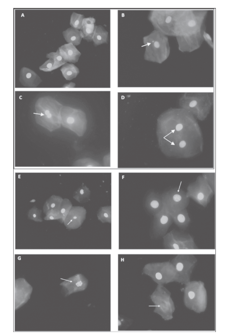
A: normal cells; B: micronuclei; C: nuclear bud; D: binucleated cells; E: pyknotic; F: condensed chromatin; G; karyorrhectic; and H: karyolytic (oil-immersion objective 60x, acridine orange stain)

Figure 2 Markers of cytotoxicity and DNA damage in buccal mucosa. México 

The number of MN was significantly higher in the Tepehuano group in comparison with Coras (p = 0.004), Huicholes (p = 0.001) and Tarahumaras (p = 0.001). The number of cells with nuclear buds in the Tepehuanos was significantly higher than that observed in the other study groups (p< 0.05). In addition, the number of binucleated cells in the Tepehuanos was significantly higher than in Tarahumaras (p < 0.05).

Regarding markers of cytotoxicity or cell death [condensed chromatin cells, karyolitic cells, karyorrhectic cells and pyknotic cells] (figure 2), no significant differences between the study groups were found in the number of karyolitic cells and pyknotic cells. Conversely, the number of condensed chromatin cells was significantly higher in Tepehuanos than in Huicholes (p = 0.02); also, the Tepehuano group presented a significantly higher number of Karyorrhectic cells than Tarahumaras, Coras and Huicholes (p < 0.05). The number of condensed chromatin cells was significantly higher in the Tepehuanos group compared to Huicholes group (p = 0.02); a significantly higher number of Karyorrhectic cells was found in Tepehuanos in comparison to Tarahumaras, Coras and Huicholes (p < 0.001).

There was no correlation between the number of MN with chronological age, gender, and number of subjects exposed to herbicides, smokers and alcohol drinkers (table III).

Table III Correlation test for general characteristics of participants and micronuclei. México 

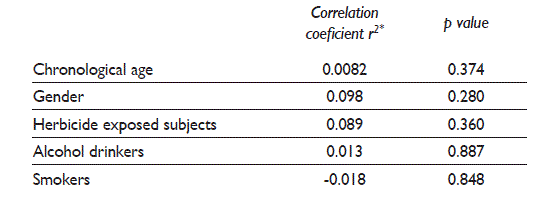
Spearman’s correlation coefficient, significance p value < 0.05 and CI95%

Note: The indigenous populations originate from the states of Durango, Chihuahua and Nayarit and the sampling took place in the period from January 2009 to December 2012

Discussion

Mexico has one of the richest ethnic, cultural and linguistic diversities in the Americas. The existing Native Mexican groups have settled in difficult-to-access areas, and retain their traditional lifestyle and language, for this reason it is important determine the impact of environment and lifestyles on DNA integrity of the Native Mexican groups.

Our results showed that the Tepehuano group presented greater number of micronuclei (DNA damage) and nuclear abnormalities (cytotoxicity) than the Huichol, Cora and Tarahumara groups (p < 0.05). The greater cytotoxic and genotoxic damage observed in 50% of Tepehuanos could be the result of a greater number of individuals exposed to herbicides than the other Amerindian groups studied.

Supporting this, some reports demonstrated that chemical agents contained in fungicides, herbicides and insecticides, increase the number of MN in exfoliated buccal cells.5,8,19,20 For example, the inhalation of glyphosate herbicide may cause DNA damage in exposed individuals.4 Also, there is evidence of cytotoxic and genotoxic effects of paraquat in human lymphocytes in vitro.21

Differences in the number of MN and NAs depend on genetic and environmental factors such as diet, age and sex.2,3,22 A higher number of MN in women compared to men was previously described,23 however in the present study such differences were not observed (data not shown).

Age can also influence the number of MN,2,24,25 since the older the subject, the greater is the number of MN.18,26 Conversely, in the present study the number of MN and NA were higher in young individuals belonging to Tepehuano group. This could be explained by the widespread use of herbicides in this group, although no correlation was found between these two variables.

In relation to smoking habits and the number of MN, the reports are very controversial, some authors described that smoking increased the frequency of MN27,28,29,30 and other authors did not observe this effect.31,32,33,34 In this study, the smoking habit was similar between the studied groups, and there was no correlation between the number of MN and number of individuals who smoke (table III).

There is evidence that ethanol increased the number of MN in exfoliated cells.35,36,37 In addition, a significant increase in the number of MN in lymphocytes of alcoholics compared with abstinent alcoholics and nonalcoholics has been reported.38 There are also reports of higher frequency of cells in karyorrhexis in alcoholics.39 Furthermore, ethanol also increases the number of chromosome breakage and sister chromatid exchange.40

In this study, the largest number of individuals that drink alcohol belong to Tarahumaras (50%), followed by Coras (36.7%), Tepehuanos (30%) and Huicholes (23.3%). The highest number of MN was found in Tepehuanos, which suggests that alcohol intake is not relevant for MN development in these populations.

Working with indigenous populations represents a big challenge because the accessibility to the communities is difficult, and there exist cultural and language barriers. To the best of our knowledge, this is the first report that determines the number of micronuclei and nuclear anomalies in Mexico’s indigenous population, however, some limitations must be considered: 1) the quantification of herbicides of biological samples was not performed; in addition, the time and duration of exposition to this contaminant was not recorded. 2) The number of cigarettes and the frequency of smoking were not obtained. 3) The frequency of use of alcohol and volume are not precise. These limitations should be considered to design additional studies in Mexican indigenous populations.

Conclusions

In relation to the four Amerindian groups studied, the Tepehuano group presented the highest number of MN and NA followed by the Tarahumara group, which may reflect the influence of ethnic diversity, habits and customs upon the DNA nuclear integrity of the Amerindian groups

Acknowledgments

We express our gratitude to all the indigenous volunteers for their collaboration despite the language barrier. This study was partly supported by Conacyt-Mexico Project(2011-C01-162368).

References

1. Ishikawa H, Tian Y, Yamauchi T. Influence of gender, age and lifestyle factors on micronuclei frequency in healthy Japanese populations. J Occup Health 2003;45(3):179-181. https://doi.org/10.1539/joh.45.179 [ Links ]

2. Fenech M, Holland N, Zeiger E, Chang WP, Burgaz S, Thomas P, et al. The HUMN and HUMNxL international collaboration projects on human micronucleus assays in lymphocytes and buccal cells-past, present and future. Mutagenesis 2011;26(1):239-245. https://doi.org/10.1093/mutage/geq051 [ Links ]

3. Bonassi S, Coskun E, Ceppi M, Lando C, Bolognesi C, Burgaz S, et al. The HUman MicroNucleus project on eXfoLiated buccal cells (HUMN XL): the role of life-style, host factors, occupational exposures, health status, and assay protocol. Mutat Res 2011;728(3):88-97. https://doi.org/10.1016/j.mrrev.2011.06.005 [ Links ]

4. Koller VJ, Fürhacker M, Nersesyan A, Mišík M, Eisenbauer M, Knasmueller S. Cytotoxic and DNA-damaging properties of glyphosate and Roundup in human-derived buccal epithelial cells. Arch Toxicol 2012;86(5):805-813. https://doi.org/10.1007/s00204-012-0804-8 [ Links ]

5. Benedetti D, Nunes E, Sarmento M, Porto C, Dos Santos CE, Dias JF, da Silva J. Genetic damage in soybean workers exposed to pesticides: evaluation with the comet and buccal micronucleus cytome assays. Mutat Res 2013;752(1):28-33. https://doi.org/10.1016/j.mrgentox.2013.01.001 [ Links ]

6. Torres-Bugarín O, Zavala-Cerna MG, Nava A, Flores-García A, Ramos-Ibarra ML. Potential uses, limitations, and basic procedures of micronuclei and nuclear abnormalities in buccal cells. Dis Markers 2014;2014:956835. https://doi.org/10.1155/2014/956835 [ Links ]

7. Corbi SC, Bastos AS, Orrico SR, Secolin R, Dos Santos RA, Takahashi CS, Scarel-Caminaga RM. Elevated micronucleus frequency in patients with type 2 diabetes, dyslipidemia and periodontitis. Mutagenesis 2014;29(6):433-439. https://doi.org/10.1093/mutage/geu043 [ Links ]

8. Ghisi N de C, de Oliveira EC, Prioli AJ. Does exposure to glyphosate lead to an increase in the micronuclei frequency? A systematic and meta-analytic review. Chemosphere 2016;145:42-54. https://doi.org/10.1016/j.chemosphere.2015.11.044 [ Links ]

9. Castañeda-Yslas IJ, Arellano-García ME, García-Zarate MA, Ruíz-Ruíz B, Zavala-Cerna MG, Torres-Bugarín O. Biomonitoring with micronuclei test in buccal cells of female farmers and children exposed to pesticides of Maneadero agricultural valley, Baja California, Mexico. J Toxicol 2016;2016:7934257. https://doi.org/10.1155/2016/7934257 [ Links ]

10. Navarrete-Linares F. Los pueblos indígenas de México. México: CDI, 2008 [cited 2016 Apr 15]. Available from: Available from: http://ru.ffyl.unam.mx/handle/10391/353Links ]

11. Comisión Nacional para el desarrollo de los pueblos indígenas. Programa Especial de los Pueblos Indígenas 2014-2018 [internet document] (cited 2016 Apr 18). Available from: Available from: http://www.cdi.gob.mx/programas/2014/programa-especial-de-los-pueblos-indigenas-2014-2018.pdfLinks ]

12. Thomas P, Holland N, Bolognesi C, Kirsch-Volders M, Bonassi S, Zeiger E, et al. Buccal micronucleus cytome assay. Nat Protoc 2009;4(6):825-837. https://doi.org/10.1038/nprot.2009.53 [ Links ]

13. Bolognesi C, Bonassi S, Knasmueller S, Fenech M, Bruzzone M, Lando C, et al. Clinical application of micronucleus test in exfoliated buccal cells: A systematic review and metanalysis. Mutat Res Rev Muta Res 2015;766:20-31. https://doi.org/10.1016/j.mrrev.2015.07.002 [ Links ]

14. Schmezer P, Pool BL, Lefevre PA, Callander RD, Ratpan F, Tinwell H, et al. Assay-specific genotoxicity of N-nitrosodibenzylamine to the rat liver in vivo. Environ Mol Mutagen 1990;15(4):190-197. https://doi.org/10.1002/em.2850150404 [ Links ]

15. Yamamoto M, Wakata A, Aoki Y, Miyamae Y, Kodama S. Chromosome loss caused by DNA fragmentation induced in main nuclei and micronuclei of human lymphoblastoid cells treated with colcemid. Mut Res 2014;762:10-16. https://doi.org/10.1016/j.mrfmmm.2014.02.002 [ Links ]

16. Rangel-Villalobos H, Martínez-Sevilla VM, Salazar-Flores J, Martínez-Cortez G, Muñoz-Valle JF, Galaviz-Hernández C, et al. Forensic parameters for 15 STRs in eight Amerindian populations from the north and west of Mexico. Forensic Sci Int Genet 2013;7(3):e62-e65. https://doi.org/10.1016/j.fsigen.2013.02.003 [ Links ]

17. Bonassi S, Fenech M, Lando C, Lin YP, Ceppi M, Chang WP, et al. HUman MicroNucleus project: international database comparison for results with the cytokinesis-block micronucleus assay in human lymphocytes: I. Effect of laboratory protocol, scoring criteria, and host factors on the frequency of micronuclei. Environ Mol Mutagen 2001;37(1):31-45. https://doi.org/10.1002/1098-2280(2001)37:1<31::AID-EM1004>3.0.CO;2-P [ Links ]

18. Bonassi S, Biasotti B, Kirsch-Volders M, Knasmueller S, Zeiger E, Burgaz S, et al. State of the art survey of the buccal micronucleus assay-a first stage in the HUMNXL project initiative. Mutagenesis 2009;24(4):295-302. https://doi.org/10.1093/mutage/gep019 [ Links ]

19. Pacheco ADO, Hackel C. Chromosome instability induced by agrochemicals among farm workers in Passo Fundo, Rio Grande do Sul, Brazil. Cad Saude Publica 2002;18(6):1675-1683. https://doi.org/10.1590/S0102-311X2002000600022 [ Links ]

20. Sycheva LP, Mozhaeva TE, Umnova NV, Zhuchenko NA, Diep VH, Tuyet HA. Cytogenetic and other cariological parameters of exfoliative buccal cells in Vietnamese children from areas where dioxin-containing herbicides were applied. Vestn Ross Akad Med Nauk 2008;(1):19-23. [ Links ]

21. Jovtchev G, Gateva S, Stergios M, Kulekova S. Cytotoxic and genotoxic effects of paraquat in Hordeum vulgare and human lymphocytes in vitro. Environmental toxicology 2010;25(3):294-303. https://doi.org/10.1002/tox.20503 [ Links ]

22. Pastor S, Creus A, Xamena N, Siffel C, Marcos R. Occupational exposure to pesticides and cytogenetic damage: results of a Hungarian population study using the micronucleus assay in lymphocytes and buccal cells. Environ Mol Mutagen 2002;40(2):101-109. https://doi.org/10.1002/em.10100 [ Links ]

23. Zalacain M, Sierrasesumaga L, Patino A. The cytogenetic assay as a measure of genetic instability induced by genotoxic agents. An Sist Sanit Navar 2005;28(2):227-236. [ Links ]

24. Tucker JD, Nath J, Hando JC. Activation status of the X chromosome in human micronucleated lymphocytes. Hum Genet 1996;97(4):471-475. https://doi.org/10.1007/BF02267069 [ Links ]

25. Fenech M. Important variables that influence base-line micronucleus frequency in cytokinesis-blocked lymphocytes-a biomarker for DNA damage in human populations. Mutat Res 1998;404(1):155-165. https://doi.org/10.1016/S0027-5107(98)00109-2 [ Links ]

26. Bukvic N, Gentile M, Susca F, Fanelli M, Serio G, Buonadonna L, et al. Sex chromosome loss, micronuclei, sister chromatid exchange and aging: a study including 16 centenarians. Mut Res 2001;498(1):159-167. https://doi.org/10.1016/S1383-5718(01)00279-0 [ Links ]

27. Konopacka M. Effect of smoking and aging on micronucleus frequencies in human exfoliated buccal cells. Neoplasma 2003;50(5):380-382. [ Links ]

28. Haveric A, Haveric S, Ibrulj S. Micronuclei frequencies in peripheral blood and buccal exfoliated cells of young smokers and non-smokers. Toxicol Mech Methods 2010;20(5):260-266. https://doi.org/10.3109/15376516.2010.482962 [ Links ]

29. Zamani AG, Durakbasi-Dursun HG, Demirel S, Acar A. Evaluation of smoking genotoxicity in Turkish young adults. Indian J Hum Genet 2011;17(1):7-12. https://doi.org/10.4103/0971-6866.82186 [ Links ]

30. Feki-Tounsi M, Khlifi R, Mhiri MN, Rebai A, Hamza-Chaffai A. Cytogenetic damage in the oral mucosa cells of bladder cancer patients exposed to tobacco in Southern Tunisia. Environ Sci Pollut Res Int 2014;21(22):12922-12927. https://doi.org/10.1007/s11356-014-3200-5 [ Links ]

31. Stich HF, San RH, Rosin MP. Adaptation of the DNA-repair and micronucleus test to human cell suspensions and exfoliated cells. Ann N Y Acad Sci 1983;407(1):93-105. https://doi.org/10.1111/j.1749-6632.1983.tb47816.x [ Links ]

32. Miloševic-Djordjevic O, Grujicic D, Vaskovic Z, Marinkovic D. High micronucleus frequency in peripheral blood lymphocytes of untreated cancer patients irrespective of gender, smoking and cancer sites. Tohoku J Exp Med 2010;220(2):115-120. https://doi.org/10.1620/tjem.220.115 [ Links ]

33. Milošević-Djordjević O, Stošić I, Grujičić D, Banković D, Arsenijević S. Cervical precancerous lesions-chromosomal instability in peripheral blood lymphocytes in relation to lesion stage, age and smoking habits. Acta Obstet Gynecol Scand 2011;90(10):1082-1087. https://doi.org/10.1111/j.1600-0412.2011.01230.x [ Links ]

34. Milosevic-Djordjevic O, Stosic I, Vuckovic M, Grujicic D, Marinkovic and D. Baseline and therapy-induced chromosome damages in peripheral blood lymphocytes of breast cancer patients assessed by the micronucleus assay. J BUON 2011;16(3):437-443. [ Links ]

35. Maffei F, Fimognari C, Castelli E, Stefanini GF, Forti GC, Hrelia P. Increased cytogenetic damage detected by FISH analysis on micronuclei in peripheral lymphocytes from alcoholics. Mutagenesis 2000;15(6):517-523. https://doi.org/10.1093/mutage/15.6.517 [ Links ]

36. Reis SR, Sadigursky M, Andrade MG, Soares LP, Espírito Santo AR, Villas Boas DS. Genotoxic effect of ethanol on oral mucosa cells. Pesqui Odontol Bras 2002;16(3):221-225. https://doi.org/10.1590/S1517-74912002000300007 [ Links ]

37. Reis SR, do Espírito Santo AR, Andrade MG, Sadigursky M. Cytologic alterations in the oral mucosa after chronic exposure to ethanol. Braz Oral Res 2006;20(2):97-102. https://doi.org/10.1590/S1806-83242006000200002 [ Links ]

38. Ishikawa H, Ishikawa T, Yamamoto H, Fukao A, Yokoyama K. Genotoxic effects of alcohol in human peripheral lymphocytes modulated by ADH1B and ALDH2 gene polymorphisms. Mutat Res 2007;615(1):134-142. https://doi.org/10.1016/j.mrfmmm.2006.11.026 [ Links ]

39. Webber LP, Pellicioli AC, Magnusson AS, Danilevicz CK, Bueno CC, Sant’Ana Filho M, et al. Nuclear changes in oral mucosa of alcoholics and crack cocaine users. Hum Exp Toxicol; 35(2):184-193. https://doi.org/10.1177/0960327115579430 [ Links ]

40. Hedner K, Wadstein J, Mitelman F. Increased sister chromatid exchange frequency in chronic alcohol users. Heredita 1994;101(2):265-266. https://doi.org/10.1111/j.1601-5223.1984.tb00926.x [ Links ]

Received: October 31, 2016; Accepted: May 08, 2017

*Corresponding author: Dr. Blanca Patricia Lazalde Ramos. Universidad Autónoma de Zacatecas. Campus UAZ Siglo XXI, Carretera Zacatecas-Guadalajara Km 6, Ejido la Escondida, Edif. L1, 3er piso. 98160 Zacatecas, México. E-mail: blancalazalde@gmail.com

Declaration of conflict of interests. The authors declare that they have no conflict of interests.

Creative Commons License This is an open-access article distributed under the terms of the Creative Commons Attribution License