Introduction
Chronic pulmonary aspergillosis (CPA) is an uncommon condition associated with diverse respiratory disorders, and is usually manifested as cavities or fibrosing changes; however, classical aspergilloma or nodular lesions may be observed in some cases.1,2 In developing areas, tuberculosis can precede or coexist with this fungal infection.1,3 The diagnosis is based on typical pulmonary images and microscopy or culture evidence of the fungus or positivity of the specific serologic test for Aspergillus.1-5 Worthy of note is the necessity of excluding diverse alternative causal agents, and pulmonary tuberculosis has been the main differential task in low income regions.1,3 The aim of this report is enhance the suspicion index about aspergillosis and briefly comment the role of galactomannan test, that may have controversial clinical impact in immunosuppressed patients with aspergillosis. The sensitivity and specificity as well as the cut-off level differ from immunocompetent ones.1,4,5
Case report
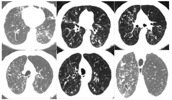
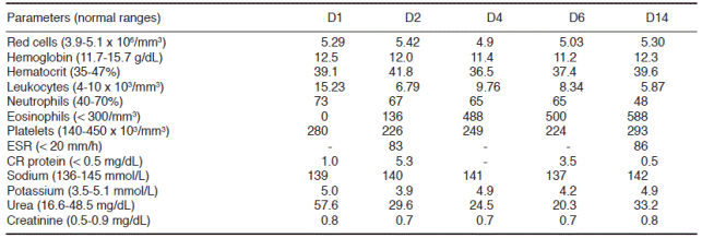
A 76-year-old woman with clinical history of bronchiectasis and depression came to emergency attention complaining of back pain after an episode of cough six hours ago. She had been with productive cough during four months and used moxifloxacin (10 days), amoxicillin and clavulanate (10 days); followed by moxifloxacin (14 days) without clinical improvement. Therefore, she was admitted for diagnostic investigation and additional treatment with piperacillin and tazobactam (4.5 g t.i.d.) during 14 days. Physical status was regular, BMI: 14.4 kg/m2, and the rest of the exam unremarkable. The chest tomography without contrast showed small scattered nodules with attenuation of soft tissue, the most conspicuous (0.8 x 0.7 cm) on the left posterior basal segment; bilateral ground-glass opacity and tree-in-bud images; and multiple bronchiectasis more evident in both bases (Figure 1). Tuberculosis and aspergillosis were then hypothesized. Routine laboratory determinations were unremarkable, except for neutrophilic leukocytosis, and moderate elevation of urea and of C-reactive protein levels (Table 1). Repeated bacilloscopy and cultures for M. tuberculosis in samples of bronchoalveolar lavages resulted negative; PCR for M. tuberculosis was also negative. Mycological evaluations of bronchoalveolar samples were negative, but ELISA test for Aspergillus antigen (galactomannan) was positive 0.7 (limit: < 0.5 index). The initial galatomannan test was performed during the use of piperacillin and tazobactam; however, the control test was negative with course of itraconazole. The anti-HIV test was negative. CA 125 and CEA were normal; and CA-19.9 was elevated 49.84 (< 34), but the evaluation of pancreatic and hepatobiliary areas ruled out malignancy. She underwent oral itraconazole and became symptomless, being referred to outpatient follow-up at Pneumology service. Furthermore, the study of image done to evaluate the response to the antifungal agents showed significant decrease in the nodes and in the ground glass changes initially observed. This evidence was also considered consistent with the diagnosis of probable aspergillosis.

Figure 1 Chest CT without contrast showing small scattered nodules up to 0.8 x 0.7 cm with attenuation of soft tissue on the left posterior basal segment; bilateral ground-glass opacity and tree-in-bud images; and multiple bronchiectasis that are more evident in both bases.
Discussion
Invasive aspergillosis, CPA, allergic bronchopulmonary aspergillosis and aspergilloma are clinical features of fungal infections caused by agents of the genus Aspergillus.1-4 Invasive aspergillosis is often late diagnosed and associated with high mortality rate, and pulmonary foci represent the major primary origin of widespread infections.1,2,5 Since 2002, the incidence of invasive pulmonary aspergillosis has growing, at least partially due to the increased utilization of the galactomannan test for diagnosis.4,5 Clinical diagnosis of aspergillosis is often challenging and depends on complementary tools. In the patient herein described, the diagnosis of CPA was established with some delay because of non-specific manifestations.4,5 here attributed to bronchiectasis sequels. Moreover, the long standing course of cough, dyspnea and wheezing in association with blood eosinophilia might propitiate the mistaken diagnosis of bronchial asthma episodes. Allergic bronchopulmonary aspergillosis can affect asthmatic people causing bronchiectasis. The hypothesis was considered, but Aspergillus was not found in bronchoalveolar lavages, nor was central bronchiectasis in absence of cystic fibrosis found in the present case study. A major concern was Aspergillus-induced asthma, but the episodes were mild, with short duration and moderate eosinophilia, and the patient never utilized corticosteroids for control. Accordingly with the literature about aspergillosis, the eosinophil count, erythrocyte sedimentation rate, and C-reactive protein levels were higher than normal range.1-3 The positive ELISA galactomannan serum test for Aspergillus (0.7) was considered significant. Galactomannan is a heat-stable hetero polysaccharide released during hyphal growth, and ELISA has higher sensitivity and earlier positivity than latex agglutination method. The test has variable sensitivity (29-100%) and both false-negative or false-positive results have been described. These are related to the species of Aspergillus, anti-Aspergillus antibodies, concomitant administration of some antibiotics, and cross-reactivity with Fusarium sp., Alternaria sp., and Mucor sp.4,5 False positivity may occur in patients using piperacillin/tazobactam or amoxicillin/clavulanate (up to six days) or calcium gluconate (up to 48 hrs) before to collections. Microorganisms were not found in bronchoalveolar lavages, and the use of amoxicillin had stopped 14 days before the collection of samples for the exams. The initial collection of sample for the galactomannan test was done during the use of piperacillin and tazobactam; but false-positive tests are lower than 0.5 and the control test after itraconazole was negative. So, the possibility of false-positive test due to the antibiotic use was considered ruled out. The older patient was immunocompetent but had chronic cough due to bronchiectasis. The main initial concerns were about pulmonary tuberculosis or other fungal infection. Microorganisms were not shown by microscopy or culture of bronchoalveolar lavages; but clinical, imaging and laboratory data were consistent with diagnosis of chronic pulmonary aspergillosis that was successfully treated by itraconazole.
Conclusions
Older immunocompetent individuals with chronic cough due to chronic pulmonary disorders are common in developing regions, and unsuspected aspergillosis may occur. The major concern is about the hypothesis of tuberculosis, due to its higher prevalence. Non-specific clinical and imaging manifestations have contributed to diagnostic pitfalls. In this setting, one must take on account the practical usefulness of galactomannan test to confirm diagnosis. The best routine procedure should be concomitantly performing the test in bronchoalveolar aspirate and serum samples; however, this was not done in the woman herein described. Without piperacillin/tazobactam use, the galactomannan test of control was negative after three weeks of the antifungal schedule. It is worth noting that the images of control showed improvement of the pulmonary changes, consistent with diagnosis of probable aspergillosis. Case reports may enhance the suspicion index about less common pulmonary conditions, contributing to establishment of early diagnosis and the early beginning of proper treatment.


 nueva página del texto (beta)


