


Serviços Personalizados
Journal
Artigo
 texto em
  Inglês (pdf)
 Artigo em XML
 Referências do artigo
 Enviar este artigo por email
Indicadores
-
 Citado por SciELO
 -
 Acessos
Links relacionados
-
 Similares em
SciELO
Similares em
SciELO
Compartilhar

 Permalink
Revista odontológica mexicana
versão impressa ISSN 1870-199X
Rev. Odont. Mex vol.22 no.1 Ciudad de México Jan./Mar. 2018
Clinical cases
Maxillary and mandibular solitary bone cyst. Case report and literature review
*Graduated of the Maxillofacial Surgery Specialty HRLALM, ISSSTE.
§Professor and Head of the Maxillofacial Surgery of the HRLALM, ISSSTE.
Solitary bone cyst is a benign lesion of unknown origin up to the present date. Its origin is mainly attribute to trauma theory, caused by intra-osseous bleeding which prevents bone repair, causing thus a cystic cavity with serous hematic content lacking epithelial lining. In most cases, the lower jaw is the most affected. It is observed in patients with ages ranging 20-30 years, generally male, it is an asymptomatic, slow-growing entity which progresses toward the cortical plates; it can cause pathological mandible fractures. Its discovery is usually incidental during a radiographic examination. It appears as a radiolucent, unilocular ormultilocular image, with well -defined borders, lacking sclerotic areas. Additional tomography and magnetic resonance studies have revealed that these cavities are not necessarily taken up by fluids: there can be presence of gas, or they could be empty. A differential diagnosis must be established to discard dentigerous cysts, ameloblastoma, keratinizing odontogenic tumor, calcifying epithelial tumor or adenomatoid odontogenic tumor.
Key words: Mandibular cysts; solitary bone cyst; traumatic bone cysts; hemorrhagic bone cyst; simple bone cyst
El quiste óseo solitario es una lesión benigna cuyo origen sigue siendo hasta hoy desconocido, siendo el traumatismo la principal teoría de su origen, atribuido a un sangrado intraóseo, el cual no permite la reparación ósea, lo que conlleva a la formación de una cavidad quística con contenido serohemático sin revestimiento epitelial siendo la mandíbula la más afectada en la mayoría de los casos; se presenta en pacientes entre 20 y 30 años, con prevalencia por género masculino, asintomática, de crecimiento lento y desplazamiento de corticales, pudiendo producir fracturas patológicas mandibulares, su hallazgo es incidental durante el examen radiográfico, observándose como una imagen radiolúcida uni- o multilocular, con bordes definidos, sin zona esclerótica. Estudios complementarios de tomografía y resonancia magnética, han mostrado que estas cavidades no necesariamente están ocupadas por líquido, sino que puede haber gas o estar vacías. Se debe de hacer diagnóstico diferencial con quiste dentígero, ameloblastoma, tumor odontogénico queratinizante, tumor odontogénico epitelial calcificante, tumor odontogénico adenomatoide.
Palabras clave: Quistes mandibulares; quiste óseo solitario; quiste óseo traumático; quiste óseo hemorrágico; quiste óseo simple
History
Solitary bone cysts are rare cavities of the mandible lacking epithelial coating. They were first described by Lucas in 1929, and later by Rushton in 1946. This condition has received different names such as traumatic bone cyst, hemorrhagic bone cyst, extravasation cyst, progressive bone cavity and single-chamber bone cyst. This lesion has a more common occurrence in the lower jaw. Causes for its origin are unknown and controversial; among them the theory of hemorrhage due to trauma is preponderant, therefore, the condition is also known as traumatic bone cyst. Defenders of this theory inform that trauma in the lower jaw causes intra-osseous hematoma which does not reorganize or repair, this causes intra-osseous liquid content which results in a cystic defect.1
Epidemiology
Solitary bone cysts are normally found in young patients, frequently in their second and third decade of life, between 20 and 30 years. They are rare in children under 5 years of age, males are affected in 60% of cases. It is mainly located in the lower jaw, 75% (body of the mandible), second place is for mandibular symphysis, nevertheless there are cases reported in in the condyles, ramus and upper jaw.1-3
Symptomatology
From the clinical standpoint, this lesion appears asymptomatically, with no inflammation data; the size of the lesion can cause cortical plate displacement causing thus bone deformities in the affected mandibular region. Forssell et al, in 1988, reported that in 30% of studies cases there was presence of pain in the affected area,4 in some cases there were reports of tooth sensitivity, paresthesia, fistulae or eruption delays in the permanent tooth without causing root resorption or pulp necrosis. Lesion size can cause permanent displacement of tooth canal and pathological fractures in the lower jaw. Due to aforementioned characteristics, inmost cases, solitary bone cysts are accidentally discovered during routine radiographic examinations of the maxillary-mandibular region.1,2,4,5
Radiographic findings
In general, the following can be observed in osteolysis areas: irregular unilocular or multilocular radiolucent lesions, with well-defined borders and lacking sclerotic lining.6 In interdental areas it is observed as a horizontal or vertical cone. In larger lesions a festoon pattern can be observed in borders in interdental spaces, with intra-lesion trabeculate and expansion of cortical bone plates.1,2,6,7
Tomography and magnetic resonance studies have revealed that the cyst cavity is not always filled with liquid, in some instances there is gas, or they can be empty. This was possible to achieve bymensof Hounsfield units, whose «gas» values are -1,000 HU and water in 0 HU. This has led to the erroneous interpretation of the term «gas content at the interior of the cyst»,8 nevertheless this can vary according to the evolution time of the cystic cavity, which is the time ion which liquid content can be absorbed due to as yet unknown causes. Odontogenic keratocyst, calcifying epithelial odontogenic cyst, adenomatoid odotogenic tumor.9
Etiology
Causes for the origins of these cysts are yet unknown and subject to controversy. This leads to the many names used to describe this condition. Nevertheless, there are different theories with respect to their origin: the theory of hemorrhage caused by trauma is one of the preponderant ones, thus, this condition is also called traumatic bone cyst. Supporters of this theory report that trauma to the mandible causes intra-osseous hematoma, which does not reorganize or repair (separate) causing thus intra-osseous liquid content resulting in a cystic defect.1,2 In 1960 Cohen proposed the theory that the cyst develops due to a lymphatic drainage failure , which conditions interstitial liquid entrapment in the area, favoring thus bone resorption and cystic cavity formation. In 1978 Mirra et al proposed that solitary bone cysts are synovial cysts, since their development is a result of synovial liquid entrapment within the bone.7
Clinical variations
According to the clinical and radiographic characteristics of these lesions, it can be observed that solitary bone cysts can appear as multilocular lesions, associated to an non-erupted tooth, as well as multiplemultilocular lesions, which conditions a differential diagnosis with dentigerous cysts and ameloblastoma.
Histopathology
From a macroscopic point of view, solitary bone cysts exhibit cystic walls composed by a thin connective tissue membrane, this membrane is of a greyish-yellow hue, friable and hemorrhagic, thus hinderingenucleation, their interior can be empty, or filled with serous matter , or serous-hematic content.
Microscopic examination reveals a cystic wall as a connective tissue membrane with numerous collagen fibers, lacking epithelial lining, with presence of fibroblasts and osteoclasts and cholesterol crystals, all of which are related to bone necrosis.1,2
Treatment
There are many treatment alternatives, the most common is cyst walls curettage, fenestration, packing of material into the cyst, aspiration; lesion recurrence after curettage was 26%.
Suei et al.10,11 suggested that prognosis of these lesions would be improved if they were to be treated with fenestration or filling of the cyst cavity (gel-foam gauze, bone), since lesions exhibited a 20% recurrence.
Cyst cavity curettage will elicit bleeding, which will be replaced by bone in approximately 12 months.1,10 Lesion follow up needs to be confirmed with full bone remodeling ( by means of X rays), this normally is achieved three years after having treated the cystic lesion.10
Case presentation
A 45 year old patient attended the Maxillofacial Surgery Service of the Regional Hospital «Lic. Adolfo Lopez Mateos» (ISSSTE) (Social Services Institute for State Employees) complaining of volume increase in the right side of the upper jaw and both sides of the lower jaw. The lesion was slow-growing, asymptomatic, indurated, of a two year evolution. Patient informed of a contusion in the right side of the face five years before, with no pathological history.
Clinical examination revealed increase in the right region of the upper jaw, measuring approximately 1.5 cm palpation of the lower jaw revealed bilateral volume increase in the region of the mandibular body and chin area. This lesion was indurated, asymptomatic to palpation, lacking infection data with a two year evolution (Figure 1).
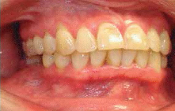
Figure 1 Upper jaw volume increase of about 1.5 cm, bilateral mandibular body and symphysis region, with no infection data.
An orthopantomography was taken; it revealed multi-lobulated radiolucent lesions in the region of right upper premolars and body of the mandible. The lesion crossed the midline up to the contra-lateral side, without causing root displacement, only a radio-opaque area could be observed around the apex of the upper canine and third right lower molar, lacking evidence of rhizolysis and exhibiting cortical bone thinning (Figure 2).

Prompted by lesion’s size and characteristics, the following was accomplished: computerized tomography, crown axial sections, and 3D reconstruction of the facial body, where the following was observed: a radiolucent multi-lobulated lesion, with presence of fluid in its interior, located in the body of the mandible and extending from lower left third molar to lower right third molar, with thinning and perforation of bone cortical plates in the lower jaw (Figure 3). On the right side of the upper jaw, a well-defined radiolucent lesion could be observed, revealing a small radio-opaque area within it, lacking data of cortical bone plate perforation (Figure 3 and 4); 3D reconstruction of the facial mass revealed expansion and perforation of bilateral mandibular vestibular cortical plate, with no data of tooth displacement (Figures 5 and 6). After practicing antisepsis of the oral region, an exploratory puncture was achieved in the lesions. This was accomplished with a 10 cm3 syringe ad 18G needle; a blood look-alike liquid with was found (Figure 7).

Figure 3 CAT, axial image of the mandible, multiple radiolucent lesions which cross the midline. Liquid content is observed in its interior as well as thinning of cortical plate.

Figure 4 CAT, well circumscribed unilocular radiolucent lesion, without maxillary sinus involvement, radio-opaque content in its interior, cortical plate thinned down.

Figure 5 3D reconstruction, face mass, evident expansion and thinning of vestibular cortical plate in the upper jaw and right side of the lower jaw.

Figure 6 Lesion 3D reconstruction; osteolyticlesion extending from left mandibular angle to right mandibular angle, perforation of vestibular cortical plate.

Surgical procedure
Iodopavidona asepsis and antisepsis were effected in face and mouth, sterile fields were placed, 2% epinephrine with xylocaine were infiltrated in nerves, lower tooth, bilaterally buccal and lingual and right area below the orbit. A hemi-circumvestibular incision was performed, with blade number 15, in the region of the body and bilaterally on mandibular symphysis, as well as in the right side of the lower jaw. Periosteum was removed, harvesting thus a muco-periosteal flap. Thinning of vestibular cortical plate was observed. Corticotomies were undertaken with a round tungsten burr. A membrane was observed within cyst cavities. This membrane was incised eliciting fluid drain. Later, cavity curettage was undertaken, and area was cleansed with 0.9% physiological solution, approach incisions were sutured with 3-0 polyglycolic acid suture. Procedure was completed uneventfully (Figures 8 and 9). Samples of liquid content, bone tissue and cavity membranes were sent to the laboratory for histopathological analysis.


Histopathological report
Lesion was of mesenchymal lineage, formed by a fibro-cellular wall with highly vascularized areas exhibiting some multi-nucleated giant cells mainly in areas of recent and old hemorrhage, presence of hemosiderin as well as scarce hematic material compatible with solitary bone cyst ( hemorrhagic bone cyst) (Figure 10).

Figure 10 Histological section of the lesion: highly vascularized areas and multinucleated giant cells can be observed.
One-year radiographic control was achieved with computerized tomography. This examination revealed decrease of cystic lesion size, bilateral bone tissue apposition in maxillary and mandibular cortical plates, involved teeth in vital circumstances, with present vitality (Figure 11).

Figure 11 Control computerized tomography and 3D reconstruction after 12 months, shows decrease of lesion size in rigth upper jaw and lower jaw.
From the clinical point of view, the patient was asymptomatic, with suitable tissue healing process as well as decrease in lesion size (Figure 12).

Discussion
Solitary bone cysts lack epithelium, radiographically they exhibit cystic appearance. They are more common in the lower jaw, in young patients, and are frequently related to trauma. Cystic cavity is frequently filled with liquid content12 of bloody appearance.
In the case here reported, solitary bone cysts were present in upper and lower jaw, the lower was the most affected in a bilateral manner.
Solitary bone cysts can be treated in many ways among which we can mention cystic wall curettage, cystic cavity exploration and placement of filling material into the cavity.
Yoshikazu10 reviewed treatment of 132 solitary bone cyst cases. Curettage of cyst cavities was the most frequently used treatment, followed by cystic cavity exploration. Recurrence was 26% and was more frequent in cases of simple lesion curettage, this situation decreases with decompression and filling material placement within the cyst cavity. Nevertheless, recurrence increases when it is associated to a bone cement dysplasia, a two year follow up is recommended in order to be able to determine remission or recurrence after three years. Radiographic imaging is considered an important tool to this end. Solitary bone cyst remission will be more reliable when suitable trabecular bone is observed and not only the decrease of a cystic cavity.
In the present case, conducted treatment was curettage and cleansing of cystic cavities, without placement of filling material, one year radiographic control showing decrease of cystic lesion size and better bilateral trabeculated pattern in lower jaw areas in the upper jaw area, better trabelulated pattern which can mean that size of lesion can play an important role in the remission of this condition.
Conclusions
Literature review concurs with this case description. The most affected area is the mandible, and to a lesser extent the maxilla. Age corresponds to young patients (third and fourth decades of life). Lesion was associated to trauma incident; decompression by means of cystic wall curettage is a suitable treatment option. Follow up must be conducted by means of radiographic imaging up to a three year span, in order to be able to determine remission or recurrence, based not only on decrease of lesion size but rather on suitable re-establishment of trabecular bone in affected area.
REFERENCES
1. Xanthinaki AA1, Choupis KI, Tosios K, Pagkalos VA, Papanikolaou SI. Traumatic bone cyst of the mandible of possible iatrogenic origin: a case report and brief review of the literature. Head & Face Medicine. 2006; 2: 40. doi:10.1186/1746-160X-2-40 [ Links ]
2. Harnet JC, Lombardi T, Klewansky P, Rieger J, Tempe MH, Clavert JM. Solitary bone cyst of the jaws: a review of the etiopathogenic hypotheses. J Oral Maxillofac Surg. 2008; 66 (11): 2345-2348. [ Links ]
3. Neville BW, Damm DD, Allen CM, Bouquot JE. Oral and maxillofacial pathology. 3rd edition, Saunders Elsevier 2009, pp. 713-715. [ Links ]
4. Forssell K, Forssell H, Happonen RP, Neva M. Simple bone cyst. Review of the literature and analysis of 23 cases. Int J Oral Maxillofac Surg. 1988; 17 (1): 21-24. [ Links ]
5. Baqain ZH, Jayakrishnan A, Farthing PM, Hardee P. Recurrence of a solitary bone cyst of the mandible: case report. Br J Oral Maxillofac Surg. 2005; 43 (4): 333-335. [ Links ]
6. Strabbing EM, Gortzak RA, Vinke JG, Saridin CP, van Merkesteyn JP. An atypical presentation of a solitary bone cyst of the mandibular ramus: a case report. J Craniomaxillofac Surg. 2011; 39 (2): 145-147. [ Links ]
7. Kuhmichel A, Bouloux GF. Multifocal traumatic bone cysts: case report and current thoughts on etiology. J Oral Maxillofac Surg. 2010; 68 (1): 208-212. [ Links ]
8. Eriksson L, Hansson LG, Åkesson L. Simple bone cyst: a discrepancy between magnetic resonance imaging and surgical observations. Oral Surg Oral Med Oral Pathol Oral Radiol Endod. 2001; 92 (6): 694-698. [ Links ]
9. Tong AC, Ng IO, Yan BS. Variations in clinical presentations of the simple bone cyst: report of cases. J Oral Maxillofac Surg. 2003; 61 (12): 1487-1491. [ Links ]
10. Suei Y, Taguchi A, Tanimoto K. Simple bone cyst of the jaws: evaluation of treatment outcome by review of 132 cases. J Oral Maxillofac Surg. 2007; 65 (5): 918-923. [ Links ]
11. Peñarrocha-Diago M, Sanchis-Bielsa JM, Bonet-Marco J, Minguez-Sanz JM. Surgical treatment and follow-up of solitary bone cyst of the mandible: a report of seven cases. Br J Oral Maxillofac Surg. 2001; 39 (3): 221-223. [ Links ]
12. Navarro VC. Tratado de cirugía oral y maxilofacial. Tomo I, editorial Aran, Segunda ed., Madrid 2009, p. 40. [ Links ]
*This article can be read in its full version in the following page: http://www.medigraphic.com/facultadodontologiaunam
Received: July 01, 2016; Accepted: January 01, 2017
 Este es un artículo publicado en acceso abierto bajo una licencia Creative Commons
