


 nueva página del texto (beta)
 Inglés (pdf)
 Artículo en XML
 Referencias del artículo
 Enviar artículo por email
 Citado por SciELO
  Similares en
SciELO
Similares en
SciELO 
 Permalink
INTRODUCTION
In the 100 m and 200 m sprint, the success of the sprint start performance depends on the capacity of the athlete to establish a large impulse over the shortest time, reaching the highest running speed as soon as possible [1] [2]. During this step, the sprinters take their positions at the blocks at the set command, and the mechanics of leaving the blocks at the sound of the gun strongly influences the acceleration at the start of a race [3] [4]. When a starter’s command is given, the athlete takes up a position with their hands just behind the line, arms vertical, and feet generally placed about a shoe length apart. In the set position, the athlete understands that the hips should rise above the line of the head. The is a misconception that they must lean forward, bringing their shoulders ahead of their hands and putting pressure on them. This position influences the execution of the low-sprint start and can drastically influence the initial running speed and acceleration achieved by the athlete. This is because when the gun goes off, it becomes impossible for the athlete to instantly drive their arms forward or backward without first lifting them off the ground, and they can lose time in the process [5]. According to Schot PK and Knutzen KM, an efficient sprint start depends on the start block positioning and joint angles of the lower limbs in the position [6]. In addition, the pushing time on the blocks and the forces generated by the front and rear legs during the pushing phase depend on the reaction time, stride length, and stride time [7]. The average external power calculated based on horizontal motion and normalized to participant characteristics, provides a single measure that accounts for the change in velocity and the time taken to achieve this change [2]. However, this parameter is more commonly adopted for measurements during early and mid-acceleration after the low-sprint start process.
In practice, an efficient sprint start integrates temporal and spatial acyclic movements into a cyclic action, whose success depends on the athlete’s ability and coach experience and is still a big challenge. Different authors have reported studies related to the optimal relationship between body position and initial acceleration. For example, Coh et al. reported the dependence of the angular velocity and maximal force of the sprinter during the start on the body positioning and associated start block settings [7]. Mero et al. found that block velocity is strongly correlated with the horizontal and vertical forces exerted on the front and rear starting blocks [8]. Gutierrez-Davila, and Prampero et al. reported biomechanical variables and their interdependencies with specific motor abilities, energy processes, anthropometric characteristics, and the central processes of motor regulation [9]. There are few previous, reports on the biomechanics of kinematic parameters such as reaction time, stride length, and stride time. All authors state that “a single optimal set position” for all athletes is not recommended because of varying physical factors; therefore, athletes must find their preferred distance between the blocks according to sensations or outcomes.
On the other hand, different technologies have been used to assess the kinematic parameters of sprint start in high-performance athletes. Bezodis et al. published in 2019 a clear review of the current biomechanics of track and field sprint starts that can be used to provide current recommendations for both researchers and practitioners [10]. Bezodis et al. proposed the use of a laser distance measurement (LDM) device to determine the measurement error in velocity data obtained during different phases of a maximal sprint, and consequently, to evaluate the usability of LDM devices to analyze the velocity profiles of sprinters [11]. Their results recommend that laser data should not be used during the first 5 m of a sprint and are likely to have limited use for assessing within-subject variation in performance during a single session. Bergamini et al. reported the use of a lower trunkmounted inertial measurement unit (IMU) to identify consistent features in the waveforms of the signals supplied by the IMU and estimate stance and stride duration during the maintenance phase of sprint running. Their results proved that the IMU is suitable for estimating stance and stride durations during sprint running, providing the opportunity to collect information in the field without constraining or limiting athletes’ and coaches’ activities [12]. Falbriard et al. reported the use of foot-worn inertial sensors to investigate different algorithms to detect initial contact (IC) and terminal contact (TC) from different features measured by foot-worn IMU kinematic signals and estimated the main inner-stride temporal parameters. The performance metrics (bias and precision) of each algorithm were assessed in comparison with a reference system (instrumented force plate treadmill), which allowed the validation of the innerstride temporal parameters over a large number of steps and a large range of running speeds. Their results showed that running speed can significantly affect the estimation bias, suggesting that speed-dependent correction should be applied to improve the accuracy of the systems [13]. Seidl et al. reported the use of a radio-based local position measurement system, RedFIR (Grün et al., 2011) to obtain spatio-temporal information during sprinting based on lightweight transmitters attached to the athletes. Based on their results, a methodology capable of automatically providing step length, step time, and ground contact time during sprinting was developed. Different transmitter positions were tested, and the accuracy of the derived spatiotemporal parameters was evaluated by comparing them to those from an optoelectronic system [14]. Nagahara et al. reported the use of two different global positioning system (GPS) units to obtain mechanical properties during sprint acceleration [15]. However, in their results, they remarked that for the current state of GPS devices’ accuracy for speed-time measurements over a maximal sprint acceleration, it is recommended that radar, laser devices, and timing gates remain the reference methods for implementing the computation methods reported by Samozino et al. [16]. Bergamini et al. validated an adapted sensor-fusion algorithm in a trunk-mounted IMU to estimate trunk inclination and angular velocity during sprint start. A Bland-Altman analysis was carried out using parameters extracted from the historical data of the estimated variables, and analysis of similar curves. Their results indicate agreement between the reference and IMU estimates, which opens a promising scenario for accurate in-field use of IMUs for sprint start performance assessment [17]. Di-Kiat et al. proposed the use of an optical motion capture system as a benchmark to validate a new approach for defining running gait. They presented a new gait event identification method, that uses foot acceleration to determine the foot-stroke (FS) and foot-off (FO) times. Temporal parameters such, as contact time and flight time can then be derived from this information [18]. This study also aimed to demonstrate that spatial parameters, such as running speed and stride length, can be estimated accurately using the method presented in [19]. However, most of the technologies described above are only applicable to one athlete, limited to straight runs, and need to be placed directly on the running track. This prohibits their use in competitions and runs including curved sections. Emergency three-dimensional (3D) kinematic analysis computer vision systems use hardware/software processing units with real-time optoelectronic video cameras to measure and quantitatively analyze 3D human segmental movement [20] [21]. Retro-reflective markers placed on the body surface are used to calculate with high precision of the body segments as well as the kinematic joints [22] [23]. The data obtained from these systems are clear and detailed, and the user is able to move freely because there are no cables attached to the computer. In addition, this allows movements in a large volume and observations of more users at one time [22] [23]. The major problem with these systems is the interference. Sometimes, the light reflected from a specific marker has insufficient intensity, which causes inaccuracy in the output. In addition, the user can sometimes cover the markers, producing an occlusion effect on the camera, which causes inaccuracy of the output. Moreover, the system can be expensive depending on the required sensors, cameras, and software [24].
Although, most kinematic parameters can be obtained from measurements with floor sensors in combination with wearable sensors, no real-time calculations and visualization of 3D segmental movements can be performed. Hence, the aim of this study was to use the Vicon Plug-in-Gait model (Vicon®, Oxford, UK) to characterize the kinematic parameters that influence the execution of low-sprint start in six high-performance athletes [25]. The main kinematic parameters are reaction time, stride length, and stride time. Average external power was not considered in this study because vertical movement and vertical velocity play an important role in the low sprint-start process. The obtained results demonstrate the potential utility of the system in assessing the kinematic parameters of a low-sprint start.
MATERIALS AND METHODS
Participants
Six high-performance athletes, three women, and three men, who compete in 100 meters passing hurdles, were evaluated. All athletes wore tight training shorts, and the women wore a sports female bra. During testing, athletes wore the footwear that they used for competition. No accessories that hindered or introduced variations in athlete performance were permitted during testing. The demographic data (age, height, body mass) are as follows: (mean ±SD, age = 17 ± 2.09 years); the height of the athletes was measured with a wall stadiometer graduated in centimetres, presenting a scale from 0 to 250cm(SECA), without shoes, straight back, front view, (Height = 1.69 ± 0.06 m) and the body mass was obtained using the AMTI platform, (Body mass = 59.77 ± 6.65 kg).
Anthropometric dimensions were measured according to the provisions of the Vicon® Plug-in Gait product guide [25] as shown in Table 1. Flexible tape and Vernier tape were used for measurements. This study conformed to the recommendations of the Declaration of Helsinki and was approved by the research and postgraduate ethics and evaluation committee of the Faculty of Sports of the Autonomous University of Baja California, México.
Table 1 The averages of the anthropometric and demographic measures of the participants.
| Variable | Male | Female |
|---|---|---|
| Age (years) | 16 ± 1 | 18 ± 2.64 |
| Height (m) | 1.73 ± 0.02 | 1.66 ± 0.07 |
| Body mass (kg) | 61.56 ±3.17 | 57.97 ± 9.52 |
| LPI (cm) | 90.6 ± 6.03 | 86.53 ± 5.85 |
| LPD (cm) | 89.66 ± 5.57 | 86.76 ± 5.56 |
| DRI (cm) | 9.56 ± 0.47 | 8.6 ± 0.34 |
| DRD (cm) | 9.46 ± 0.55 | 8.63 ± 0.35 |
| DTI (cm) | 6.73 ± 0.35 | 6.7 ± 0.36 |
| DTD (cm) | 6.76 ± 0.46 | 6.86 ± 0.45 |
| DHI (cm) | 6.7 ± 0.52 | 5.26 ± 0.30 |
| DHD (cm) | 5.53 ± 0.57 | 5.3 ± 0.79 |
| DCI (cm) | 6.73 ± 0.057 | 5.76 ± 0.20 |
| DCD (cm) | 6.9 ± 0.10 | 5.96 ± 0.15 |
| DMI (cm) | 5.58 ± 0.27 | 4.73 ± 0.25 |
| DMD (cm | 5.56 ± 0.32 | 4.76 ± 0.11 |
| EMI (cm) | 2.73 ± 0.11 | 2.16 ± 0.1 |
| EMD (cm) | 2.7 ± 0.1 | 2.1 ± 0.1 |
LPI = Left Leg Length, LPD = Right Leg Length, DRI = Left Knee Diameter, DRD = Right Knee Diameter, DTI = Left Ankle Diameter, DTD = Right Ankle Diameter, DHI = Left Shoulder Displacement, DHD = Right Shoulder Displacement, DCI = Left Elbow Diameter, DCD = Right Elbow Diameter, DMI = Left Wrist Diameter, DMD = Right Wrist Diameter, EMI = Left hand Thickness, EMD = Right Hand Thickness.
Marker placement and Motion Capture
The evaluations were performed at the Biomechanics Laboratory of the Faculty of Sports at the Autonomous University of Baja California, Mexico. 39 passive reflective markers, 24 mm in diameter, were placed on each participant as shown in Figure 1. The position of each reflective marker followed the standard Plug-in-Gait model, as described in Table 2. To characterize complete body movement, the markers were divided into upper-body and lower-body markers. The upper body markers contained four head markers, two for the front side and two for the backside. The torso markers contained five markers, two for vertebrae, a clavicle marker, a sternum marker and a scapula marker. The arm markers contain a marker for the shoulder and, markers for the upper arms, elbow, forearm, wrist, and fingers. Similarly, the lower body markers contained five pelvis markers, two for the left anterior superior side and two for the right anterior superior side of the iliac spine. The leg contained five markers, a knee marker, thigh marker, ankle marker, and tibial marker. The foot contained a toe and heel marker.
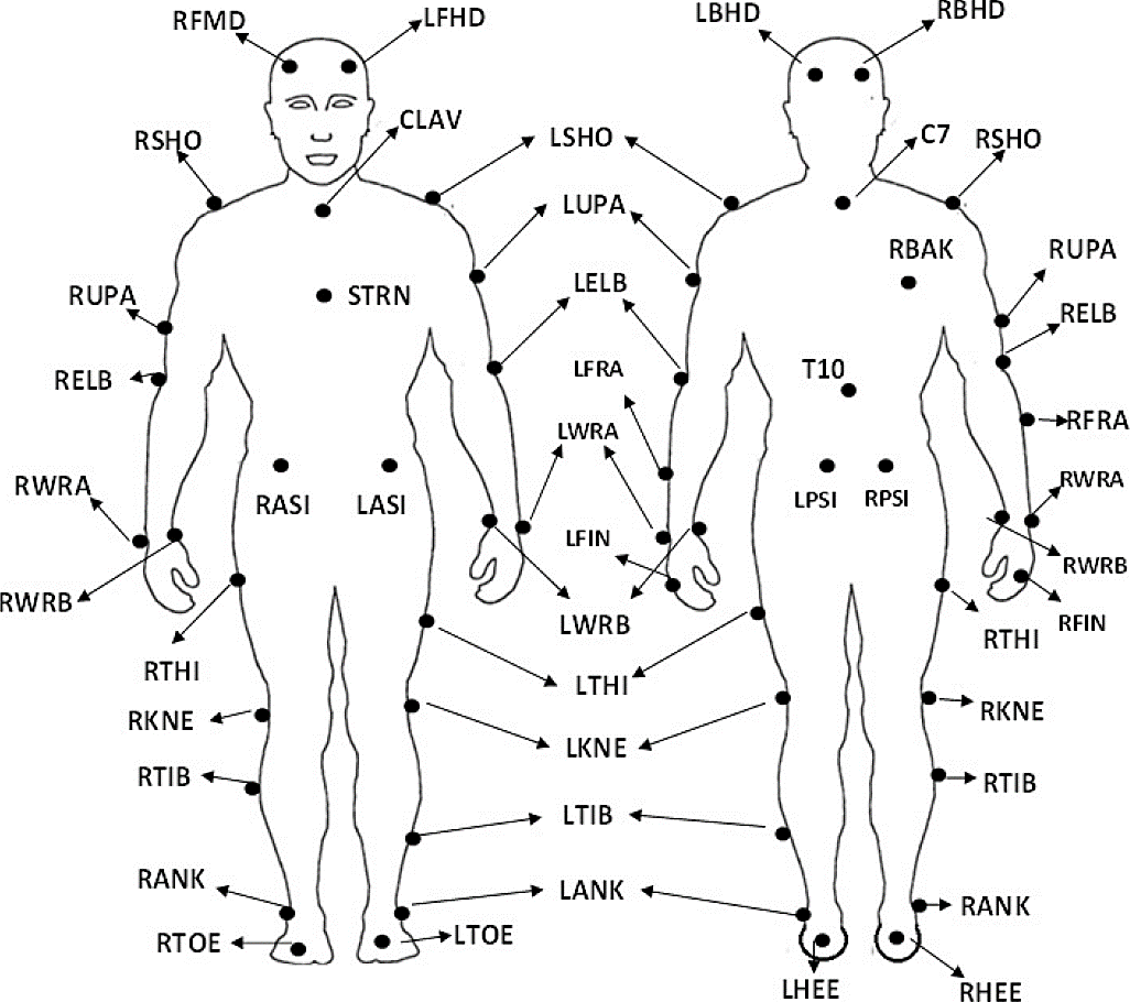
Figure 1 Skeleton model illustrating the placement of the passive reflective markers (anterior and posterior view).
Table 2 Plug-in-Gait model, markers placement.
| Upper Body | ||
| Head Markers | ||
| LFHD | Left front head | Located approximately over the left template |
| RFHD | Right front head | Located approximately over the right template |
| LBHD | Left-back head | Placed on the back of the head |
| RBHD | Right-back head | Placed on the back of the head |
| Torso Markers | ||
| C7 | 7th Cervical Vertebrae | Spinous process of the 7th cervical vertebrae |
| T10 | 10th Thoracic Vertebrae | Spinous process of the 10th thoracic vertebrae |
| CLAV | Clavicle | Jugular Notch where the clavicles meet the sternum |
| STRN | Sternum | Xiphoid process of the Sternum |
| RBAK | Right Back | Placed in the middle of the right scapula |
| Arms Markers | ||
| LSHO | Left shoulder marker | Placed on the Acromio-clavicular joint |
| LUPA | Left upper arm marker | Placed on the upper arm |
| LELB | Left elbow | Placed on the lateral epicondyle |
| LFRA | Left forearm marker | Placed on the lower arm |
| LWRA | Left wrist marker A | Left wrist bar thumb side |
| LWRB | Left wrist marker B | Left wrist bar pinkie side |
| LFIN | Left fingers | Placed on the dorsum of the hand |
| RSHO | Right shoulder marker | Placed on the Acromio-clavicular joint |
| RUPA | Right upper arm marker | On the lateral area in the lower third of the arm |
| RELB | Right elbow | On the lateral epicondyle |
| RFRA | Right forearm marker | On the lateral area in the upper third of the forearm |
| RWRA | Right wrist marker A | Next to the thumb on the wrist |
| RWRB | Right wrist marker B | Next to the pinky on the wrist |
| RFIN | Right fingers | Placed on the dorsum of the hand |
| Lower Body | ||
| Pelvis | ||
| LASI | Left ASIS | Placed directly over the left anterior superior iliac spine |
| RASI | Right ASIS | Placed directly over the right anterior superior iliac spine |
| LPSI | Left PSIS | Placed directly over the left posterior superior iliac spine |
| RPSI | Right PSIS | Placed directly over the right posterior superior iliac spine |
| Leg Markers | ||
| LKNE | Left knee | Placed on the lateral epicondyle of the left knee |
| LTHI | Left thigh | Placed over the lower lateral 1/3 surface of the thigh |
| LANK | Left ankle | Placed on the lateral malleolus along an imaginary line that passes through the transmalleolar axis. |
| LTIB | Left tibial wand marker | Similar to the thigh markers, these are placed over the lower 1/3 of the shank to determine the alignment of the ankle flexion axis |
| RKNE | Right knee | Placed on the lateral epicondyle of the right knee |
| RTHI | Right thigh | Placed on the upper lateral 1/3 surface of the thigh |
| RANK | Right ankle | Placed on the lateral malleolus along an imaginary line that passes through the transmalleolar axis. |
| RTIB | Right tibial wand marker | Placed on the upper 1/3 of the lateral surface of the stem |
| Foot Markers | ||
| LTOE | Left toe | Placed over the second metatarsal head, on the mid-foot side of the equinus break between fore-foot and mid-foot |
| LHEE | Left heel | Placed on the calcaneous at the same height above the plantar surface of the foot as the toe marker |
| RTOE | Right toe | Placed over the second metatarsal head, on the mid-foot side of the equinus break between fore-foot and mid-foot |
| RHEE | Right heel | Placed on the calcaneous at the same height above the plantar surface of the foot as the toe marker |
The three-dimensional motion capture system used contained eleven optoelectronics infrared cameras (Bonita B10) up to one megapixel (1024 x 1024) high resolution, which accurately captures up to 0. 5 mm for a 4 m x 4 m volume, with variable focal length, and speed of 250 frame rate (fps), and two video cameras (Bonita 720c) of a 1280 x 720 HD resolution, with an impressive 120 Hz fully synchronized frame rate. The cameras were distributed in the capture volume to measure all possible details in the athlete’s movement in 3D. In addition, the system includes a Giganet camera switch (POE) in an Ethernet network, a Vicon lock for analog signal observation, a host PC with Vicon Nexus 2 software, and two force platforms at 1000 Hz (AMTI, Waterdown, MA, USA). Figure 2 illustrates the setup of the capture system. In addition, all demographic and anthropometric dimensions of the participants were captured using the Nexus 2 software.

Figure 3 shows a general diagram of the Vicon Nexus 2 system. Before starting the test, it was necessary to calibrate the video and infrared cameras using the T-rod tool. The T-rod tool carries five LEDs, and this tool moves at the area work of 6 x 4m. Is was then necessary to place this at the origin point on the floor, according to the method described [25] in Figure 3(a). When the markers were placed, the athlete was notified to enter into a static position, as shown in Figure 3(b), to perform static capture. The system then detected the markers, as shown in Figure 3(c), and performed the reconstruction model, as shown in Figure 3(d). Once the reconstruction model was obtained, the athlete performed the movement, and the trajectories of the markers were labeled and filtered using a Butterworth low-pass filter with a frequency of 100Hz.

Description of the applied exercise
In this study, the reaction time (RT) is defined as the time between the sound emitted by the timing system and the time the foot of the athlete leaves the starting block. The wireless Brower Timing Systems TS-T17 was used to measure the RT. To avoid the risk of injury and achieve maximum performance, the athletes realized a standardized dynamic warm-up before the tests. To capture the movement of the athletes during the start of running three critical steps were performed. The first step is positioning the athlete at the starting block. Once the athlete is placed in the correct position, the athlete hears the first beep emitted by the Brower Timing Systems, which means being prepared. The second step is when a second beep is emitted, which indicates being ready for the athlete, and the third step is when a third beep is emitted and the start is executed, as shown in Figure 4. Once the start is executed, the RT is obtained, the first impact of the reaction force from the ground in three steps 1) moment of release of the foot from the block, 2) maximum extension of the leg behind and 3) the first contact of the foot with the ground. At that moment, the stride length and stride time were determined by the motion capture system.

Data processing
The 3D trajectories of the passive reflective markers were corrected using a low pass filter (100Hz Butterworth filter) and then imported into Matlab R2019b (The MathWorks, Inc).
Stride length
Stride length is the distance from the point where the toe leaves the starting block until it touches the ground again (after the swing phase). The TOE marker was used to calculate stride length using Equation (1)[26].
where L is the stride length.
Stride time
Stride time was calculated using the total number of frames and the elapsed time between frames. The square where the foot takes off is considered to be the square with which it first impacts the ground. In this case, the time between frames is 0.01 s. Therefore, it was calculated using Equation (2) as follows [26].
Where T is the stride time and NF is the number of frames.
Statistical analysis
R software was used for statistical analysis [27]. Descriptive statistics (mean ± SD) were determined for each variable. The Shapiro-Wilk test was applied because the sample size was less than fifty which complies with the normal principle (p> 0.05). In this sense, the parametric test was used, the Student's t-test was used for independent samples.
RESULTS AND DISCUSSION
Table 3 shows the results of the three variables evaluated in men. In this group, M1 obtained the shortest reaction time of 0.19 s and a stride length of 1.36 m with the right leg behind, while with the left leg behind, which is the leg that normally performs the start; he obtained the longest reaction time of 0.32s. Besides, the group of men obtained a mean of 0.276 with a standard deviation of 0.04 in reaction time using the left leg behind while a mean of 0.256 with a standard deviation of 0.05 in reaction time was obtained with the right leg behind. Similarly, Table 4 shows the results of the 3 variables evaluated in women. In this group, F1 obtained the best reaction time of 0.24s and a stride length of 1.11 m with the left leg behind, while with the left leg behind, which is not the leg that normally performs the start, she obtained the longest reaction time of 0.32s. In addition, the group of women obtained a mean of 0.28 with a standard deviation of 0.05 in reaction time using the left leg behind while a mean of 0.263 with a standard deviation of 0.03 in reaction time was obtained using the right leg behind. From Tables 3 and 4, it is observed that men performed better in reaction times, as well as greater stride length, than to women. Table 5 presents the descriptive statistics of each variable evaluated in men and women.
Table 3 Obtained results of the 3 evaluated variables for male.
| Participant | Reaction time (LLB) | Stride length (m) | Stride time (S) | Reaction time (RLB) | Stride right (m) | Stride time (S) |
|---|---|---|---|---|---|---|
| M1 | 0.32 | 1.357 | 0.34 | 0.19 | 1.369 | 0.36 |
| M1 | 0.32 | 1.120 | 0.34 | 0.19 | 1.130 | 0.36 |
| M1 | 0.35 | 1.070 | 0.37 | 0.20 | 1.140 | 0.38 |
| M2 | 0.28 | 0.950 | 0.28 | 0.30 | 1.074 | 0.30 |
| M2 | 0.30 | 1.020 | 0.32 | 0.32 | 1.070 | 0.30 |
| M2 | 0.28 | 0.950 | 0.29 | 0.31 | 1.100 | 0.34 |
| M3 | 0.23 | 1.227 | 0.31 | 0.28 | 1.264 | 0.28 |
| M3 | 0.23 | 1.120 | 0.30 | 0.28 | 1.020 | 0.27 |
| M3 | 0.25 | 1.100 | 0.30 | 0.30 | 1.120 | 0.30 |
Table 4 Obtained results of the 3 evaluated variables for female.
| Participant | Reaction time (LLB) | Stride length (m) | Stride time (S) | Reaction time (RLB) | Stride right (m) | Stride time (S) |
|---|---|---|---|---|---|---|
| F1 | 0.34 | 1.010 | 0.32 | 0.32 | 0.870 | 0.30 |
| F1 | 0.36 | 1.100 | 0.35 | 0.35 | 1.010 | 0.30 |
| F1 | 0.24 | 1.110 | 0.32 | 0.32 | 1.047 | 0.31 |
| F2 | 0.25 | 1.115 | 0.28 | 0.28 | 1.106 | 0.32 |
| F2 | 0.25 | 1.110 | 0.27 | 0.27 | 1.010 | 0.33 |
| F2 | 0.25 | 0.980 | 0.28 | 0.28 | 1.080 | 0.33 |
| F3 | 0.25 | 1.100 | 0.34 | 0.27 | 1.010 | 0.33 |
| F3 | 0.25 | 1.200 | 0.34 | 0.26 | 1.288 | 0.31 |
| F3 | 0.25 | 0.980 | 0.33 | 0.26 | 1.080 | 0.30 |
Table 5 Descriptive statistics for each variable evaluated.
| Variable | Participants | Mean | SD |
|---|---|---|---|
| Reaction Time (LLB) | Male | .28 | .046 |
| Female | .27 | ..036 | |
| Stride length (m) | Male | 1.10 | .112 |
| Female | 1.07 | .013 | |
| Stride time (S) | Male | .31 | .029 |
| Female | .31 | .032 | |
| Reaction time (RLB) | Male | .26 | .061 |
| Female | .29 | .035 | |
| Stride right (m) | Male | 1.14 | .066 |
| Female | 1.05 | .075 | |
| Stride time (S) | Male | 0.32 | 0.42 |
| Female | 0.31 | 0.12 |
The Student's t-test for independent samples indicates that Ho is acceptable, that is, there are no significant differences between men and women.
Table 6 shows the results obtained from the Student´s T-test, degrees of freedom, and p-values >0.05.
Table 6 Independent samples of Student´s t-test results.
| Variable | t | gl | p |
|---|---|---|---|
| Reaction Time (LLB) | .401 | 4 | .709 |
| Stride length (m) | .357 | 4 | .739 |
| Stride time (S) | .079 | 4 | .941 |
| Reaction time (RLB) | -.647 | 4 | .553 |
| Stride right (m) | 1.506 | 4 | .207 |
| Stride time (S) | .261 | 4 | .807 |
Figure 5 shows the left toe speed obtained for the male and female participants. It is observed that all participants reached their highest left toe speed between 13 and 20 s. It is also observed that male participants (M2) reached the highest speed of 7.5 m/s at 16s while the female participants (F3) reached the lowest speed of 7.1 m/s at 18 s.

Similarly, Figure 6 shows the right-toe speed obtained for male and female participants.

It is observed that the participants reached the highest speed on the right toe between 15 and 25 s. It is also observed that male (M2) reached the highest speed of 7.96 m/s at 16 s while the male (M1) reached the lowest speed of 6.9 m/s at 17 s. Besides, the female participant (F3) obtained the lowest speed of 6.9 m/s at 17 s while the participant (F1) obtained the maximum speed of 7.7 m/s at 18 s. From Figure 5 and Figure 6 it is observed that the best speeds are obtained on the right toe, which is in contrast with the common practice that indicates the left toe behind is the toe that participants normally perform the start.
In this study, the Helen-Hayes PGM for snatch output motion analysis was used to determine the reaction time, stride length, and stride time of both legs of the six athletes. The Vicon® Plug-in-Gait model (PGM) is one of the most widely used models for evaluating different kinematic and kinetic parameters of different motor or sports gestures [28] [29] [30]. Remi K. reported that stride length, determined when the rear leg moves forward in the frontal plane, ranges from 100 to 120 cm [31]. Considering this range, in the group of men, (M2) in two of the three repetitions of the left leg behind, obtained 0.950 m, while with the right leg behind, all values were within the range. In the group of women (F2) and (F3) with the left leg behind in the last repetition did not obtain (100-120cm). With respect to the right leg behind (F1) was the only one, where the value was not within the reported Remi K. range.
Slawinski J et al. used an optoelectronic motion analysis system containing 12 digital cameras (250 Hz) to characterize four repetitions of sprint snatches of six elite sprinters and six well-trained sprinters. The average RT of the elite sprinters was 0.151 ± 0.016 s, and that of the six well-trained sprinters was 0.158 ± 0.033 s [32].
In comparison with the results obtained by Slawinski J et al., the sprinters characterized in this study obtained a higher performance. For three repetitions, they obtained an average RT of 0.2767 ± 0.045 s using the right leg back, and an average RT of 0.2778 ± 0.042 s using the left leg back. There is a difference in their RT averages, however, it is not significant considering that they are elite and well-trained sprinters. Despite the small sample size, this study aimed to characterize the snatch output, using a motion analysis system, and make pertinent corrections to obtain the RT, stride length, and stride time for each leg of the athletes.
CONCLUSIONS
In this study, a 3D motion capture system was used to characterize the kinematic parameters that influence the execution of a low-sprint start in high-performance athletes. The Vicon 3D capture system is precise and highly accurate for performing biomechanical evaluations of body motor gestures. The high reliability obtained data is not just empirical, but also numerical ones. Although a small number of samples were used, the results provide evidence of the effectiveness of using 3D capture technology to quantify kinematic parameters of low-sprint starts. The characterized kinematic parameters can be used to identify improvements for the athlete, such as errors in the execution of the start to avoid possible injuries in the athlete. The Student's t-test for independent samples indicates that Ho is accepted, and there are no significant differences between men and women (p-value > 0.05), for all the variables. However, with this technology, it was found that three athletes obtained better times with the nondominant leg. In addition, this study illustrated the importance of coaches and the athletes understanding of the use of 3D motion capture system technology and its scope. This technology can be part of their evaluations to avoid possible injuries, detect errors in the execution of precision movements, and improve performance.

