


Servicios Personalizados
Revista
Articulo
 texto en
  Inglés (pdf)
 Artículo en XML
 Referencias del artículo
 Enviar artículo por email
Indicadores
-
 Citado por SciELO
 -
 Accesos
Links relacionados
-
 Similares en
SciELO
Similares en
SciELO
Compartir

 Permalink
Revista mexicana de fitopatología
versión On-line ISSN 2007-8080versión impresa ISSN 0185-3309
Rev. mex. fitopatol vol.36 no.2 Texcoco may./ago. 2018
https://doi.org/10.18781/r.mex.fit.1711-5
Scientific articles
Response of ten yellow mango cultivars to powdery mildew (Erysiphe quercicola) damage in Mexico
1Instituto de Fitosanidad, Colegio de Postgraduados, km 36.5, Carretera México-Texcoco, Montecillo, Texcoco, Estado de México, CP. 56230, México
2Unidad Académica de Ciencias Agropecuarias y Ambientales, Universidad Autónoma de Guerrero, Carretera Iguala-Tuxpan km 2.5, CP. 40101, Iguala, Guerrero, México
3Laboratorio de Fitopatología, Instituto Politécnico Nacional-CIIDIR, Calle Hornos 1003, Colonia Noche Buena, Municipio Santa Cruz Xoxocotlán, CP. 71230, Oaxaca, Oaxaca, México.
Mango powdery mildew (Erysiphe quercicola) causes up to 90% production losses, so it is necessary to estimate the tolerance to this pathogen of the new germplasm introduced or recently generated to increase the export potential of Mexico. The objective of this study was to determine the response to powdery mildew damage by means of an optimized inoculation technique to induce the disease in attached leaves of 10 new yellow mango cultivars for Mexico. Two inoculation methods were evaluated. The best was to spray conidia at 4.6 x 10⁵ spores mL-1 on the adaxial and abaxial leaves surfaces, at ± 300-450 lux and suspended in polysorbate 20 + surfactant based on ethoxilate alcohols at 2%. The inoculation test showed that the cv. Alphonso was moderately tolerant and Neelum and Fairchild were slightly tolerant. In contrast, Nam Doc Mai, Rosigold, Ataulfo Zafiro, Cotaxtla and Kesar were susceptible and Mallika and Ivory were highly susceptible. The most tolerant cultivars had lower values of incidence, maximum severity, area under the disease progress curve, apparent infection rate and conidia density per cm2 of damaged leaf area (LSD, P≤0.05).
Key words: leaves; susceptibility; germplasm; severity
La cenicilla del mango (Erysiphe quercicola) causa pérdidas de producción de hasta 90%, por lo que es necesario estimar la tolerancia a este patógeno en el nuevo germoplasma introducido o generado recientemente para incrementar el potencial de exportación de México. El objetivo de este estudio fue determinar la respuesta al daño por cenicilla mediante una técnica de inoculación optimizada para inducir la enfermedad en hojas adheridas de 10 cultivares de mango amarillo nuevos para México. Se evaluaron dos métodos de inoculación. El mejor fue por aspersión de conidios a una concentración de 4.6 × 10⁵ esporas mL-1, en las superficies adaxial y abaxial de las hojas, a ± 300 - 450 lux y suspendidos en polisorbato 20 + surfactante a base de alcoholes etoxilados al 2%. La prueba de inoculación mostró que el cv. Alphonso fue moderadamente tolerante y Neelum y Fairchild ligeramente tolerantes. En contraste, Nam Doc Mai, Rosigold, Ataúlfo Zafiro, Cotaxtla y Kesar fueron susceptibles y Mallika e Ivory altamente susceptibles. Los cultivares más tolerantes presentaron valores menores de incidencia, severidad máxima, área bajo la curva del progreso de la enfermedad, tasa de infección aparente y densidad de conidios por cm2 de área foliar dañada.
Palabras clave: hojas; susceptibilidad; germoplasma; severidad
Mexico is the seventh largest mango (Mangifera indica L.) producing country in the world, with an annual volume of more than 1.8 Mt, and is the main exporter, accounting for 24% of global mango exports (FAO, 2016). Between 1992 and 2006, Mexico lost approximately 27.6% of its competitiveness as a mango exporter to the United States due to the increased commercial share of India, Thailand, Peru, Brazil and Ecuador (Hernández and Martínez, 2009). This growing problem is associated with the limited supply of yellow mangoes on the international market, since Mexico trades only Ataulfo cv. fruits, whose productivity is low mainly due to diseases, parthenocarpic fruits, genetic mixtures, as well as marked seasonality and alternate bearing (Villegas and Mora, 2011). To mitigate such limitations, in 2011 and 2012, the Colegio de Postgraduados introduced eight new yellow mango cultivars from Florida, USA, that have export potential. Also, the Instituto Nacional de Investigaciones Forestales Agrícolas y Pecuarias (INIFAP) registered two new local cultivars of the Ataulfo clone in 2009 (A. Diamante) and 2012 (A. Zafiro).
Mango powdery mildew caused by Erysiphe quercicola (anamorph: Pseudoidium anacardii) (Braun and Cook, 2012; Félix et al., 2013; Tam, 2017) is one of the most important diseases that affects mango because of its high level of severity, endemism and cosmopolitan distribution (Raheel et al., 2008) that cause 80-90% of the mango production losses (Gupta, 1989b; Shoeman et al., 1995; Nasir et al., 2014). In mango exporting states such as Michoacan, it may affect 60% of commercial trees, the equivalent of 30,000 to 50,000 tons of fruit (Arias et al., 2004); in Sinaloa, the disease causes 70% losses during the flowering stage (Félix et al., 2017). The fungus damages leaves, flowers and young fruits. The infected tissue is covered with white powder due to mycelial growth and sporulation. The first lesions on leaves are reddish in color, but at advanced stages, the fungus deforms the leaf lamina and produces abundant sporulation, necrosis and severe defoliation. In the reproductive tissue, it causes fall of flowers, extensive necrosis of inflorescences and abortion of young fruits (Sinha et al., 2001; 2002; Nasir et al., 2014).
The effect of foliar infections on the frequency and severity of epidemics on inflorescences is widely documented, because the fungus survives in the form of mycelium in buds and leaves during the growth season, or from previous years, when environmental conditions do not favor infection, or when there is no reproductive tissue (Schoeman et al., 1995; Misra, 2001; Nasir et al., 2014). When there are no flowers, early infection on young leaves perpetuates inoculum availability and favors the beginning of the infection on panicles (Munshi et al., 1988; Misra et al., 2012). Also, during flowering, sporulation on leaves (with conidia attached to conidiophores) increases inoculum development and preserves its viability from 10 to 19 (Gupta, 1989a; Nelson, 2008) or 40 more days (Khalid et al., 2000) compared with free conidia deposited on other susceptible host organs, which favors longer lasting and more intense epidemics, given that conidia require only from 5 to 7 h to germinate, and from 5 to 8 days to induce the first symptoms (Misra, 2001). In turn, the multiple vegetative and floral flows that are simultaneously formed during the same growth season also stimulate massive inoculum increase and dispersion, and the frequency of epidemics (Misra, 2001; Guillén et al., 2003). In Tecpan de Galeana, Mexico, Pérez et al. (2017a) reported three periods of vegetative epidemic development with the same number of growth flows during a Manililla cv. crop cycle. Although the final incidence was lower (1.65-1.80%) than that registered on inflorescences (6.15-6.90%), the authors showed the relevance of plant infections for inducing epidemics in reproductive tissue.
Since young vegetative flows in highly susceptible cultivars are severely affected by E. quercicola, using tolerant cultivars is the best management option for this pathogen (Nelson, 2008). In current mango breeding programs, varietal tolerance to pathogens is estimated based on natural infection recorded in the field. Using this approach, Galli et al. (2009) and (2012) characterized 17 cultivars for tolerance to powdery mildew and anthracnose using five severity classes in Pindorama, Brazil. Similarly, Nelson (2008) found that Alphonso, Zill and Kent cvs. were highly susceptible; Rosa and Haden moderately susceptible, and Sensation and Tommy Atkins, slightly susceptible to O. mangiferae in Hawaii, USA. Other similar studies to estimate powdery mildew tolerance were conducted by Palti et al. (1974), (1976), Peterson (1984), Akhtar et al. (1999), Galli et al. (2008), Nofal and Haggag (2006) and Naqvi et al. (2014). However, these methodological approaches have serious drawbacks, since the level of incidence and severity of powdery mildew in the field depends on factors such as seasonality, distribution and density of inoculum, availability of susceptible tissue and environmental conditions (Nasir et al., 2014) or inductive management conditions such as frequency and intensity of irrigation (Guillén et al., 2003).
To prevent infection escape and ensure the pathogen’s optimal parasitic expression, it is necessary to use standardized techniques that efficiently reproduce the disease. Given that E. quercicola is an obligate parasite, most of the studies conducted to document its distribution, importance, biology, epidemiology, management and genetic resistance have been based on naturally infected plants (Nasir et al., 2014), and for this reason there is little information about the artificial induction of mango powdery mildew. Adikaram et al. (2002) reported that by depositing O. mangiferae spores on Pedilanthus tithymaloides leaves using a paint brush, they produced successful infections and promoted the expected synthesis of new anthocyanins and chlorophyll degradation. In previous studies conducted on mango leaves, the highest levels of O. mangiferae infection were observed in leaves of 8-12 day-old that were inoculated at dusk (300-350 Lux) at 4.6 x 10⁵ spores mL-1 (Pérez, 2017).
Based on the different susceptibility to powdery mildew that all mango cultivars shows (Palti et al., 1974; Nasir et al ., 2014), the great influence of E. quercicola foliar infection on the disease cycle and epidemic development (Misra, 2001; Pérez et al., 2017a), as well as the need to promote adequate selection and agronomic use of the new cultivars introduced or developed in Mexico, according to their sanitary reaction to the pathogen, this study was conducted to determine the response to E. quercicola at the vegetative stage of ten new yellow mango cultivars that have export potential using nursery plants and an optimized inoculation technique.
MATERIALS AND METHODS
Study area
The study was conducted in 2015 and 2016 at the laboratory and greenhouse of the Postgrado de la Unidad Académica de Ciencias Agropecuarias of the Universidad Autónoma de Guerrero, Iguala, Guerrero, México (18°20’39.505” N; 99°29’52.796”O; 738 masl). For the experiment, 18-month old mango plants grafted with Nam Doc Mai, Rosigold, Neelum, Mallika, Cotaxtla, Kesar, Fairchild, Alphonso, Ivory, Ataulfo Zafiro and Manililla cultivars were placed in 18 L plastic pots in a nursery covered with 600-gauge transparent plastic and polypropylene mesh-shade at 50%. The plants were fertilized every five days using 1.0 g of a 100-60-60 (NPK) formulation and watered to the point of saturation every other day. Readings of temperature, relative humidity and photoperiod in the nursery were recorded every two hours using a datalogger Hobo® U12.
Experiment inoculum
Leaves and inflorescences colonized by E. quercicola were collected from 20 year-old Manililla cv. mango orchards in Guerrero, Mexico, and placed in paper bags for temporary conservation and transportation. Tissue was dehydrated for seven days under laboratory conditions (470 ± 10 lux, 24-26°C and 30% HR). One portion of dehydrated tissue was kept in paper bags as inoculum reservoir. From the remaining tissue, conidia were recovered using a fine hair paint brush (Rodin® No. 5). Then, they were suspended in glass tubes containing 30 mL of sterile distilled water and kept at 24 ± 2ºC for later use as an inoculum source.
Inoculation treatments
Vegetative sprouts with initial disease development were identified on Manililla cv. plants highly susceptible to E. quercicola and with similar growth and vigor characteristics. When leaves were 8-12 days-old, they were disinfected with NaClO (0.5%) for 30 s using a manual sprayer, rinsed three times with distilled water and dried for 10 min. Three inoculation treatments were evaluated: T1) spraying conidia suspended in polysorbate 20 (Tween 20®) at 5% + surfactant based on ethoxylated alcohols (Inex-A®) at 2%; T2) spraying a conidia mixture containing calcium caseinate (Rennet Casein® 90 mesh, Charotar Casein Company, IndiaMartTM); and T3) spraying sterile distilled water (control). A base mixture was prepared from each treatment. For T1, we used 1 g of inoculum (mycelium, conidia and conidiophores) in 2 mL-1 of the solution; for T2, 1 g of inoculum in 2 g of casein, and then both were adjusted to a concentration of 4.6 × 10⁵ spores mL-1 in a Neubauer chamber. Inoculation was done at 18:00 h (± 300-450 lux) on the adaxial and abaxial areas of disinfested leaves that remained attached to the plant. Fifty (50) leaves per treatment were inoculated (5 leaves per plant; 10 plants per treatment). The experiment was replicated three times in 2015.
Varietal tolerance
For this experiment, 18 month-old mango plants grafted with Nam Doc Mai, Rosigold, Neelum, Mallika, Cotaxtla, Kesar, Fairchild, Ivory, Alphonso and Ataulfo Zafiro cultivars were used. To replicate the symptoms, the most effective inoculation treatment from previous experiments, adjusted to 4.6 × 10⁵ spores mL-1, was applied to the adaxial and abaxial areas of leaves at a range of ± 300-450 lux. Fifty (50) 8-12 day-old leaves attached to the plant were inoculated per cultivar (5 leaves per plant; 10 plants per cultivar). The experiment was replicated three times a year in 2015 and 2016.
Variables and statistical analysis
The development of lesions on leaves, with and without sporulation, was observed every 24 h in both experiments (inoculation and varietal tolerance treatments), and this information was used to determine the incidence (Inc) and incubation period (Pi) to maximum severity (Y max ). Based on the progressive Y max values, the apparent infection rate (b -1 ) was calculated using the lineal regression model (Achicanoy, 2000), as well as the area under the disease progress curve (AUDPC) using the trapezoidal integration method (Campbell and Benson, 1994). Also, in the varietal tolerance experiment, the conidia concentration (Cc) per cm2 of infected leaf area was quantified according to Sholberg et al. (2001), and the tolerance was estimated using a five-class scale based on the symptom pattern and degree of severity observed in commercial mango orchards located in Iguala and Huamuxtitlan, Guerrero, Mexico, as follows: 1) healthy leaf or with lesions ≤ 1.0 mm without mycelial colonization (resistant), 2) reddish lesions with little mycelium and 5.0 mm maximum diameter (moderately tolerant), 3) reddish to dark lesions 5-10 mm in diameter, and white mycelium (slightly tolerant), 4) reddish to dark lesions 10-15 mm in diameter, and abundant mycelium (susceptible), 5) coalescing dark lesions >15 mm in diameter and abundant mycelium (highly susceptible) (Figure 1). A completely randomized design with 10 replications was used in each experiment. Calculations of b -1 , AUDPC, variance analyses (Proc GLM) and mean comparisons (LSD, 5%) for Pi, Inc, Ymax, Cc, AUDPC and b -1 were performed using the SAS software v.9.4 (SAS Institute Inc., 2016).

RESULTS
Inoculation treatments
The conidia suspension in polysorbate 20 + surfactant based on ethoxylated alcohols, sprayed at a concentration of 4.6 × 10⁵ spore mL-1 on both surfaces of 8-12 day-old leaves and incubated at ± 300-450 lux induced higher incidence, severity and AUDPC values with the shortest incubation period in Manila cv., which is highly susceptible to powdery mildew (Table 1) (LSD, P≤0.05).
Table 1 Incubation period, incidence and severity of powdery mildew caused by E. quercicola in 8-12 day-old mango (Mangifera indica L.) leaves Manililla cv. infected using two inoculation methods. Iguala, Guerrero, Mexico. 2015.
| Tratamientow | Parámetros fitosanitariosx | ||||
| Pi (ddi) | Inc (%) | Y max | ABCPE | b-1 | |
| T1 | 5.0z | 90.1 | 4.8 | 107 | 0.4025 |
| T2 | 7.2 | 72.3 | 3.5 | 43.7 | 0.3875 |
| T3 | 0 | 0 | 0 | 0 | 0 |
| DMSy | 1.8 | 17.5 | 0.9 | 17.6 | 0.0973 |
| R2 | 88 | 90 | 90 | 91 | 89 |
wT1= Polisorbato 20 + surfactante a base de alcoholes detoxilados, T2= caseína, T3= agua destilada estéril (control). xPi= periodo de incubación, Inc= incidencia, Y max = severidad máxima, ABCPE= área bajo la curva del progreso de la enfermedad, b -1 = tasa de infección aparente; yDMS= Diferencia mínima significativa de acuerdo con la prueba de Fischer (P≤0.05); zValores promedio de 50 hojas por tratamiento en tres réplicas experimentales / wT1= Polysorbate 20 + surfactant based on ethoxylated alcohols, T2= casein, T3= sterile distilled water (control). xPi= incubation period, days after inoculation, Inc= incidence, Y max = maximum severity, ABCPE = area under the disease progress curve (AUDPC), b -1 = apparent infection rate; yDMS= Least Significant Difference (LSD), according to Fischer’s Test (P≤0.05); zAverage values of 50 leaves per treatment in three experiment replications.
Varietal tolerance
Based on Y max , AUDPC and b -1 parameters, the infected cultivars by E. quercicola were grouped into four levels of susceptibility. Alphonso was moderately tolerant (MT) followed by Neelum and Fairchild, which were slightly tolerant (LT). Nam Doc Mai, Rosigold, Ataulfo Zafiro, Cotaxtla and Kesar were susceptible (S), and Ivory and Mallika, highly susceptible (AS) (Figure 2). Although none of the cultivars was resistant, the least susceptible ones (MT and LT) consistently showed lower incidence, Y max , AUDPC, b -1 and Cc values. Pi ranged between 4 and 6 days, a period that did not show a pattern consistent with the level of susceptibility of the evaluated cultivars. The most susceptible cultivars (S and AS) showed 20.6-22.0% higher incidence, 33-38% higher Y max , 95.5-140% higher AUDPC, 183-223% higher development rates (b -1 ) and conidial concentration per damaged area (cm2) that was 226-300% higher compared to cultivars from the MT and LT groups. In this study, the polyembryonic cultivars Nam Doc Mai, Kesar, Cotaxtla, Ivory and Ataulfo Zafiro were more susceptible than the monoembryonic types (Table 2) (LSD, P ≤ 0.05).
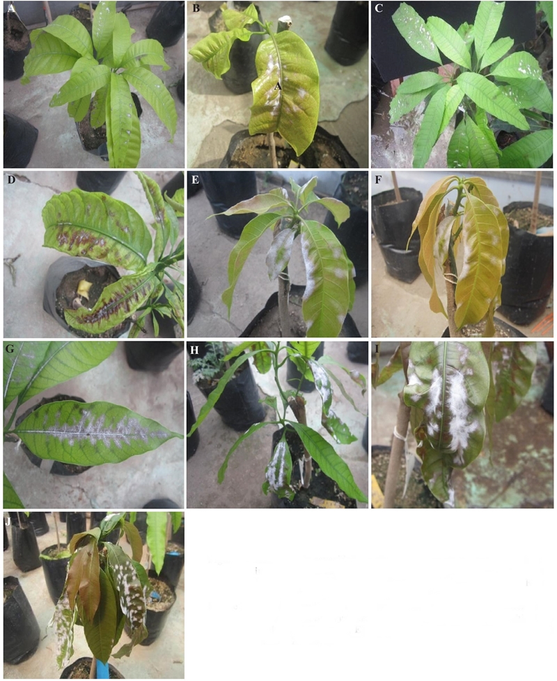
Figure 2 Severity reaction of ten yellow mango cultivars to experimental foliar infection by Erysiphe quercicola. A) Alphonso, B) Fairchield, C) Neelum, D) Rosigold, E) Nam Doc Mai, F) Kesar, G) Cotaxtla, H) Ataulfo Zafiro, I) Mallika, J) Ivory.
Table 2 Tolerance of 10 mango cultivars inoculated with Erysiphe quercicola in 8-12 day-old leaves based on some phytosanitary parameters in Iguala, Guerrero, Mexico, during the 2015 and 2016 cycles.
| Parámetros fitosanitariosw | ||||||||
| Cultivar | Pi (ddi) | Inc (%) | Y max | Cc (104 /cm 2) | ABCPE | b -1 | Reacción | Semillax |
| Alphonso | 6.3z | 70.1 | 2.7 | 1213.5 | 38.1 | 0.121 | MT | M |
| Fairchild | 4.7 | 75.3 | 3.0 | 1305.1 | 43.7 | 0.132 | LT | M |
| Neelum | 4.6 | 83.1 | 3.4 | 3685.2 | 50.2 | 0.265 | LT | M |
| Rosigold | 6.1 | 86.2 | 3.6 | 3961.4 | 74.5 | 0.347 | S | M |
| Nam Doc Mai | 5.3 | 84.0 | 3.8 | 4283.6 | 83.4 | 0.305 | S | P |
| Kesar | 4.2 | 91.0 | 4.1 | 4600.3 | 90.3 | 0.368 | S | P |
| A. Zafiro | 4.8 | 90.8 | 4.3 | 5221.4 | 96.1 | 0.378 | S | P |
| Cotaxtla | 4.9 | 91.2 | 4.4 | 5183.6 | 97.5 | 0.371 | S | P |
| Mallika | 5.1 | 90.5 | 4.7 | 5981.7 | 100.6 | 0.389 | AS | M |
| Ivory | 4.6 | 93.3 | 4.7 | 6321.6 | 105.8 | 0.423 | AS | P |
| DMSy | 1.1 | 9.3 | 0.9 | 1115.8 | 17.7 | 0.101 | - | - |
| R2 | 91 | 89 | 90 | 88 | 89 | 87 | - | - |
wPi= período de incubación, Inc= incidencia, Sev= severidad, Cc= concentración de conidios por área dañada, Y max = severidad máxima, ABCPE= área bajo la curva del progreso de la enfermedad, b -1 = tasa de infección aparente; xM= monoembriónica, P= poliembriónica; yDMS=Diferencia mínima significativa de acuerdo con la prueba de Fischer (P≤0.05); zMedia de 50 hojas por tratamiento en seis réplicas experimentales / wPi= incubation period, days after inoculation, Inc= incidence, Sev= severity, Cc= conidia concentration per damaged area, Y max = maximum severity, ABCPE = area under the disease progress curve (AUDPC), b -1 = apparent infection rate; xM= monoembryonic, P= polyembrionic; yDMS= Least Significant Difference (LSD), according to Fischer’s Test (P≤0.05); zaverage of 50 leaves per treatment in six experiment replications.
DISCUSSION
Spraying conidial inoculum of E. quercicola suspended in a mixture of polysorbate 20 (5%) + surfactant based on ethoxylated alcohols (2%) was more effective than casein to induce the disease with a higher level of incidence (90.1%), severity (Ymax = 4.8, AUDPC = 107) and speed (Pi = 5 ddi) in the Manilla cv. (Table 1). Since it proved to be more effective, this inoculation method was used to evaluate the response of cultivars to infection in this study. No studies were found on the effect of polysorbate 20 on E. quercicola conidial germination. However, because of its surfactant, adherent and dispersant properties, polysorbate 20 has been used in some studies to prevent spore agglutination and enable uniform spore dispersion. For example, Jansen et al. (2005) achieved 100% successful infections by inoculating wheat caryopses with Fusarium graminearum at a concentration of 1500 macroconidia mL-1 suspended in polysorbate 20. Similarly, Frias et al. (1995) favored germination and obtained from 97 to 100% infection by inoculating cacao (Teobroma cacao) seedlings with Crinipellis perniciosa basidiospores suspended in a polysorbate 20 solution containing 3% glycerol. Similarly, Jia et al. (2003) observed that polysorbate 20 (0.25%) combined with 16 and 18 h light-darkness periods increased the levels of expected spores adherence and maximum severity of Magnaporthe grisea inoculated on rice (Oryza sativa) leaves.
In this study, we evaluated the reaction of 10 mango cultivars inoculated with E. quercicola using an optimized technique that included the control of inoculum density and effective deposition on foliar tissue, moisturizing additives to prevent conidia dehydration, as well as susceptible host phenology stages and optimal incubation conditions. The lack of this kind of studies limits the precise estimation of varietal tolerance to E. quercicola, since evaluating tolerance based on natural epidemics, as it is usually done (Akthar et al., 1999; Galli et al., 2008; Nelson, 2008), may lead to wrong conclusions because the occurrence and intensity will be affected by the presence, density and variable distribution of the inoculum, heterogeneous environmental conditions and the availability of susceptible tissue, among other factors (Shoeman et al., 1995; Nasir et al., 2014).
Our results indicated that all the cultivars were susceptible to E. quercicola and grouped into four susceptibility levels as reported by Naqvi et al. (2014), who tested 25 mango cultivars and found that although none of them was resistant, they exhibited levels of tolerance. In our study, cv. Alphonso showed the highest tolerance to E. quercicola, in contrast to Nofal and Haggag (2006), who indicated that this cultivar was highly susceptible. However, Neelum and Fairchild showed slight tolerance, as reported by Nasir et al. (2014). The most susceptible cultivars were Mallika and Ivory, and this result coincides with that obtained by Palti et al. (1974) and Nofal and Haggag (2006). Also, the present study reports for the first time the degree of vegetative susceptibility to E. quercicola in new mango cultivars for Mexico (Nam Doc Mai, Rosigold, Ataulfo Zafiro and Kesar), which have export potential. It is important to consider that the cultivars with the lowest susceptibility consistently showed lower incidence, Y max , accumulated severity (AUDPC), rate of disease development and sporulation density, and that the ranges of magnitude of those values were in agreement with each susceptibility group (MT, LT, S and AS) (Table 2).
It is important to note that the previously cited tolerance comparisons were based on natural epidemics (Galli et al., 2008, 2009; Naqvi et al., 2014), in which conditions to promote the maximum expression of the pathogen virulence and disease development were not controlled, so the level of severity may have been underestimated or overestimated. Nasir et al., (2014) documented the differentiated virulence of O. mangiferae in terms of the level of environmental humidity and length of the driest environmental periods. For this reason, it is important to use effective and standardized methods to reproduce the disease under controlled conditions (laboratory, glass houses or other protected spaces), as well as integrate the greater number of phytosanitary variables and use severity scales based on the symptomatic pattern and maximum severity degree observed in commercial orchards (Figure 1) located in particular mango-producing regions to estimate the varietal tolerance more accurately.
All the polyembryonic cultivars (Nam Doc Mai, Kesar, Cotaxtla, Ivory and Ataulfo Zafiro) showed greater susceptibility when they grouped as S and AS. However, since monoembryonic cultivars (Rosigold and Mallika) were also grouped into those categories (Table 2), this relationship must be verified when doing future comparative studies on a greater number of cultivars of each embryonic type. It is important to provide evidence of the participation of this factor, since it suggests that polyembryony in mango promotes susceptibility to foliar diseases because it causes peroxidase activity and ethylene synthesis to decrease (Campbell, 1961) in contrast to soil pathogens, where the resistance increases (Pinto et al., 2002).
Powdery mildew develops in a wide temperature range (10-31 °C) (Ploetz and Freeman, 2009), although it is more severe in dry and cold environments (50-70% RH, ≈ 20-22°C) (Gupta, 1989b; Schoeman et al., 1995). Conidia can germinate in a range from 9 to 32°C (being the optimum 23°C) with relative humidity as low as 20%, so disease development does not usually depend on environmental moisture (Ploetz and Freeman, 2009). Mango-producing areas in the Mexico’s Pacific basin have climatic gradients (dry, humid and subhumid tropics) with different inductivity to powdery mildew. For example, in dry tropical regions such as the Apatzingan Valley (Michoacan, Mexico), powdery mildew shows high levels of severity, damages leaves and reproductive tissue, and causes up to 80% of fruits to fall. In this region, Arias et al. (2004) classified as high-risk areas for this disease those at altitudes higher than 550 m where night temperatures can reach <17 °C. In agreement with this assumption, Pérez et al. (2017b) reported the development of low-severity epidemics (2.98%) and only on inflorescences in orchards located in Arcelia, Guerrero (dry tropics), a municipality located at 330 masl, with ≥ 30 °C temperature and ≤ 60% RH during the study. In contrast, in subhumid tropical areas such as Tecpan de Galeana (Guerrero, Mexico), the recurrent presence of cold sea winds favors the appearance of multiple vegetative and floral flows that extend the phenological stages of higher susceptibility, and promote multiple powdery mildew epidemics (Pérez et al., 2017a) of varying intensity. In view of the above, this study supports the selection of agroecological regions that are more adequate for growing new yellow mango cultivars for the export market. For example, cultivars in high demand in the US and Canadian markets, such as Nam Doc Mai, Alphonso and Kesar, will have a better productive performance and higher profitability in regions where E. quercicola shows less parasitic aptitude and the financial costs for phytosanitary maintenance will be lower. However, the farmer should consider factors such as the susceptibility of the new cultivars to other important pathogens and their agronomic adaptation to weather and soil types when making his decision.
Except for Cotaxtla, the other cultivars evaluated in this study were recently established in experimental orchards in Guerrero, Oaxaca and Chiapas, Mexico, so they are at a young stage. Once they mature, we will continue our studies on varietal response to E. quercicola infection in reproductive tissues.
CONCLUSIONS
In this study, it was determined that the best method for inoculating E. quercicola in mango was to spray conidia at 4.6 × 10⁵ spore mL-1 on both leaf areas, at dusk and suspended in polysorbate 20 + surfactant based on 2% ethoxylated alcohols to favor its dispersion and prevent dehydration. Although the 10 mango cultivars evaluated in this study were susceptible to E. quercicola, Alphonso, Neelum and Fairchield were the most tolerant to foliar infection, while Mallika and Ivory were the most susceptible.
ACKNOWLEDGMENTS
The authors wish to thank to the authorities of the Postgrado de la Unidad Académica de Ciencias Agropecuarias, Universidad Autónoma de Guerrero for the facilities provided to carry out this work.
REFERENCES
Achicanoy LH. 2000. Descripción cuantitativa de epidemias de plantas. Revista de la Facultad Nacional de Agricultura de Medellín 53(1):941-968. Disponible en línea: http://www.bdigital.unal.edu.co/26473/1/24069-84222-1-PB.pdf [ Links ]
Adikaram NKB, Mlewa G and Weerahewa D. 2002. Changes in pigment composition, acid metabolism, etc. in Pedilanthus tithymaloides leaf following powdery mildew infection. Journal of the National Science Foundation of Sri Lanka 30:1-11. DOI: 10.4038/jnsfsr.v30i1-2.2556 [ Links ]
Akhtar KP, Khan MA, Kazmi MR, Hussain RI and Fatima B. 1999. Preventive control of powdery mildew disease of mango. Agricultural Sciences 4(1):23-28. Disponible en línea: https://www.researchgate.net/publication/289519342_Preventive_control_of_powdery_mildew_of_mango [ Links ]
Arias SJF, Espinosa AJ, Rico PHR, Miranda SMA y Chávez CX. 2004. La cenicilla Oidium mangiferae Berthet del mango en Michoacán. INIFAP, CIRPAC. Campo Experimental Valle de Apatzingán. Folleto Técnico Núm. 1. Apatzingán, Michoacán, México. Disponible en línea: http://biblioteca.inifap.gob.mx:8080/jspui/bitstream/handle/123456789/1288/cenicilla_1288.pdf?sequence=1 [ Links ]
Braun U and Cook RTA. 2012. Taxonomic manual of the Erysiphales (Powdery Mildews). CBS-KNAW Fungal Biodiversity Centre, Utrecht, The Netherlands. 707 p. Disponible en línea: https://www.nhbs.com/taxonomic-manual-of-the-erysiphales-powdery-mildews-book [ Links ]
Campbell CW. 1961. Comparison of yield of poliembrionic and monoembrionic mangos. Proceedings of the Florida State Horticultural Society 74: 363-365. Disponible en línea: http://fshs.org/proceedings-o/1961-vol-74/363-365%20(CAMPBELL).pdf [ Links ]
Campbell CL and Benson DM. 1994. Spatial aspects of the development of root disease epidemics. Pp. 195-243. In: Campbell CL and Benson DM. (eds.). Epidemiology and management of root diseases. Berlin: Springer-Verlag. 339p. https://doi.org/10.1007/978-3-642-85063-9_7 [ Links ]
FAO. 2016. Organización de las Naciones Unidas para la Alimentación y la Agricultura. Disponible en línea: http://faostat.fao.org/DesktopDefault.aspx?PageID=339&lang=es [ Links ]
Félix GR, Herrera RG, Martinez VC, Longoria ERM, Maldonado MIE, Quiroz FFR, Martinez AJC, Garcia PLM and Espinoza MS. 2013. First report of powdery mildew (Pseudoidium anacardii) of mango trees in Sinaloa, Mexico. Plant Disease 97(7):994. http://dx.doi.org/10.1094/PDIS-11-12-1014-PDN [ Links ]
Félix GR, Maldonado MIE, Beltran PH, Apodaca SMA, Espinoza MS, Martínez VMC, Longoria ERM and Olivas PNG. 2017. Powdery mildews in agricultural crops of Sinaloa: Current status on their identification and future research lines. Revista Mexicana de Fitopatología 35:106-129. DOI: 10.18781/R.MEX.FIT.1607-4 [ Links ]
Frias GA, Purdy LH and Schmidt RA. 1995. An inoculation method for evaluating resistance of cacao to Crinipellis perniciosa. Plant Disease 79:787-791. Disponible en línea: http://www.apsnet.org/publications/PlantDisease/BackIssues/Documents/1995Articles/PlantDisease79n08_787.PDF [ Links ]
Galli JA, Fischer IH and Palharini MCA. 2012. Pre and post-harvest diseases in mango varieties cultivated in organic system. Revista Brasileira de Fruticultura 34(3):734-743. http://dx.doi.org/10.1590/S0100-29452012000300012 [ Links ]
Galli JA, Silveira LCP, Michelotto MD and Martins ALM. 2008. Powdery mildew (Oidium mangiferae Bert.) infection in mango varieties. Bioscience Journal 24(2):43-46. Disponible en línea: http://www.seer.ufu.br/index.php/biosciencejournal/article/view/6744/4451 [ Links ]
Galli JA, Silveira LCP, Michelotto MD and Martins ALM. 2009. Evaluation of anthracnose incidence, development and nutritional status of mango trees varieties for organic cultivation in north center region of São Paulo state. Revista Brasileira de Fruticultura 31(3):701-709. Disponible en línea: http://www.scielo.br/pdf/rbf/v31n3/a12v31n3.pdf [ Links ]
Guillén SD, Téliz OD, Mora AG, Mora AA, Sánchez GP y González HV. 2003. Desarrollo temporal de epidemias de cenicilla (Oidium mangiferae Berthet) en huertos de mango (Mangifera indica L.) en Michoacán, México. Revista Mexicana de Fitopatología 21(2):181-188. Disponible en línea: http://www.redalyc.org/html/612/61221213/index.html [ Links ]
Gupta JH. 1976. Reaction of mango varieties to powdery mildew (Oidium mangiferae) in Uttar Pradesh. Progressive Horticulture 8:63-64. [ Links ]
Gupta JH. 1989. Longevity of conidia of Oidium mangiferae causing powdery mildew of mango. Indian Journal Mycology and Plant Pathology 19:123-124. [ Links ]
Gupta JH. 1989b. Perpetuation and epidemiology of powdery mildew of mango. Acta Horticulture 231:528-533. https://doi.org/10.17660/ActaHortic.1989.231.4 [ Links ]
Hernández SD y Martínez DMA. 2009. Procedimiento para el análisis de equilibrio parcial de las exportaciones mexicanas de mango (Mangifera indica) a EE.UU. Revista Fitotecnia Mexicana 32(3):251-256. Disponible en línea: http://www.revistafitotecniamexicana.org/documentos/32-3/10a.pdf [ Links ]
Jansen C, Wettstein D, Shäfer W, Kogel KH, Felk A and Maier FJ. 2005. Infection patterns in barley and wheat spikes inoculated with wild-type and trichodiene synthase gene disrupted Fusarium graminearum. Proceedings of the National Academy of Sciences, USA 102(46):16892-16897. Disponible en línea: http://www.pnas.org/content/102/46/16892.full.pdf [ Links ]
Jia Y, Valent B and Lee FN. 2003. Determination of host responses to Magnaporthe grisea on detached rice leaves using a spot inoculation method. Plant Disease 87:129-133. Disponible en línea: https://www.researchgate.net/publication/249303304_Determination_of_Host_Responses_to_Magnaporthe_grisea_on_Detached_Rice_Leaves_Using_a_Spot_Inoculation_Method [ Links ]
Khalid P, Akhtar and Alam SS. 2000. Powdery mildew of mango: a review. Pakistan Journal of Biological Sciences 3 (7):1119-1122. Disponible en línea: http://scialert.net/qredirect.php?doi=pjbs.2000.1119.1122&linkid=pdf [ Links ]
Misra AK. 2001. Powdery mildew - A serious disease of mango. Journal of Applied Horticulture 3(1):63-68. Disponible en línea: https://www.researchgate.net/publication/281590887_Powdery_mildew_-A_serious_disease_of_mango [ Links ]
Misra AK, Shukla PK and Pandey BK. 2012. Diseases of Mango. Pp. 278-335. In: Diseases of fruit crops. Misra AK, Chowdappa P, Sharma P and Khetrapal RK. (eds.). Indian Phytopathological Society. New Delhi. 342p. Disponible en línea: https://www.researchgate.net/publication/311886856_Diseases_of_Mango [ Links ]
Munshi GD, Jhooty JS and Jasmit K. 1988. Perennation of powdery mildew of mango as leaf infections. Indian Journal Mycologyl and Plant Pathology 18: 68-69. [ Links ]
Naqvi SAH, Perveen R, Manzoor SA, Umar HMI, Iqbal MT, Liaquat F, Majid T and Irshad A. 2014. Evaluation of various mango varieties against the infection dynamics of powdery mildew (Oidium mangiferae Bert.). American Journal of Plant Sciences 5:2372-2377. http://dx.doi.org/10.4236/ajps.2014.515250 [ Links ]
Nasir M, Mughal SM, Mukhtar T and Awan MZ. 2014. Powdery mildew of mango: a review of ecology, biology, epidemiology and management. Crop Protection 64:19-26. https://doi.org/10.1016/j.cropro.2014.06.003 [ Links ]
Nelson SC. 2008. Mango powdery mildew. Cooperative Extension Service. College of Tropical Agriculture and Human Resources. University of Hawaii at Manoa. Plant Disease: PD-46. Disponible en línea: https://www.ctahr.hawaii.edu/oc/freepubs/pdf/PD-46.pdf [ Links ]
Nofal MA and Haggag WM. 2006. Integrated management of powdery mildew of mango in Egypt. Crop Protection 25:480-486. https://doi.org/10.1016/j.cropro.2005.08.003 [ Links ]
Palti J, Pinkays Y and Chorin M. 1974. Powdery mildew of mango. Plant Disease Report 58:45-49. [ Links ]
Pérez RA. 2017. Tolerancia de cultivares de mango (Mangifera indica L.) a la cenicilla (Oidium mangiferae Berthet.) en México. Tesis de Doctorado. Colegio de Postgraduados. Postgrado de Fitosanidad, Fitopatología. Montecillo, Texcoco, Edo. de México. 78p. [ Links ]
Pérez RA, Monteon OA, Mora AA, Hernandez CE. 2017a. Epidemiology and strategies for chemical management of powdery mildew in mango. Pesquisa Agropecuaria Brasileira 52(9):715-723. Disponible en línea: http://www.scielo.br/pdf/pab/v52n9/1678-3921-pab-52-09-00715.pdf [ Links ]
Pérez RA, Pérez RM, Talavera VA, García EP y Durán TY. 2017b. Manejo Químico de la cenicilla del mango y su interacción con factores ambientales. Revista Mexicana de Fitosanidad 1(3):30-37. Disponible en línea: http://revimexfito.com.mx/files1/Remefi%201_3/REMEFI_30-37_2017.pdf [ Links ]
Peterson RA. 1984. Mango diseases. Pp: 233-247. In: Proceedings of the First Australian Mango Research Workshop, Cairns, Queensland. CSIRO. 392p. Disponible en línea: http://catalog.lib.msu.edu/record=b1711286~S39a [ Links ]
Pinto ACQ, Costa JG and Santos CAF. 2002. Principais cultivares. Pp: 95-116. In: GENÚ PJ de C and Pinto AC de Q (eds.). A cultura da mangueira. Brasilia: Embrapa, Informação Tecnológica. 116p. Disponible en línea: https://www.passeidireto.com/arquivo/1095467/a-cultura-da-mangueira [ Links ]
Ploetz RC and Freeman S. 2009. Foliar, floral and Soilborne Diseases. Pp. 231-302. In: Litz RE (2nd ed.). The mango: botany, production and uses. CAB International, Wallingford, United Kingdom. 669p. https://dx.doi.org/10.1079%2F9781845934897.0000 [ Links ]
Raheel M, Anwar SA, Javed N, Ilyas MB, Iqbal M and Zia A. 2008. Management of powdery mildew of mango by foliar spray fungicides. Pakistan Journal of Phytopathology 21:173-174. Disponible en línea: https://www.researchgate.net/publication/235816522_Management_of_powdery_mildew_of_mango_by_foliar_spray_fungicides [ Links ]
SAS Institute. 2016. SAS 9.4 OLAP server: User´s Guide, SAS Institute. Fifth Edition. Cary, North Caroline, USA. Disponible en línea: https://www.sas.com/es_mx/software/sas9.html [ Links ]
Schoeman MH, Manicom BQ and Wingfield MJ. 1995. Epidemiology of powdery mildew on mango blossoms. Plant Disease 79:524-528. DOI: 10.1094/PD-79-0524 [ Links ]
Sholberg PL, Lane WD, Haag P, Bedford K and Lashuk L. 2001. A novel technique for evaluation of apple (Malus x domestica Borkh) cultivars for susceptibility to powdery mildew. Canadian Journal of Plant Science 81(2):289.296. https://doi.org/10.4141/P00-072 [ Links ]
Sinha P, Prajneshu R and Varma A. 2001. Studies on determining favourable factors for the germination of conidia of Oidium mangiferae. Indian Phytopathology 54(2):197-200. Disponible en línea: https://www.cabdirect.org/cabdirect/abstract/20013126011 [ Links ]
Sinha P, Prajneshu R and Varma A. 2002. Growth models for powdery mildew development of mango. Annals of Plant Protection Sciences 10(1):84-87. Disponible en línea: http://www.indianjournals.com/ijor.aspx?target=ijor:apps&volume=10&issue=1&article=020 [ Links ]
Tam LTT. 2017. Identification powdery mildew Erysiphe quercicola damaging on mango in Hanoi, Vietnam. Journal of Bacteriology and Mycology 4(6):00111. DOI: 10.15406/jbmoa.2017.04.00111 [ Links ]
Villegas MA y Mora AA. 2011. Avances de la fruticultura en México. Revista Brasileira de Fruticultura Vol. Especial: 179-186. Disponible en línea: http://www.scielo.br/pdf/rbf/v33nspe1/a21v33nspe1.pdf. [ Links ]
Received: November 30, 2017; Accepted: January 28, 2018
 Este es un artículo publicado en acceso abierto bajo una licencia Creative Commons
