


Servicios Personalizados
Revista
Articulo
 texto en
  Inglés (pdf)
 Artículo en XML
 Referencias del artículo
 Enviar artículo por email
Indicadores
-
 Citado por SciELO
 -
 Accesos
Links relacionados
-
 Similares en
SciELO
Similares en
SciELO
Compartir

 Permalink
Revista mexicana de fitopatología
versión On-line ISSN 2007-8080versión impresa ISSN 0185-3309
Rev. mex. fitopatol vol.36 no.3 Texcoco oct./dic. 2018
https://doi.org/10.18781/r.mex.fit.1805-2
Scientific articles
Effect of alcoholic extracts of garlic (Allium sativum) and clove (Syzygium aromaticum) on the development of Mycosphaerella fijiensis Morelet
1 Instituto de Biociencias. Universidad Autónoma de Chiapas. Boulevard Príncipe Akishino s/n, Colonia Solidaridad 2000, CP. 30798, Tapachula, Chiapas.
Black Sigatoka is the most destructive disease of the leaves of bananas. The increased resistance of the pathogen and the contamination caused by chemical fungicides have guided the search for control alternatives. The objective of the work was to determine the effect of the combination of extracts of garlic (Allium sativum) and clove (Syzygium aromaticum) in the development of Mycosphaerella fijiensis. The phytopathogen was grown in potato-dextrose-agar (PDA) media added with different concentrations of extracts. A factorial design of two factors at four levels was established. At higher concentration of garlic extract, less capacity to inhibit M. fijiensis colony. In contrast, the clove extract showed greater inhibition capacity at the higher concentration. The greatest inhibition of the growth, 39.6%, was with the mixture of 150 mg of garlic extract·ml-1 and 36 mg of clove extract·ml-1.
Key words: phytopathogen; banana; extract; plants; aromatic
La Sigatoka Negra es la enfermedad más destructiva del área foliar de plátanos y bananos. La incrementada resistencia del patógeno, y la contaminación provocada por los fungicidas químicos, han guiado a la búsqueda de alternativas para su control. El objetivo del trabajo fue determinar el efecto de la combinación de los extractos de ajo (Allium sativum) y clavo (Syzygium aromaticum) en el desarrollo de Mycosphaerella fijiensis. Después de aislar a M. fijiensis, fue cultivado en medios agar-dextrosa-papa (ADP) adicionados con diferentes concentraciones de los extractos etanólicos de las plantas mencionadas. Los tratamientos utilizados fueron el resultado de establecer un diseño factorial de dos variables a cuatro niveles. Los resultados mostraron que, a mayor concentración de ajo, menor capacidad de inhibir el desarrollo de la colonia de M. fijiensis. Contrariamente el extracto de clavo mostró mayor capacidad de inhibición a mayor concentración. Sin embargo, la mayor inhibición del desarrollo de la colonia del fitopatógeno, 39.6%, fue observada en ADP adicionada con la mezcla de 150 mg de extracto de ajo·ml-1 y 36 mg de extracto de clavo·ml-1. Se discute el posible mecanismo de acción de los extractos.
Palabras clave: fitopatógeno; banano; extracto; plantas; aromáticas
Black Sigatoka (BS) is a foliar disease of bananas and plantains caused by Mycosphaerella fijiensis Morelet. The disease was first detected in the Fiji Islands but is currently present in all banana and plantain production areas worldwide (Etebu and Young-Harry, 2011). When areas of leaves become infected, they darken, which obstructs photosynthesis, and, if the disease is not controlled, the leaves will eventually die. When limited by photosynthates, bananas and plaintains ripen prematurely and production diminishes (Ewané et al., 2013). Chemical fungicides are usually applied to control BS. In the Americas, the annual frequency of chemical fungicide applications ranges from 35 to 45 times (Ploetz, 2000). However, the increased resistance of M. fijiensis to chemical fungicides, as well as their residual effects, range of action and phytotoxicity, have made it necessary to find alternative methods for controlling BS (Bastos and Albuquerque, 2004).
Plants are the organisms that have been most extensively studied in order to counteract the harmful effects of phytopathogenic fungi. For this reason, a wide range of plants (Castillo et al., 2012) and plant products (Malik et al., 2016) have been reported to have fungicidal activity. Herbs and spices (aromatic plants) are the plants that have been studied the most. Both essential oils and extracts obtained from those plants using different solvents and processes have shown an antifungal effect against phytopathogens of the following genera: Colletotrichum (Sundaramoorthy et al., 2014; Radwan et al., 2014; Garcia, 2011), Aspergillus (Tijjani et al., 2014; Askun et al., 2008), Phoma (Touba et al., 2012), Fusarium (Taskeen-Un-Nisa et al.,2011), Penicillium (Daniel et al., 2015; Ikeura et al., 2011), Botrytis (Daniel et al., 2015; Sesan et al., 2015) and Alternaria (Nashwa and Abo-Elyousr, 2012). Essential oils and extracts have been reported to have antifungal activity both on mycelial growth and spore germination (Hernández et al., 2007); and [the activity] can be biocidal or biostatic (Landero et al., 2013).
As for M. fijiensis, Gutiérrez-Jiménez et al. (2017) reported that essential clove oil (50 to 5000 ppm in agar-dextrose-potato (ADP) medium) inhibited the colony’s radial growth by 3.5% to 10.5%. Sharanamma (2012) reported that adding 5%, 10% and 15% of garlic extract (Allum sativum) (1 ggarlic·mlH2O -1) to the ADP medium inhibited the colony’s growth by 14.8%, 18.5% and 23.0%, respectively. De Hora (2009) reported that the pathogen’s spore germination was inhibited between 92% and 86% on average when it was grown in ADP to which 10% or 30% of essential clove (Syzygium aromaticum) oil or essential eucalyptus oil (Eucalypto globulus) were added, for the first value, and thyme (Thymus vulgaris) for the second.
Based on the information above, we would assume that a combination of extracts and/or essential oils of aromatic plants could be an alternative for reducing M. fijiensis development. However, since using a combination of two or more extracts and/or essential oils for this purpose has been underexplored, the objective of the present research was to determine the effect of a combination of garlic (Allium sativum) and clove (Syzygium aromaticum) extracts on Mycosphaerella fijiensis development.
MATERIALS AND METHODS
Mycosphaerella fijiensis isolates
Mycosphaerella fijiensis was isolated from banana leaves with BS symptoms, following the procedure described by Conde-Ferráez et al. (2008). The leaves were washed with water and detergent, and then subjected to aseptic processing (they were immersed in 5% sodium hypochlorite for 5 min, in 70% ethanol for 5 min, and then rinsed with sterilized water). The leaves were placed in plastic bags containing cotton swabs saturated with sterile distilled water, and incubated at 26 °C for 48 h in darkness. Once the incubation period ended, circles 20 cm in diameter from infected leaves were stuck to filter paper sterilized and moistened with sterile water, taking care to ensure that the leaves’ underside faced the paper. This array was placed on the lid of a Petri dish in such a way that the leaf surface faced the culture medium (ADP) at the base of the Petri dish. The Petri dish was incubated at 27 °C for 12 h. Once the period of incubation ended, M. fijiensis hyaline ascospores were spotted under a laminar flow hood using an stereoscopic microscope.
Morphological ascospore identification and characterization of the M. fijiensis colony
To morphologically identify M. fijiensis ascospores, we followed the procedure suggested by Pérez (2002). The ascospores were hyaline and spherical with a slightly constricted septum. To characterize the M. fijiensis colony, the ascospores were grown in ADP medium, following the procedure of Sepúlveda (2015) and Manzo-Sánchez et al. (2001). The colony’s morphology was quasi-circular, white above and black below, and presented exudates.
Preparing garlic (Allium sativum) and clove (Syzygium aromaticum) extracts.
The garlic extract was obtained in a Soxhlet extractor applying the reflux technique. For extraction, 45 g of fresh garlic were macerated in a porcelain mortar and placed on filter paper to form a “thimble-shaped” structure that was placed in the Soxhlet. For the reflux procedure, 60% ethanol (200 ml) was used. When the boiling point of 60% ethanol was reached, the reflux was repeated for six cycles. To obtain the clove extract, 100 g of cloves previously ground to <2mm particles were suspended in one liter of 96% ethanol. The mixture was kept in darkness for 28 days, and shaken manually every three days. The extracts were concentrated by low-pressure evaporation at 37 °C in a Rotavapor (Buchi R300) until dry. The solids obtained were kept in amber jars at 6 °C until they were used. To use the extracts at the required concentration, the solids were diluted in absolute ethanol.
Treatment design.
A two-factor (garlic and clove extracts) 42 factorial design was used at four levels (extract concentration), which resulted in 16 treatments (Table 1).
Table 1 Treatments used to determine the capacity of alcoholic clove and garlic extracts to inhibit the growth of M. fijiensis.
| Tratamiento | Extracto de ajo (µg·ml-1) |
Extracto de clavo (µg·ml-1) |
| 1 | 0 | 0 |
| 2 | 0 | 12 |
| 3 | 0 | 24 |
| 4 | 0 | 36 |
| 5 | 150 | 0 |
| 6 | 150 | 12 |
| 7 | 150 | 24 |
| 8 | 150 | 36 |
| 9 | 300 | 0 |
| 10 | 300 | 12 |
| 11 | 300 | 24 |
| 12 | 300 | 36 |
| 13 | 450 | 0 |
| 14 | 450 | 12 |
| 15 | 450 | 24 |
| 16 | 450 | 36 |
Determining M. fijiensis growth inhibition and colony growth rate.
Mycosphaerella fijiensis growth inhibition was calculated after analyzing the area gained by the colony. For this purpose, Petri dishes (15 cm in diameter) were filled with ADP, to which the required amount of garlic and/or clove extract for each treatment was added. A disk ≈3 mm in diameter containing mycelium from the M. fijiensis colony, which had been previously grown in ADP culture medium, was placed in each Petri dish (four dishes per treatment). The fungus was cultured at 30 °C. Every seven days, and for a 28-day period, and the colony’s diameter was measured with a digital calibrator (Mitutoyo, Digimatic, at 0.01 mm resolution). Having obtained the diameter of the colony, the growth area was calculated by applying the circle formula [A (mm2) = π r2)]; to calculate the treatment’s inhibition (%), we used the following formula:
Inhibition (%) = [(AET1 - AETN) (AET1)-1] (100), where
AET1 = Effective growth area of the control treatment
(mm2) = AT1n - AT10
AETN= Effective growth area of the N treatment
(mm2) = ATNn - ATN0
AT1n = Growth area of the control treatment at any time (mm2)
AT10 = Growth area of the control treatment at t = 0 (mm2)
ATN0 = Growth area of any treatment at any time (mm2)
ATN0 = Growth area of any treatment at t = 0 (mm2).
The rate at which the colony grew (µ=week-1) was calculated as the slope of the linear ratio between the conversion of the nominal value of AETN to Ln and the time (weeks).
Statistical analysis.
The values of the colony area were subjected to an analysis of variance, and where differences were found, we used the Tukey’s test (α<0.05). The analysis and tests were conducted with the InfoStat program Profesional version 2011.
RESULTS
Table 2 shows the values of the growth area of M. fijiensis colonies in the different treatments and sampling times. These results show that the inoculum was of similar size (week 0), and that the growth of the colony during the first week was similar too. As of the second week and up to the final week, the area of the colony was different. When the present research was concluded (week 4), the area of the colonies in all the treatments in which garlic and/or clove were used was statistically smaller than the area in the treatment with no extracts (Treatment 1). Also, the area of the colony in Treatment 1 was 1.6 times larger than the area in treatments 9 and 10. The analysis of variance for the values of M. fijiensis growth area showed that there were significant differences between them, for clove and garlic extracts individually, and when they were combined (Table 3).
Table 2 Area of M. fijiensis colony after four weeks of growth in ADP medium to which garlic and/or clove extract were added.
| Tratamiento | Semana 0 | Semana 1 | Semana 2 | Semana 3 | Semana 4 |
| 1 | 7.23 R | 26.89 P | 100.88 LM | 193.98 CDE | 264.73 A |
| 2 | 6.52 R | 29.38 P | 97.52 MN | 150.94 GHIJK | 201.95 BCD |
| 3 | 8.45 QR | 28.26 P | 97.09 MN | 160.40 FGH | 210.77 B |
| 4 | 7.36 R | 27.65 P | 85.36 MN | 137.74 K | 194.97 BCDE |
| 5 | 7.42 R | 28.32 P` | 88.76 MN | 151.82 GHIJK | 194.39 BCDE |
| 6 | 7.76 R | 31.06 P | 100.49 LM | 163.20 FG | 200.00 BCDE |
| 7 | 7.23 R | 24.36 PQ | 82.05 N | 142.66 IJK | 168.83 F |
| 8 | 8.04 QR | 30.67 P | 52.38 Q | 88.07 MN | 163.61 FG |
| 9 | 7.15 R | 27.03 P | 92.01 MN | 145.49 HIJK | 185.31 E |
| 10 | 6.91 R | 34.95 P | 90.39 MN | 155.77 FGHIJ | 207.61 BC |
| 11 | 6.75 R | 25.22 P | 87.79 MN | 155.31 FGHIJ | 200.79 BCDE |
| 12 | 7.31 R | 34.26 P | 92.44 MN | 141.89 JK | 203.32 BCD |
| 13 | 7.24 R | 30.87 P | 115.01 L | 163.47 FG | 204.14 BCD |
| 14 | 6.84 R | 22.12 PQR | 85.02 MN | 148.97 GHIJK | 210.16 BC |
| 15 | 6.59 R | 25.30 P | 90.93 MN | 159.60 FGH | 192.00 DE |
| 16 | 6.93 R | 25.17 P | 86.14 MN | 159.00 FGHI | 191.87 DE |
Different letters indicate significant values [Tukey a< 0.05; significant minimum difference (DMS) =16.05; standard error = 5.91]. The composition of the culture medium for the treatment was ADP to which garlic and/or clove extract was added, in accordance with the statistical design.
Table 3 Analysis of variance of the values of the area of an M. fijiensis colony grown in ADP medium to which garlic and/or clove extract was added.
| Fuente de variación | gl | F | Valor de P |
| Ajo | 3 | 22.89 | <0.0001 |
| Clavo | 3 | 23.71 | <0.0001 |
| Ajo * Clavo | 9 | 10.5 | <0.0001 |
| Error | 260 |
Regarding the individual effect of the extracts, Figure 1 shows that as the garlic extract concentration increased, the average growth area of the colony increased (less inhibition). In contrast, the higher the concentration of clove extract, the greater the inhibition. In both cases, the differences were significant (Tukey α< 0.05; DMS = 3.57).
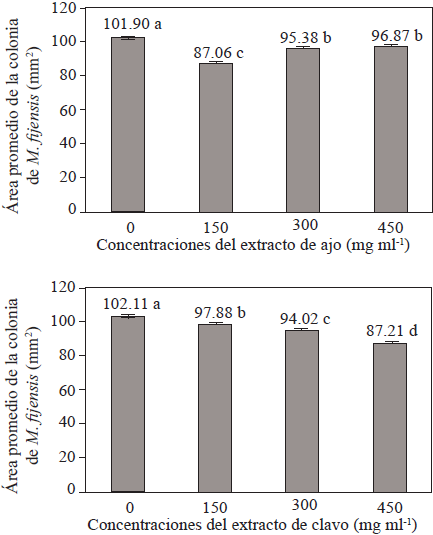
Figure 1 Average growth area of the M. fijiensis colony in ADP culture medium to which diffferent concentrations of garlic (above) and clove (below) were added.
Figure 2 shows the percentage of colony inhibition after four weeks growing in the culture media of the different treatments. All treatments proved to be able to inhibit the development of the M. fijiensis colony (ranging from 21.0% to 39.6%). The highest inhibition percentage was observed in a culture medium containing the smallest amount of garlic extract and the greatest amount of clove extract (Treatment 8).
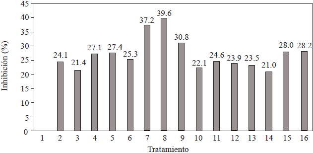
Figure 2 Growth inhibition of the M. fijiensis colony (% area) after four weeks of cultivation in the different treatments shown in Table 1.
When data from the area of the colony (mm2) were converted to Ln for each week of culture, a similar performance was observed in all treatments. Figure 3 shows the extreme treatments, that is, the treatment with the largest areas (Treatment 1) and the one with the smallest areas (Treatment 8). The values of the regression line for all the treatments are shown in Table 4. As can be seen in the table, the slope of the line (m) or colony growth rate (week-1) for all the treatments ranged from 0.7081 week-1 to 0.9177 week-1. Therefore, the colony growth rate of Treatment 10 was 23% slower compared to the growth rate of Treatment 1. As for the intercept, it had an average value of 2.3589 ±0.07675 and, except for Treatment 13, all the determination coefficients were higher than 0.9.
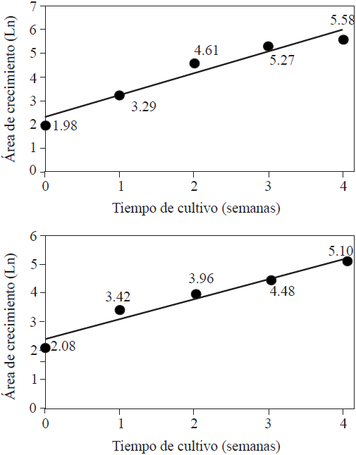
Figure 3 Correlation between the Ln of the M. fijiensis colony in the treatment without garlic and/or clove extracts (Treatment 1, above) and the treatment in which 150 µg of garlic extract ml-1 and 36 µg of clove extract ml-1 were used (Treatment 8; below), and growing time (weeks).
Table 4 Slope values (m) or growth rate (week-1), intercept (b) and determination coefficient (r2) after linearizing Ln data from the colony area (mm2) and the cultivation week.
| Tratamiento | m | B | r2 |
| 1 | 0.92 | 2.31 | 0.94 |
| 2 | 0.85 | 2.33 | 0.91 |
| 3 | 0.82 | 2.46 | 0.93 |
| 4 | 0.82 | 2.36 | 0.93 |
| 5 | 0.82 | 2.38 | 0.92 |
| 6 | 0.82 | 2.47 | 0.91 |
| 7 | 0.81 | 2.32 | 0.92 |
| 8 | 0.71 | 2.39 | 0.95 |
| 9 | 0.82 | 2.36 | 0.91 |
| 10 | 0.83 | 2.42 | 0.91 |
| 11 | 0.86 | 2.27 | 0.93 |
| 12 | 0.81 | 2.45 | 0.91 |
| 13 | 0.83 | 2.44 | 0.89 |
| 14 | 0.88 | 2.21 | 0.94 |
| 15 | 0.86 | 2.27 | 0.92 |
| 16 | 0.85 | 2.29 | 0.9243 |
DISCUSSION
Like many phytopathogenic fungi, M. fijiensis was sensitive to the components of ethanolic garlic and clove extracts. The type of compound and/or its concentration, or flavonoids, phenols, terpenes, essential oils, alcaloids, lectins, polypeptides, among others, could be responsible for this action (Hernández et al., 2007). Oxidation of various compounds caused by phenols, lipophilic action of essential oils, insertion of alcaloids into the DNA, formation of ionic channels in the cell envelope mediated by lectins and competitive inhibition of receptor polysaccharides by polypeptide adherence, are some of the mechanisms that may explain the fungicidal activity (Cowan, 1999; Nonsee et al., 2011; Rahnama et al., 2012).
The higher sensitivity of M. fijiensis to the antifungal action of clove (Figure 1) may also be due to the composition and/or concentration of the compounds it contains. In this regard, essential clove oil, which is miscible in ethanol, contains, among other compounds, eugenol, eugenyl acetate, caryophyllene and a-humulene (Costa et al., 2011; Moura et al., 2012). Garlic is rich in alicin, diallyldisulfide and diallyltrisulfide (Campa-Siqueiros et al., 2017; Chong et al., 2015).
In the beginning, the expectation was that increased concentrations of clove and garlic extracts would increase the level of antifungal activity. This did happen when clove extract was used alone, but exactly the opposite happened when garlic extract was used (Figure 1), a fact that corroborated the results reported by Landero et al. (2013). This might be explained as being a model of competitive inhibition of some enzyme, where as the garlic extract concentration increases, one of its components takes the place of the one that has antifungal activity. Therefore, when the extract is diluted (lower concentration), the competitive component of the antifungal is not able to occupy all the “active sites;” as a result, the inhibition of the development of the M. fijiensis colony was lower. The results of the final phase (data not shown) support what we previously proposed, because when the garlic extract concentration was reduced to 100 mg·ml-1, the antifungal activity increased. In the future, it will be necessary to determine which competitive component (or components) is/are involved and where the action takes place (site(s)).
As for the antifungal activity of the clove extract (Figure 1), it was typical of molecules whose activity increases as their concentration increases. However, it had a maximum value (Figure 4), and, again, this may suggest that it contains an active fungicidal component and that the carrier system has a limited concentration in the M. fijiensis cell envelope.
The fact that no significant differences in the area of M. fijiensis growth were found during the first two weeks of treatment (Table 2) may indicate that a) the action site of the antifungal treatment is intracellular, which means that first it has to be introduced through a transportation mechanism, possibly an active one, and b) the active component is a compound derived from the metabolic process of one of the extract components. In both cases, transportation through the cell envelope seems to be the limiting stage because, once the critical concentration of the antifungal is reached, its impact is expressed as a reduction of growth rate (Figure 3).
The maximum inhibition value expressed when using a mixture of clove and garlic extracts in Treatment 8 (Figure 2) shows a potential synergy between the components of both extracts, since in the treatments containing only garlic (Treatments 5, 9 and 13) or only clove (Treatments 2, 3 and 4) at different concentrations, the average inhibition activity was 27.2% and 24.2%, respectively. Based on the above, it could be assumed that the components of both extracts act independently, a fact that remains to be proven.
Regardless of the above, the maximum inhibition of M. fijiensis population growth caused by the garlic and clove mixture (Treatment 8) obtained in the present study was four times higher than the inhibition reported by Gutiérrez-Jiménez et al. (2017), who used Petri dishes containing ADP to which 50, 100, 500, 1000 and 5000 ppm of essential clove oil were added, and 2.7 to 1.8 times higher than the inhibition reported by Sharanamma (2012), who used Petri dishes containing ADP to which 5%, 10% and 15% of aqueous garlic extract (1 ggarlic·mlH2O -1) was added.
On the other hand, the maximum inhibition is within the range reported for garlic extracts used for controlling diverse phytopathogenic fungi, such as Colletotrichum sp. (García, 2011; Landero et al., 2013; Sundaramoorthy et al., 2014; Cruz et al., 2013; Hernández et al., 2007), Rhizoctonia sp. (Chávez and Aquino, 2012), Fusarium sp. (Taskeen-Un-Nisa et al., 2011; Chávez and Aquino, 2012), Sclerotium sp. (Chávez and Aquino, 2012), Botrytis sp (Daniel et al., 2015; Sesan et al., 2015), Alternaria sp. (Nashwa and Abo-Elyousr, 2012), Aspergillus (Tijjani et al., 2014), and Penicillium (Ikeura et al., 2011), or for clove extracts: Colletotrichum sp. (Radwan et al., 2014).
CONCLUSIONS
As the concentration of alcoholic garlic extract in the ADP medium increases, the inhibition of M. fijiensis colony growth decreases.
As the concentration of alcoholic clove extract in the ADP medium increased, the level of inhibition of M. fijiensis colony growth also increased.
The mixture of alcoholic garlic and clove extracts has a positive synergistic effect that inhibits M. fijiensis colony growth.
In the ADP medium to which 150 mg·ml-1 of alcoholic garlic extract and 36 mg·ml-1 of alcoholic clove extract were added, M. fijiensis growth colony was reduced by 39.6%, so this could be an alternative for controlling the pathogen in the field.
LITERATURA CITADA
Askun T, Tumen G, Satil G and Kilic T. 2008. Effects of some Lamiaceae species methanol extracts on potential mycotoxin producer fungi. Pharmaceutical Biology 46:688-694. https://doi.org/10.1080/13880200802215792. [ Links ]
Bastos CN y Albuquerque PSB. 2004. Efeito do óleo de Piper aduncum no controle em pós-colheita de Colletotrichum musae em banana. Fitopatologia Brasileira 29:555-557. http://dx.doi.org/10.1590/S0100-41582004000500016 [ Links ]
Campa-Siqueiros P, Vallejo-Cohen S, Corrales-Maldonado C, Martínez- Téllez MA y Vargas-Arispuro I. 2017. Reducción en la incidencia de la pudrición gris en uva de mesa por el efecto de volátiles de un extracto de ajo. Revista Mexicana de Fitopatología 35:493-508. http://dx.doi.org/10.18781/R.MEX.FIT.1707-1 [ Links ]
Castillo F, Hernández D, Gallegos G, Rodríguez R and Aguilar CN. 2012. Antifungal properties of bioactive compounds from plants. Pp:81-106. In: Dhanasekaran D, Thajuddin N and Panneerselvam A (ed). Fungicides for Plant and Animal Diseases. InTech Rijeka, Croatia. 258p. Disponible en línea: https://www.intechopen.com/books/fungicides-for-plant-and-animal-diseases/antifungal-properties-of-bioactive-compounds-from-plants [ Links ]
Chávez AR y Aquino AS. 2012. Control de los hongos del suelo Rhizoctonia sp., Fusarium sp. y Sclerotium sp. con extractos vegetales. Investigación Agraria 14:17-23. Disponible en linea: http://scielo.iics.una.py/scielo.php?script=sci_arttext&pid=S2305-06832012000100003 [ Links ]
Chong K, Zamora MP, Tilakawardane DA, Buckley NE, Rego JA and Liu Y. 2015. Investigation of allicin stability in aqueous garlic extract by high performance liquid chromatography method. Journal of Scientific Research and Reports 4:590-598. DOI: 10.9734/JSRR/2015/14301 [ Links ]
Conde-Ferráez L, Grijalva-Arango R, Raigoza-Flores NE and James-Kay AC. 2008. A simple method to obtain single conidium isolates directly from banana (Musa sp.) leaves infected with Mycosphaerella fijiensis Morelet. Revista Mexicana de Fitopatología 26:76-78. Disponible en línea: http://www.redalyc.org/articulo.oa?id=61226112 [ Links ]
Costa ART, Amaral MRZ, Martins PM, Paula JAM, Fiuzza TS, Tresvenzol LMF, Paula JR y Bara MTF. 2011. Ação de óleo esencial de Syzygium aromaticum (L) Merr. y L. M. Perry sobre as hifas de alguns funguos fitopatogénicos. The Brazilian Journal of Medicinal Plants 13:240-245. http://dx.doi.org/10.1590/S1516-05722011000200018 [ Links ]
Cowan MM. 1999. Plant products as antimicrobial agents. Clinical Microbiology Reviews 10:564-582. Disponible en línea: http://cmr.asm.org/content/12/4/564 [ Links ]
Cruz MES, Schwan-Estrada KRF, Clemente E, Itako AT, Stangarlin JR and Cruz MJS. 2013. Plant extracts for controlling the post-harvest anthracnose of banana fruit. The Brazilian Journal of Medicinal Plants 15:727-733. http://dx.doi.org/10.1590/S1516-05722013000500013 [ Links ]
Daniel CK, Lennox CL and Vries FA. 2015. In-vitro effects of garlic extracts on pathogenic fungi Botrytis cinerea, Penicillium expansum and Neofabraea alba. South African Journal of Science 111:1-8. https://doi.org/10.17159/sajs.2015/20140240 [ Links ]
De Hora BR. 2009. Ação de óleos essenciais no controle de sigatoka-negra (Mycosphaerella fijiensis Morelet) de bananeiras (Musa sp). Dissertação (mestrado). Universidade Estadual Paulista, Instituto de Biociências, Botucatu, Brasil. 68p. Disponible en línea: http://hdl.handle.net/11449/88138 [ Links ]
Etebu E and Young-Harry W. 2011. Control of Black Sigatoka disease: Challenges and prospects. African Journal of Agricultural Research 6:508-514. Disponible en línea: http://www.academicjournals.org/article/article1380878095_Etebu%20and%20Young-Harry.pdf [ Links ]
Ewané CA, Chillet M, Castelan F, Brostaux Y, Lassois L, Essoh J, Hubert O, Chilin-Chales Y, Lepoivre P and De Lapeyre L. 2013. Impact of the extension of black leaf streak disease on banana susceptibility to post-harvest diseases. Fruits 68:351-365. Disponible en línea: https://www.pubhort.org/fruits/2013/05/fruits130081.htm [ Links ]
Garcia L. 2011. A comparative study on the antifungal effects of tamarind (Tamarindus indica) and garlic (Allium sativum) extracts on banana anthracnose. Journal of Nature Studies 10:96-107. Disponible en línea: http://www.pssnonline.org/wp-content/uploads/2012/05/96-107-Garcia.pdf [ Links ]
Gutiérrez-Jiménez E, Pedroza-Sandoval A, Martínez-Bolaños L, Samaniego-Gaxiola JA and García-González F. 2017. Effect of natural oils against Mycosphaerella fijiensis under in vitro conditions and detection of active plant chemicals. Revista Mexicana de Fitopatología 36: 141-150. http://dx.doi.org/10.18781/R.MEX.FIT.1707-4 [ Links ]
Hernández AN, Bautista S y Velázquez MG. 2007. Prospectiva de extractos vegetales para controlar enfermedades postcosecha hortofrutícolas. Revista Fitotecnia Mexicana 30:119-123. http://www.redalyc.org/pdf/610/61030202.pdf [ Links ]
Ikeura H, Somsak N, Kobayashi F, Kanlayanarat S and Hayata Y. 2011. Application of selected plant extracts to inhibit growth of Penicillium expansum on apple fruits. Plant Pathology Journal 10:79-84. DOI: 10.3923/ppj.2011.79.84 [ Links ]
Landero N, Nieto D, Téliz D, Alatorre R, Orozco M y Ortiz CF. 2013. Potencial antifúngico de extractos de cuatro especies vegetales sobre el crecimiento de Colletotrichum gloeosporioides en papaya (Carica papaya) en poscosecha. Revista Venezolana de Ciencia y Tecnología de Alimentos 4:47-62. Disponible en línea: http://oaji.net/articles/2017/4924-1495453202.pdf [ Links ]
Malik AA, Ahmed N, Babita, Chauhan H and Gupta P. 2016. Plant extracts in post harvest disease management of fruits and vegetables: A review. Journal of Food Processing and Technology 7:592-597. DOI: 10.4172/2157-7110.1000592 [ Links ]
Manzo-Sánchez G, Orozco-Santos M y Guzmán-González S. 2001. Caracterización morfológica de Mycosphaerella fijiensis Morelet de la región Pacífico-Centro de México y su desarrollo en medios liquidos. Revista Mexicana de Fitopatologia 19:66-71. http://dx.doi.org/10.18781/R.MEX.FIT.1507.8 [ Links ]
Moura J, Sarmento FQ, de Oliveira F, Pereira J, Nogueira V y de Oliveira E. 2012. Actividad antifúngica del aceite esencial de Eugenia caryophyllata sobre cepas de Candida tropicalis de aislados clínicos. Boletín Latinoamericano y del Caribe de Especias Medicinales y Aromáticas 11:208-217. Disponible en línea: http://www.redalyc.org/pdf/856/85622739002.pdf [ Links ]
Nashwa SMA and Abo-Elyousr KAM. 2012. Evaluation of various plant extracts against the early blight disease of tomato plants under greenhouse and field conditions. Plant Protection Science 48:74-79. https://doi.org/10.17221/14/2011-PPS [ Links ]
Nonsee K, Supitchaya C and Thawien W. 2011. Antimicrobial activity and the properties of edible hydroxypropyl methylcellulose based films incorporated with encapsulated clove (Eugenia caryophyllata Thumb.) oil. International Food Research Journal 18:1531-1541. Disponible en línea: http://www.ifrj.upm.edu.my/18%20(04)%202011/(46)IFRJ-2011-043.pdf [ Links ]
Pérez L. 2002. Morfología de las especies de Mycosphaerella asociadas a manchas de las hojas en Musa spp. Fitosanidad 6:3-9. Disponible en línea: http://www.redalyc.org/pdf/2091/209118291001.pdf [ Links ]
Ploetz R. 2000. Black sigatoka. Pesticide Outlook 1:19-23. DOI: 10.1039/B006308H [ Links ]
Radwan MM, Tabanca N, Wedge DE, Tarawneh AH and Cutler SJ. 2014. Antifungal compounds from turmeric and nutmeg with activity against plant pathogens. Fitoterapia 99:341-346. https://doi.org/10.1016/j.fitote.2014.08.021 [ Links ]
Rahnama M, Najimi M and Ali S. 2012. Antibacterial effects of Myristica fragans, Zataria multiflota Boiss, Syzygium aromaticum, and Zingiber officinale Roci essential oils, alone and in combination with nisin on Listeria monocytogenes. Comparative Clinical Pathology 21:1313-1316. Disponible en línea: https://link.springer.com/article/10.1007/Fs00580-011-1287-3 [ Links ]
Sepúlveda L. 2016. Caracterización fenotípica de Mycosphaerella fijiensis y su relación con la sensibilidad a fungicidas en Colombia. Revista Mexicana de Fitopatologia 34:1-21. http://dx.doi.org/10.18781/R.MEX.FIT.1507.8 [ Links ]
Sesan TE, Enache E, Iacomi BM, Oprea M, Oancea F and Iacomi C. 2015. Antifungal activity of some plant extracts against Botrytis cinerea Pers. in the blackcurrant crop (Ribes nugrum L.). Acta Scientiarum Polonorum. Hortorum Cultus 14:29-43. DOI: 10.24326/asphc.2017.6.15 [ Links ]
Sharanamma AR. 2012. Studies of sigatoka leaf spotof banana caused by Cercospora musae. Zimm. [MSc. Dissertation.]. University of Agricultural Sciences. Dharwad. India. Disponible en línea: http://krishikosh.egranth.ac.in/bitstream/1/86799/1/th10491.pdf [ Links ]
Sundaramoorthy S, Usharani S and George AP. 2014. Antifungal activity of plant products for the management of fruit rot infection in chillies. Plant Pathology Journal 13:87-99. DOI: 10.3923/ppj.2014.87.99 [ Links ]
Taskeen-Un-Nisa, Wani AH, Bhat MY, Pala SA and Mir RA. 2011. In vitro inhibitory effect of fungicides and botanicals on mycelial growth and spore germination of Fusarium oxysporum. Journal of Biopesticides 4:53-56. Disponible en línea: https://pdfs.semanticscholar.org/408b/76e3145fbbd6f407129a9c3f56b3359bbe4f.pdf [ Links ]
Tijjani A, Adebitan SA, Gurama AU, Haruna SG and Safiya T. 2014. Effect of some selected plant extracts on Aspergillus flavus, a causal agent of fruit disease of tomato (Solanum lycopersicum) in Bauchi State. International Journal of Biosciences 4:244-252. http://dx.doi.org/10.12692/ijb/4.12.244-252 [ Links ]
Touba EP, Zakaria M and Tahereh E. 2012. Antifungal activity of cold and hot water extracts of spices against fungal pathogens of Roselle (Hibiscus sabdariffa) in vitro. Microbial Pathogenesis 52:125-129. https://doi.org/10.1016/j.micpath.2011.11.001 [ Links ]
Received: May 16, 2018; Accepted: July 17, 2018
 Este es un artículo publicado en acceso abierto bajo una licencia Creative Commons
