SciELO - Scientific Electronic Library Online

 
vol.92 número4Trasplante de páncreas: revisión índice de autoresíndice de materiabúsqueda de artículos
Home Pagelista alfabética de revistas  

Servicios Personalizados

Revista

Articulo

Indicadores

Links relacionados

Compartir


Cirugía y cirujanos

versión On-line ISSN 2444-054Xversión impresa ISSN 0009-7411

Cir. cir. vol.92 no.4 Ciudad de México jul./ago. 2024  Epub 09-Ago-2024

https://doi.org/10.24875/ciru.22000631 

Letters to the editor

Modified Klingler technique: a universal tool for surgical training in neurosurgery residents and specialists

Técnica de Klingler modificada: una herramienta universal para el adiestramiento quirúrgico en residentes y especialistas de neurocirugía

Manuel de J. Uribe-Miranda1  * 

Mateo A. Rodríguez-Delgado2 

Jetzabel A. Amozurrutia-Hernández1 

1Department of Neuroanatomy, Cuauhtémoc University School of Medicine, San Luis Potosí, San Luis Potosí, Mexico

2Laboratory of Morphophysiology, Juan N. Corpas University Foundation, Bogotá Colombia


Dear Editor,

The neurosurgical training of residents and neurosurgery specialists in our country, and the rest of the world, is through dissection in cadavers fixed with formaldehyde, and human brains preserved with the Klingler technique, a technique that in recent years has become a fundamental tool in neurosurgical teaching. In the same way, this technique allows dissection, investigation, and exposure of white matter fibers; such as association fibers, projection fibers, and commissural fibers belonging to the corpus callosum and other commissures. The Klingler technique was created by the German neuroanatomist Joseph Klingler in 1935 as a unique method for the preservation and dissection of white matter fibers, nuclei, and brain stem1. This method consists of 5 stages. Obtaining human brains without alterations in the parenchyma and, with a post-mortem between 24 and 48 h; fixation with 10% formaldehyde for a minimum period of 60 days; dissection of arachnoids and vascular elements with the help of forceps and microdissection scissors; the freezing process for 10 days at a temperature between 15 and 18 degrees; and finally the thawing process for 24 h at room temperature2,3 (Fig. 1). About 10% formaldehyde perfectly penetrates the gray matter, which when frozen forms microcrystals that separate both substances, allowing easy dissection using a wooden spatula2-4. On the other hand, the dissection of white matter fibers using the Klingler technique is not a new technique, its study and understanding are still extremely useful since it gives us the possibility of better understanding and manipulating the internal configuration of the brain, obtaining better neurosurgical training.

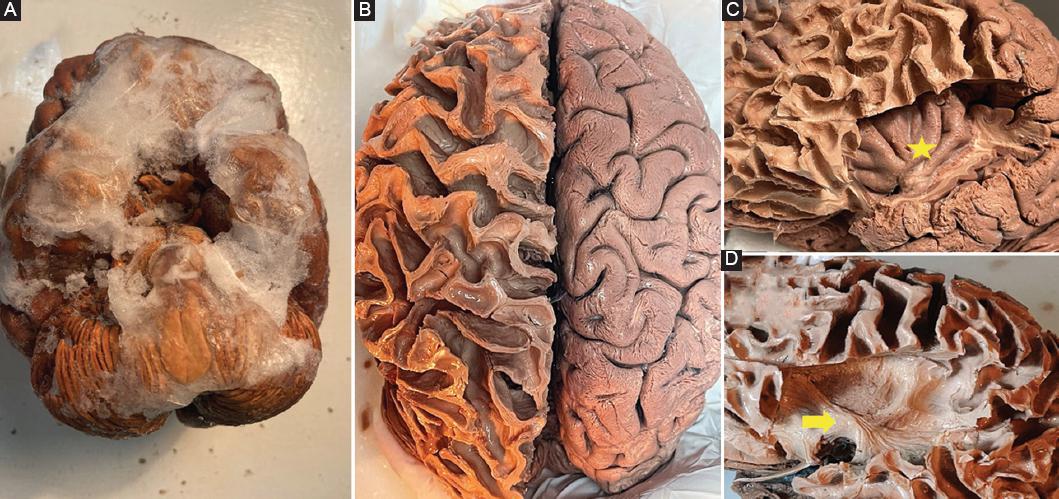
Figure 1 A: ventral side of the brain, previously fixed with 10% formaldehyde and frozen. B: dorsal side of the brain, left hemisphere decorticated and exposing white matter fibers of short association. C: lateral view of the brain, exposing the lobe of the insula (yellow star). D: dissection of the lateral aspect of the brain, exposing the uncinate fasciculus (yellow arrow). 

Without forgetting that to achieve optimal results, a minimum of three fundamentals are needed: (1) possess extensive knowledge of neuroanatomy, (2) possess excellent training and manual dexterity, and (3) patience and perseverance1.

Acknowledgments

Special thanks to the Universidad Cuauhtémoc Plantel San Luis Potosí.

References

1. Pérez JC, Baldoncini M, Ledesma L. Atlas-manual of Encephalon and Encephalic White Matter Dissection Klingler Method. Mexico City:INAV 2.0;2014. Available from:https://matiasbaldoncini.com/images/publicaciones/17_atlas_compressed_1.pdf [Last accessed 2021 Nov 22]. [ Links ]

2. Pérez JC, Gallegos SP, Garduño P, Reyes G, Valderrama MR, Herrera I, et al. Standardization of Klingler method and its tridimensional visualization. Rev Hosp Jua Mex. 2008;75:99-108. [ Links ]

3. Uribe M, Zamarripa C, Salazar J. Basic and Low-Cost Three-Dimensional Model in Cow Brain Using the Klingler Technique. Vol. 13. Ayacucho:Revista Argentina;2022. 19-23. Available at:https://www.revista-anatomia.com.ar/archivos-parciales/2022-1-revista-argentina-de-anatomia-online-b.pdf [Last accessed on 2022 May 27]. [ Links ]

4. Rubino P, Baldoncini M, Conesa H. Cerebral White Matter Dissection Importance for Neurosurgical training. Vol. 3. Ayacucho:Revista Argentina;2012. 130-6. Available from:https://www.revista-anatomia.com.ar/archivos-parciales/2012-4-revista-argentina-de-anatomia-online-f.pdf [Last accessed on 2022 May 29]. [ Links ]

FundingThe authors declare that no funding is available.

Ethical responsibilities

Protection of humans and animals. The authors declare that no experiments on humans or animals have been performed for this research.

Confidentiality of data. The authors declare that no patient data appear in this article.

Right to privacy and informed consent. The authors declare that no patient data appear in this article.

Use of artificial intelligence for generating text. The authors declare that they have not used any type of generative artificial intelligence for the writing of this manuscript or for the creation of images, graphics, tables, or their corresponding captions.

Received: December 18, 2022; Accepted: January 09, 2023

* Correspondence: Manuel de J. Uribe-Miranda E-mail: mdjum93@gmail.com

Conflicts of interest

The authors declare that they have no conflicts of interest.

Creative Commons License Instituto Nacional de Cardiología Ignacio Chávez. Published by Permanyer. This is an open ccess article under the CC BY-NC-ND license